Associate Professor ampActing HOD Dept of Surgery EPIDEMIOLOGY Collectively US India and China account for almost one third of the global breast cancer burden India has a long way to go ID: 912656
Download Presentation The PPT/PDF document "BREAST CANCER Dr. Farhanul Huda" is the property of its rightful owner. Permission is granted to download and print the materials on this web site for personal, non-commercial use only, and to display it on your personal computer provided you do not modify the materials and that you retain all copyright notices contained in the materials. By downloading content from our website, you accept the terms of this agreement.
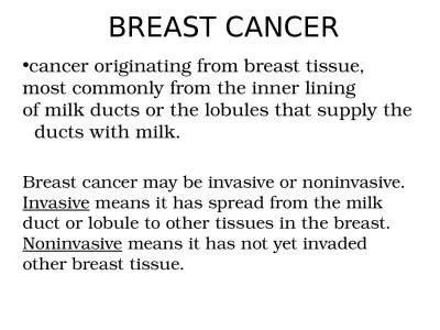
Slide1
BREAST CANCER
Dr. Farhanul Huda
Associate Professor &Acting HOD
Dept.
of Surgery
Slide2EPIDEMIOLOGY
Slide3Collectively, US, India and China account for almost one third of the global breast cancer burden.
India has a long way to go!
See the images below and listen to the discussion and you will understand why.
Slide4Slide5Slide6Slide7?
Why is the mortality so high?
more patients turn up in later stages.
What are the reasons for late presentations?
lack of awareness,
shyness on part of patients,
social stigma,
ignorance of doctors
So what do we learn today?
Slide8WHO prediction for breast cancer in India
For the years 2015, there will be an estimated 1,55,000 new cases of breast cancer and about 76000 women in India are expected to die of the disease. The gap only seems to be widening, which means, we need to work aggressively on early detection.
Slide9RISK FACTORS
Slide10Three main groups:
Major
Intermediate and
Minor
Slide11Major risk factors
Gender
100 times more common in women than in men.
Age
Very rare before the age of 20 and rare below 30 years.
The incidence of breast cancer doubles every 10 years until the menopause.
Previous breast cancer
Family history and genetic predisposition
Slide12Intermediate risk factors
Diet and alcohol intake
Endocrine factors
Increased duration of exposure to endogenous estrogens.
Early age of menarche (age< 12), late age of menopause (> 55), and late age at first pregnancy (> 30),
nulliparity,HRT,OCPs
.
Lifetime number of menstrual cycles.
Irradiation
Slide13Minor and controversial risk factors
Body size
Stress
Slide14Genetics of breast cancer
BRCA 1
BRCA 2
Slide15BRCA-1 is located on chromosome 17q.
BRCA-1–associated breast cancers are invasive ductal carcinomas, are poorly differentiated, and are hormone receptor–negative.
BRCA-2 is located on chromosome 13q .
BRCA-2–associated breast cancers are invasive ductal carcinomas, are well differentiated and express hormone receptors.
Slide16PATHOLOGY
Slide17Why?
Paramount importance in establishing the diagnosis of the
tumour
.
It also helps determine the patient's prognosis
There are many methods of pathologically classifying breast cancer; most are based on whether the
tumour
is
invasive or non-invasive
and whether it is derived from the
duct
system or the
lobule.
Ductal
carcinoma of the breast
Most common form of breast cancer accounting for 85 to 90 per cent of all cases.
Slide19Lobular carcinoma of the breast
subdivided into
in situ and invasive forms
Slide20Clinical scenarios
Slide21A 38 years old lady (with a history of breast cancer in her sister) presented with a 4 cm lump in her right breast which turned out to be a cancer and had a few enlarged
axillary
nodes. She had noticed the lump only a few months back. However, on evaluating all past records, doctor found one mammogram done 2 years back (was advised by her gynecologist), just for screening; she did not have any lump or other symptom then. In that mammogram, there was a small area of stippled
microcalcification
, which was very suspicious (Stippled
microcalcifications
are
pathognomonic
for cancer) . The radiologist had also mentioned it in the report. But since there was no palpable lump, her gynecologist told her, not to worry. She didn't do anything for that for the next 2 years, and finally, was detected with cancer in the same site, in a minimum of clinical stage 2B. Finally after surgery, 5 (out of 27) nodes were positive for cancer and this placed her in stage
3A.
So please understand here, the gynecologist advised the mammogram, but did not
not
know how to interpret or act, and the lady, who would have otherwise been detected with cancer of stage 1 and would have had more than 90% chance of 10 years survival, now ended up with stage 3A and will have about 60% chance of 5 year survival. So two years of wait have definitely decreased her life by 5 years.
Slide22A 32 years old lady presented with a history of heaviness in breast before the periods as well as pain in the breast for a few days before the periods. On clinical examination, breasts were normal, except for slightly engorged. Again here, her family doctor had advised her mammography (I wouldn't have advised her mammography, if at all needed, I would have gone for an ultrasound of the breast first). On the ultrasound which was done with the mammogram, there were multiple cysts of varying sizes in both the breasts, from few
millimetres
to 8 to 9
millimetres
. She was overtly worried about cancer, and had already taken opinion from one surgeon and one gynecologist. One had advised surgery (!!) and the other had given some non specific medications. All the doctor did was to reassure her, that this was nothing to worry about (She was visibly more worried about the cancer than the symptoms of pain and heaviness she had). The doctor assured her that this was not cancer, this did not require surgery, this occurs in many women of her age - some have more symptoms while some have less symptoms, and that over a period of time, it will all settle. Gave her some symptomatic medications and some vitamin supplements and believe me, after three months, she was almost settled of symptoms and was very happy. Not that medications worked or something, but it was the re assurance that worked.
Slide23CLINICAL FEATURES
Slide24A lump
Changes in the skin may be the sole presenting symptom.
Puckering .
Peu
d'orange
.
Ulceration .
Nipple distortion and inversion .
A
unifocal
or bloodstained nipple discharge.
Slide25Diagnosis
Fine-needle aspiration cytology
Core biopsy
Mammography
Slide26TNM definitions
Primary
Tumour
Tx – Primary tumour cannot be assessed
To – No evidence of primary tumor
Tis – Carcinoma in situ
T1 – Tumor 2 cm or less
T2 – 2 – 5 cm tumor
T3 – Tumor 5 cm and above
T4 – Extn. to chest wall / skin
Slide27Regional lymph node involvement - clinical
NX – Regional lymph nodes cannot be
assessed.
No – No regional lymph nodes.
N1 – Movable ipsilateral axillary nodes.
N2 – Fixed ipsilateral axillary nodes.s
N3 – Ipsilateral internal mammary nodes
Slide28Regional lymph node involvement - pathological
pN
X
–
Regional
lymph nodes cannot be assessed.
pNo
– No regional lymph node metastasis.
pN1 – Movable
ipsilateral
axillary
node metastasis.
pN1a –
Micrometastases
(< 0.2 cm )
pN1b – Metastases ( > 0.2 cm )
i
) 1 – 3 nodes
ii) 4 or more nodes
iii) extending beyond the capsule (< 2 cm)
iv)Metastases to nodes ( > 2 cm )
pN2 - Fixed
ipsilateral
axillary
nodes
pN3 –
Ipsilateral
internal mammary nodes
Slide29Distant Metastases
Mx – Distant metastases cannot be assessed.
Mo – No distant metastases.
M1 – Distant metastases ( ipsilateral
supraclavicular lymph nodes )
Slide30AJCC / UICC Stage grouping
St 0 - Tis No Mo
St 1 – T1 No Mo
St 2a
To N1 Mo
T1 N1 Mo
T2 No Mo
St 2b
T2 N1 Mo
T3 No Mo
Slide31AJCC / UICC Stage grouping
St 3a
To N2 Mo
T1 N2 Mo
T2 N2 Mo
T3 N1 Mo
T3 N2 Mo
St 3b
T4 any N Mo
any T N3 Mo
St 4
any T any N M1
STAGING
The Manchester system (1940)
Stage I
.
Tumour
confined to breast. Any skin involvement covers an area less than the size of the
tumour
.
Stage II
.
Tumour
confined to breast. Palpable, mobile
axillary
nodes.
Stage III
.
Tumour
extends beyond the breast tissue because of skin fixation in an area greater than the size of the
tumour
or because of ulceration.
Tumour
fixity underlying fascia.
Stage IV
. Fixed
axillary
nodes,
supraclavicular
nodal involvement, satellite nodules or distant metastases.
Slide33MANAGEMENT
Slide34Management of non-invasivebreast cancer
Stage 0
Slide35LCIS
Because LCIS is considered a marker for increased risk rather than an inevitable precursor of invasive disease, the current treatment of LCIS is
observation with or without
tamoxifen
.
The goal of treatment is to prevent or detect at an early stage the invasive cancer.
There is no benefit to excising LCIS, as the disease diffusely involves both breasts and the risk of invasive cancer is equal for both breasts. The use of
tamoxifen
as a risk-reduction strategy
should be considered in women with a diagnosis of LCIS.
Slide36DCIS
Women with DCIS and evidence of widespread disease (two or more quadrants) require mastectomy.
For women with limited disease, lumpectomy and radiation therapy are recommended.
Low-grade DCIS of the solid,
cribriform
, or papillary subtype, which is less than 0.5 cm in diameter, may be managed by lumpectomy alone.
Adjuvant
tamoxifen
therapy is considered for all DCIS patients.
Slide37Simple mastectomy
95% cure rate
Rarely relapse, due to micro-invasive cancer
No need for
axillary
dissection
Wide excision alone
—30% recurrence at 5 years
Wide excision + radiotherapy
—15% recurrence at 5 years
Slide38Early Invasive Breast Cancer
Stage I,
IIa
, or
IIb
T1–3, N0–1 tumors.
Slide39Treatment of the breast and
axilla
Pathological staging to direct adjuvant therapy
Adjuvant therapy—endocrine, chemotherapy, radiotherapy
Follow-up
Slide40Breast surgery
Quadrantectomy
removes the primary cancer with a margin of 2.0 cm of normal breast tissue.
Lumpectomy
is the removal of the
tumour
mass with a limited portion of normal tissue (1 cm).
MRM
Slide41INDICATIONS OF BCS
T1,T2lesions, N0/N1,M0 disease.
Tumor>4cm in a large breast.
Single clinical and mammographic lesion.
Patient should be willing
tomaccept
the chances of recurrence.
Slide42CONTRA INDICATIONS OF BCS
T4,N2 Lesions
Patients choice
Multifocal/
Multicentric
disease
Tumor size high as compared to breast size.
Extensive calcification on mammography
Pregnancy
Persistent positive margins
Patient’s contraindication to radiotherapy.
Slide43Treatment of the axilla
Surgery
—sentinel node biopsy:
—removal of first node which contains secondary deposit
—use either blue dye or 99MTc colloid
—negative sentinel node avoids clearance
Slide44Loco-regional radiotherapy
Reduce the risk of local recurrence after BCS
Irradiation of axilla—not required if clearance performed
Radiation to
axilla
may cause
lymphodema
and brachial neuropathy
Slide45Adjuvant endocrine therapy
60% of breast cancers are
oestrogen
receptor positive
Ovarian ablation
Side-effects of tamoxifen—menopausal symptoms
—endometrial cancer, 4-fold increase in risk
LHRH agonists
Slide46Adjuvant chemotherapy
CMF (cyclophosphamide, methotrexate, 5FU)
Anthracycline
regimes may be better
Taxanes
based regimes
Slide47Management of locally advancedbreast cancer
Stage
IIIa
or
IIIb
Slide48The probability of metastatic disease is high (>70%).
A combination of
neoadjuvant
chemotherapy, surgery and radiotherapy is commonly used.
Slide49Management of metastatic
breast cancer
Aim is palliation
If hormone-sensitive, bony disease—may survive years .
Visceral, ER-negative disease has bad prognosis
Usual sites—lung, liver, bone, brain
Rare sites—choroid, pituitary
Combination of endocrine therapy, chemotherapy, radiotherapy and symptomatic
tt
is given.
Slide50SENTINAL LYMPH NODE BIOPSY
Slide51SENTINEL NODE
CONCEPT
Based on the hypothesis lymph flow is orderly
, predictable
&
tumor
cells spread sequentially
Sentinel node is the first node encountered by the
tumour
cells
The sentinel node is in the direct pathway of the primary
tumour
Slide52Advantages of sentinel node biopsy
Minimally Invasive
Low Cost
low morbidity
Nodal metastasis outside
axilla
detected
obviates the need for ALND without compromising staging & local control
Slide53Disadvantages of Sentinel node Biopsy
Has a False negative rate of 6% (ALND3%)
Not useful in clinically involved
axilla
Not useful in pregnancy & lactation
Cannot be done in multifocal /
multicentric
breast carcinomas
Cannot be done in patients with previous breast surgery on the same side
Slide54Technique
Blue dye
isosulfan
blue (or)
technitium
labelled
colloidal albumin with gamma camera and probe can be used
Slide55Sub dermal injection
A single dose of 0.2 ml of the dye is injected at the
tumour
site sub-
dermally
one day prior to surgery
Peri
tumour
injection
Dye injected at four sites.
Larger volumes are given
Removal of dye or tracer is slower due to
scanty lymph supply of breast parenchyma
imaged 1 to 2 hrs after injection
Slide56SENTINEL LYMPH NODE DISSECTION
WITH DYE TECHNIQUE
Blue lymphatics leading to SLN are traced
Discolouration
of breast and blue urine
ISOTOPE TECHNIQUE
Probe guided surgery is superior
Useful for intra-operative
localisation
After removal of SLN probe is reapplied to site and radioactivity measured for confirmation
Slide57PIT FALLS IN SENTINEL NODE DISSECTION
6% FALSE NEGATIVE
SKIP PHENOMENON & CHANGED FLOW DIRECTION
INFILTRATION BY CARCINOMA
FATTY DEGENERATION
UPPER OUTER QUADRANT -CLOSE PROXIMITY TO SENTINEL NODE. SHINE THROUGH PHENOMENON-Breast to be retracted when probing
Slide58Special problems
Slide59SPECIAL PROBLEMS IN BREAST CANCER – PAGETS DISEASE
Rare before 30 years
, peak
between 50 & 60
Can occur in the male
Erythematous exudative or scaly lesion
appears first on the nipple spreads to areola
Does not involve surrounding skin
Nipple retraction & nipple pigmentation & mass
Slide60D
D
for
Pagets
disease
Chronic Eczema
Malignant melanoma
Syphilitic chancre
Bowens disease
Mammary
ductectasia
Slide61Mammography
Mass , sub areolar micro calcification
or only thickening of nipple areola
complex
Biopsy
Full thickness nipple biopsy or
exfoliative
scrape cytology
Slide62PAGETS TREATMENT
1) with palpable mass-
segmentectomy
with 1.5 cm margin
with ALND with PO-RT
2)if resection margins positive or
muticentric
or solid or
comedo
type or high grade with necrosis
completion mastectomy is done
Slide63Pagets
without palpable mass
Biopsy of nipple areola complex positive
first step: On
mammo
no occult mass.no
microcalcification
—do
segmentectomy
of nipple areola complex +RT without axillary dissection
Mammography +
ve
Stereotactic needle
localisation
of occult mass
or
microcalcification
with frozen section biopsy and proceed
Tamoxifen
Slide64BREAST CANCER IN PREGNANCY&
LACTATION
DELAY IN DIAGNOSIS
1
) firm ,nodular &hypertrophied breast
2) small
tumours
can be missed
3) present at advanced stage
4) high proportion of ER-
ve
5) bad prognosis
Slide65BREAST CANCER IN PREGNANCY
Mammography
FALSE NEGATIVE rate is high
due to high radiographic density of
pregnant breast
Slide66BREAST CANCER IN PREGNANCY
Alkaline phosphatase is elevated in pregnancy
Chest X-ray is allowed with proper shielding
Bone scan
A) Stage 1 & 2-Bone mets uncommon
scan not done
B)Stage 3 Especially with bone pain
Bone scan done in later stages of pregnancy or after pregnancy
Slide67BREAST CANCER IN PREGNANCY
Treatment
Modified Radical Mastectomy
is the choice irrespective of the trimester
In the first & second trimester breast
conservation with radiotherapy should not be
done due to radiation induced anomalies
in
foetus
Slide68Study questions
Slide69A 57-year-old woman undergoes core-needle biopsy of a breast mass. The
pathologic diagnosis is infiltrating
ductal carcinoma of the breast
.
How will you stage
this cancer
?
What are the important prognostic factors?
Slide70A 49-year-old woman presents with a breast mass. You are examining the
affected breast
.
◆
How would the following clinical
findings
affect the woman’s prognosis?
1. Red
edematous
breast with an underlying mass
Edema
of the skin overlying the
mass
Puckering of
the skin overlying the
mass
Retraction of the
nipple
A 1.5-cm mass fi
xed
to the deeper
tissues
A lymph node palpable in the supraclavicular
area
A hard, fi
xed
lymph node in the ipsilateral
axilla
Arm
edema
Slide71A 60-year-old woman has breast cancer and undergoes preliminary staging. The lesion
is 1.5
cm in diameter, and no axillary nodes are palpable. A metastatic workup is negative
.
What stage is this woman’s cancer
?
What are this woman’s surgical options, both for sampling the lymph
nodes and
treating the primary
tumor
?
Slide72A 38-year-old woman is scheduled for a mastectomy and sentinel node biopsy.
She is
concerned about her appearance and would like to know her options for
breast reconstruction.
What options should you offer?
Slide73A 38-year-old woman presents with a 3-month history of a progressively
enlarging breast
mass. At the time she sees you, she has a 6-
7-cm
fi
xed
mass, with
erythema and
edema
on the upper, outer aspect of her right breast. Clinically, her
axilla is
positive with enlarged,
firm
lymph nodes
.
What is the suspected diagnosis
?
What histologic features are typical of this condition
?
The
surgeon
confirms
the physical
findings
and obtains a punch biopsy of the
mass. Pathology
reveals
inflammatory
carcinoma.
Estrogen
and progesterone
receptors are
negative
.
What is the recommended treatment?
Slide74A 55-year-old woman has a
modified
radical mastectomy for a stage II carcinoma
of the
breast
.
A small, 0.5-cm nodule in the suture line 5 years
after surgery.
A mammographic abnormality in the opposite
breast
Elevated liver function
studies
A fracture of the femur