/
ardiac output monitoringThomas Lawson and Andrew Hutton Correspondence ardiac output monitoringThomas Lawson and Andrew Hutton Correspondence

ardiac output monitoringThomas Lawson and Andrew Hutton Correspondence - PDF document

deena
deena . @deena
Follow
342 views
Uploaded On 2022-08-21

ardiac output monitoringThomas Lawson and Andrew Hutton Correspondence - PPT Presentation

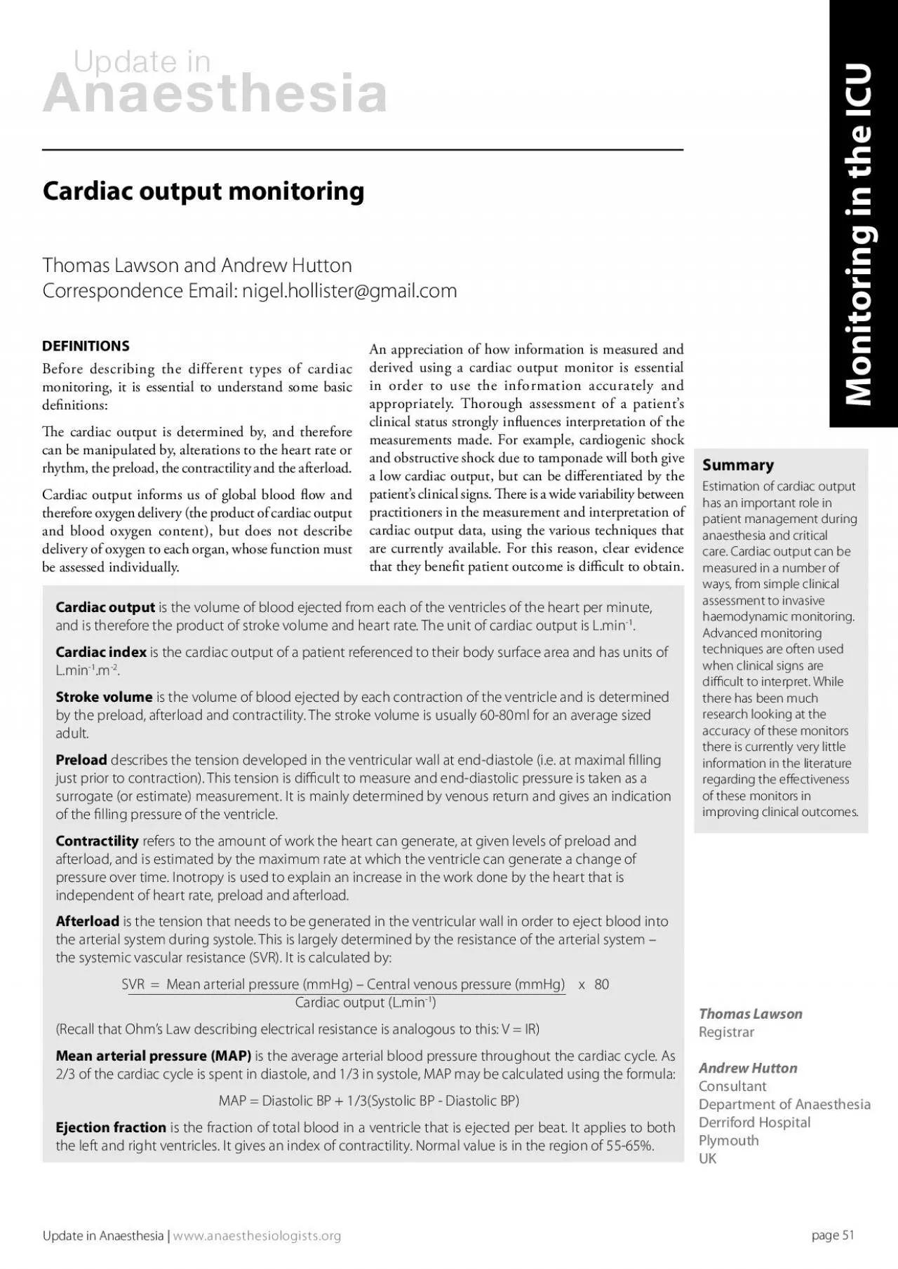
Before describing the different types of cardiac 30e cardiac output is determined by and therefore can be manipulated by alterations to the heart rate or rhythm the preload the contractility an ID: 939111

output cardiac volume blood cardiac output blood volume oxygen 146 pulmonary patient uid pressure catheter artery measured venous pulse

Share:

Link:

Embed:

Download Presentation from below link

Download Pdf The PPT/PDF document "ardiac output monitoringThomas Lawson an..." is the property of its rightful owner. Permission is granted to download and print the materials on this web site for personal, non-commercial use only, and to display it on your personal computer provided you do not modify the materials and that you retain all copyright notices contained in the materials. By downloading content from our website, you accept the terms of this agreement.


Presentation Transcript

ardiac output monitoringThomas Lawson and Andrew Hutton Correspondence Email: nigel.hollister@gmail.com Before describing the different types of cardiac e cardiac output is determined by, and therefore can be manipulated by, alterations to the heart rate or rhythm, the preload, the contractility and the afterload.Cardiac output informs us of global blood ow and SummaryEstimation of cardiac output has an important role in patient management during anaesthesia and critical care. Cardiac output can be measured in a number of ways, from simple clinical assessment to invasive haemodynamic monitoring. Advanced monitoring techniques are often used Monitoring in the An appreciation of how information is measured and ardiac output is the volume of blood ejected from each of the ventricles of the heart per minute, and is therefore the product of stroke volume and heart rate. The unit of cardiac output is L.minardiac index is the cardiac output of a patient referenced to their body surface area and has units of Stroke volume is the volume of blood ejected by each contraction of the ventricle and is determined CLINICALINDICATOCADIACe interpretation of data from invasive haemodynamic monitoring is made in light of the clinical examination. No single clinical sign can be used to make an accurate assessment of cardiac output. Heart rate, blood pressure, pulse strength at various sites, patient colour, respiratory rate and core to peripheral temperature gradient all give an indication to a patient’s haemodynamic status. Note that although blood pressure is often used as an indicator of cardiac output, it is frequently unhelpful. Blood pressure may be maintained by intense peripheral vasoconstriction in the face of a perilously low cardiac A patient’s ability to compensate for a haemodynamic insult is highly variable, depending on age, premorbid status and other comorbidities. An example is the rise in the diastolic pressure in early hypovolaemic shock, associated with peripheral vasoconstriction that is usually only seen in young, t individuals. In addition, clinical parameters such urine output, capillary rell time and cognitive function give a guide to end organ perfusion. Change in heart rate, blood pressure and central venous pressure in response to a straight leg raise is useful to predict a patient’s response to a uid bolus.Measurement of lactate and base decit in arterial blood and, in particular, the trend of these variables over time gives non-specic information about a patient’s organ perfusion. Lactate is produced by anaerobic metabolism, and is an indicator of tissue hypoperfusion. It is measured on most modern blood gas machines. It can be used to monitor therapy, as it will fall as oxygen delivery improves, and as liver perfusion (which enables lactate metabolism) increases.e oxygen saturation in central venous blood (ScvO) also gives a global indication of haemodynamic status, is useful in directing uid and is a reliable surrogate of mixed venous oxygen saturation (see under pulmonary artery otation catheters, below). improves with stretching of the ventricular muscle bres, to a certain optimal point beyond which further stretching impairs performance (see Figure 1). To apply this strategy we would like to know the left ventricular end-diastolic volume (LVEDV) and monitor changes in the LVEDV as we give 

1;uid boluses. e best surrogate estimate of LVEDV we have is to use a pulmonary artery catheter (PAC) to measure pulmonary artery occlusion (‘wedge’) pressure, which gives us an estimate of left atrial pressure, which is in turn and estimate of LVEDP, which is a surrogate of LVEDV (and makes assumptions about normal compliance of the left ventricle). is is not a reliable measure of lling, particularly given the eects of ventilation, applied PEEP and the anatomical location of the catheter tip in dierent lobar pulmonary artery branches. ermodilution using the PAC does provide an accurate measurement of cardiac output, which can be measured continuously given the correct equipment, however use is diminishing in many parts of the world, due to concerns over safety and lack of robust evidence to support their use.Update in Anaesthesia | www.anaesthesiologists.orgLearning point – blood pressure is a poor indicator of cardiac OVECADIACONITOMortality in sepsis increases by 15-20% for each ‘organ failure’ that a patient develops. Organ failure results when delivery of oxygen is inadequate for the organ’s requirements. Since the 1980s, research has suggested that optimisation of oxygen delivery (a product of cardiac output and blood oxygen content) in high risk surgical patients prevents organ failure and improves mortality.investigated early in critical illness, and prior to, during and after surgery. Although no single study provides categorical evidence, the weight of evidence suggests that therapies directed at enhancement of oxygen delivery (goal-directed therapy) should be our aim. ere is also increasing evidence that, while hypovolaemic septic patients need uid to optimise their cardiovascular delivery of oxygen, excessive e major factor limiting this eld of clinical practice has been development of a monitoring device that will reliably and accurately guide our use of uid therapy - to recognise where uid is needed and give enough, but not too much. Measurement of ‘lling’ is dicult. We aim to apply Starling’s Law, where cardiac performance Figure 1.Simplied explanation of Starling’s Law.Currently, the main focus of research and development is towards less invasive monitors with inherently lower risks of use. Broadly these are monitors that use Doppler analysis of the aortic blood velocity (viewed from the oesophagus) or monitors that analyse the shape of the arterial waveform (‘pulse contour analysis’). Some of the cardiac output monitors that rely on arterial waveform analysis, use thermo- or indicator dilution to obtain an accurate estimate of cardiac output, which can then be used to calibrate continuous analysis of the waveform, transduced from a modied arterial catheter. In order to make these easier to set up and use, more recent models calibrate their pulse contour analysis using population data, based on age, weight and height. e disadvantage is that the population data is derived from healthy volunteers, and so is not validated for patients with abnormal vascular resistance, which undoubtedly has a major eect on derived indices such as stroke volume. e oesophageal Doppler also uses population data to estimate aortic diameter.However, even if we are sceptical about the absolute numbers generated, these monitors can be reliably use

d to observe trends in stroke volume, and the eect of interventions such as uid administration. e key feature is to determine whether the patient is uid responsive; meaning Update in Anaesthesia | www.anaesthesiologists.orgthat a bolus of uid augments their cardiovascular performance (for example their stroke volume), thereby improving oxygen delivery. Fluid responsiveness implies that we have moved the patient up the Starling curve.A current and future area of development is the use of stroke volume variation (SVV) or pulse pressure variation (PPV) that is measured from the transduced arterial waveform. We have long observed that hypovolaemia causes an exaggerated swing in systolic pressure during the respiratory cycle; SVV and PPV quantify this swing or variation as a single number. Again, it is a change in the number, rather than the absolute value that is useful in assessing the uid responsivenesss of your patient.From a pragmatic perspective, these monitors are most useful when observing the eect of a single intervention (such as uid administration) in isolation - this is often dicult during the changing stimuli of surgery, or when the physiological response to sepsis is changing rapidly. Measurements are most plausible when interventions and pre- and post- stroke volume, SVV or PPV measurements are performed during a ‘lull’ in other stimulating activity.ULTOCAltrasoundUltrasound is any high-frequency sound wave. Ultrasound is used medically to create a 2 dimensional image by using a probe to transmit high-frequency sound waves (1-5MHz) into the body, and to detect the waves as they are reected o the boundaries between tissues interfaces. By using a mathematical model involving the speed of sound and the intensity and timing of each echo’s reection, the distance from the probe to the tissue boundaries is calculated, and used to create a oppler ultrasoundWhen sound waves are reected from a moving object, their frequency is altered. is is the Doppler eect. By using an ultrasound probe to visualise directional blood ow, the phase shift (i.e. the change in frequency before and after reection o moving red blood cells) can be determined. is, together with the cross-sectional area of the blood vessel being observed (measured or estimated) can be used to determine ow, where:Flow = area x velocityTheory of techniqueA Doppler probe is inserted into the distal oesophagus (Figure 2) and is directed to measure the blood ow in the descending aorta at about 35 to 40cm from the incisors. e monitor calculates cardiac output using descending aorta diameter, which is either obtained from an age-related nomogram or measured directly in newer machines. e ventricular ejection time, corrected for heart rate (the corrected ow time, FTc), gives an indication of preload and the peak ow velocity (PV) estimates the contractility of the ventricle. Newer probes incorporating M-mode Doppler measurement may improve accuracy and reliability.Practical applicatione technique is straight-forward, easily learned and relatively non-invasive. e disposable probes are easy to insert, however some expertise must be gained in recognition of intracardiac and pulmonary artery signals. Continuous measurement is possible, although frequent positional adjustments are needed. Some use

r variability is inevitable. e cardiac output data is best used as a trend to guide the eectiveness of interventions such as uid challenges.Wave form interpretationA full description of the use of oesophageal Doppler is beyond the scope of this article but guidance can be obtained from the NHS Technology Adoption Centre at http://www.ntac.nhs.uk/searchresent.aspx?search=cardioQAdvantages• Minimallyinvasive• Minimalinterference• Quicklyinserted• Littlerequired• �erelativelyportable• Paediatricprobesareavailable.Disadvantages• Mayrequire• User• Interferencefrominstruments• Dependsprobe• Probevesselsintracardiac/intrapulmonary• Assumespercentagecardiac(approxenters the descending aorta. May therefore be inaccurate in a hypovolaemic patient where ow may be redirected to the cerebral circulation.• Contraindicatedpresencevarices.ransthoracic echocardiographyEchocardiography is cardiac ultrasound and can be used to estimate cardiac output by direct visualisation of the contracting heart in real time. Echocardiography is becoming widely accepted as one of the safest and most reliable cardiac output monitors in the critically ill. A focused echocardiogram can be performed in a matter of minutes and assist in determining the cause of haemodynamic instability. Using transthoracic echocardiography four views are obtained (parasternal long axis, parasternal short axis, apical, and subcostal), and it is possible to make an assessment of ventricular function and size of cardiac Update in Anaesthesia | www.anaesthesiologists.orgransoesophageal echocardiographyTheory of techniqueA specialized probe is inserted into the oesophagus, providing real-time, high resolution ultrasound images. Both qualitative and quantitative values for cardiac output are available, using a two dimensional cross-sectional area measurement, a Doppler ow measurement at that point and the heart rate. Practical applicationA multiplane transducer is inserted into the oesophagus and stomach, where various standardized views are gained. AdvantagesA large amount of haemodynamic information is available beyond just cardiac output. Figure 2 Image of descending aortic waveform obtained using an oesophageal Doppler probe, CardioQ® (Courtesy of Deltex Medical).Summary of variables obtained from oesophageal Doppler D Interpretation Peak velocity The highest detectable aortic ow – can be used as a measure of afterload, vascular resistance and contractilitySlope of upstroke Mean acceleration Measure of contractilityWidth of base Flow time Left ventricular ejection time, i.e. duration of aortic blood ow. When corrected for heart rate gives an index of preload (e.g. if base is narrow suggests hypovolaemia)Area under waveformStroke distance Distance a column of blood travels along the aorta during each curve ventricular systoleStroke distance Stroke volume Aortic cross-sectional areaAfterload Shown by a reduction in waveform height and base Disadvantages e probes are still expensive and the machinery is large and bulky. Various levels of examination skill are required and these take time and resources to learn. A full study can take over twenty minutes. Some form of local pharyngeal anaesthesia or sedation is required to tolerate the probe. ere is a risk of trauma from the probe, although the risks are low in

patients with no oesophageal disease. e probes generate a degree of heat and are therefore not suited to continuous measurement. As the technology advances and costs decrease, TOE may nd more applications in theatre and the ICU.DILUTIONETese techniques require:• amarkerremainwithin the circulatory system, and is minimally metabolised. • aveinmarkera peripheral artery (from which the arterial content of the substance can be measured) must be cannulated. As long as blood ow between the injection and measuring sites is constant, ow (i.e. cardiac output) can be calculated from the area under a concentration versus time graph, using a modied Stewart-Hamilton equation.Advantages of dilution methods• LessinvasivePAFCbelow).Disadvantages of dilution methods• Cancardiacventilated• Speci�cheart-lungrequiredstroke volume variation (SVV) and pulse pressure variation (PPV)• Invasivemortality.• User• Canunderestimatecardiaclowilution Monitoring – idco® and COidcoplus®Theory of techniqueis technique combines the techniques of lithium dilution (Lidco and Lidcoplus) and pulse contour analysis (PulseCO). A small dose of lithium is injected into a peripheral vein and an ion selective electrode is attached to a peripheral arterial line. e area under the curve of a plot of lithium concentration against time allows calculation of the cardiac output. is information is then used to calibrate the PulseCO which provides ‘beat-to-beat’ cardiac output measurement, using pulse of the arterial waveform.Practical applicatione convenience of this system is that it uses catheters which are likely to be in place or are likely to be needed in a critically ill patient. e system requires some familiarity to set up, but is relatively quick. e total dose of lithium is small and is clinically insignicant. Calibration is recommended every 8 hours, or after any signicant change in the patient’s clinical condition. AdvantagesA gure for stroke volume variation is produced and provides an indicator of volume responsiveness to uid therapy. Disadvantageshave recently received vecuronium or atacurium. e monitor performs poorly in the presence of atrial brillation and other tachyarrhythmias. e system is prone to technical diculties related to damping and resonance within the measurement system (see page 38).hermodilution pulse contour monitoring – CCOTheory of techniqueis technique utilises thermodilution in combination with Pulse Contour Analysis (PulseCO) to measure cardiac output, and correlates well with the PAFC (below). ‘Stroke volume variation’ (the mean dierence between the highest and lowest arterial pressure wave peaks over 30 seconds) gives an indication of the blood volume status of e system is calibrated using intermittent cold transpulmonary thermodilution, where cold uid is injected through a central venous catheter and traverses the pulmonary circulation. A curve of blood thermodilution is measured in a systemic artery and, in addition to cardiac output, other data is derived. e calculated extra-vascular (EVLW) gives an indication of the water content of the lungs and is increased in left ventricular failure, pneumonia and sepsis. e normal range is 3-10ml.kg-1 and values greater than 14ml.kg-1 are associated with an increased

mortality. e intra-thoracic blood volume index gives an indication of blood volume status (normal value 850-1000ml.mPiCCOplus replaced the original PiCCO machines in 2002 and has subsequently been replaced by PICCO with improved displays, automated features and the use of room temperature injectate for Update in Anaesthesia | www.anaesthesiologists.orgFigure 3Concentration-time graph for determining cardiac output. Practical applicationA specialised arterial catheter, inserted into either the brachial artery or femoral artery is required, along with either a thoracic or femoral central line. Some centres use treatment algorithms based on these variables, to guide use of uid and inotropes in an attempt to maximise intravascular lling, without increasing the EVLW and causing pulmonary oedema. e use of EVLW as an endpoint for resuscitation has not been validated.Advantagese arterial line can be simultaneously used for blood pressure monitoring and for blood sampling. e system is relatively easy to set up and calibrate. It can also be used to estimate preload using global-end-diastolic volume and index (GEDI), intra-thoracic blood volume (ITBV) and pulmonary vascular permeability index (PVPI) which gives a ratio of EVLW to pulmonary blood volume. Note that pleural eusions do not aect measurements.Disadvantagese arterial catheter is relatively large gauge and expensive, although few complications have been reported. Recalibration is required every 12 hours, or following a major change in the patient’s clinical condition. Variations in speed of injection and thermistor positioning may aect results. Results can be aected by arrhythmias, shunting, positive pressure ventilation and tricuspid regurgitation.ulse contour analysisProAQT (Pulsion), Vigileo (Edwards Lifesciences) and LIDCOrapid (LIDCO) are all similar, minimally invasive cardiac output monitors. ey all work by pulse contour analysis using a specialised transducer on any arterial line. Parameters obtained may include: continuous cardiac output, stroke volume, stroke volume variation (SVV) and pulse pressure variation (PPV). In order to obtain SVRi (the systemic vascular resistance index), the patient needs CVP monitoring. To obtain values for PPV and SVV, the patient should be ventilated with a xed tidal volume and so is less useful when weaning respiratory support in intensive care. dP max gives an indication of contractility.ulmonary artery otation catheters (PAe use of PAFCs has been hotly debated in recent years and use in the United Kingdom is currently low. e PAC-Man trial showed no improvement in survival for patients randomised to have a PAFC inserted, compared to those who were not.Theory of techniqueA exible balloon-tipped, ow-directed catheter is inserted via a wide-bore catheter sited in a central vein. e catheter is ‘oated’ through the right atrium and ventricle to enter the pulmonary trunk. From this position it can intermittently be ‘wedged’ in one of the pulmonary arteries. e catheter allows a number of variables to be measured and others to be derived.e measured variables are pulmonary artery pressure, pulmonary capillary wedge pressure (PCWP), cardiac output and mixed venous oxygen saturation. Traditionally, cardiac output is measured by thermodilution of 10ml iced water, injected through

the proximal lumen of the catheter. Measurement of the fall in blood temperature against time from injection, as the cooled blood passes the distal end of the catheter, allows calculation of the cardiac output of the right (and therefore the left ventricle). Semi-continuous cardiac output measurements are now available which use warming coils in the right ventricular portion of the catheter. A sequence of heating and recording gives an averaged cardiac output after a short delay. Practical applicatione catheter is inserted with reference to certain waveforms seen in the right atrium, right ventricle, pulmonary outow tract and when wedged in the pulmonary artery. Insertion may take several attempts and is more dicult in patients with a low cardiac output.AdvantagesMeasurement of cardiac output is probably the most reliable of the variables measured using a PAFC and is therefore a valuable guide to interventions introduced to increase cardiac output. e numerous assumptions made in interpretation of the PCWP as a measure of preload or ventricular lling make the PCWP a less reliable measurement. Some units use the mixed venous oxygen saturation, measured using a sample taken slowly from the pulmonary artery aperture of the catheter, as a further indicator of a patient’s overall tissue perfusion (see below). Interpretation of SvO readings.nterpretation Increased O delivery e.g. high FiO Decreased O utilization e.g. sepsis causing shunt Maybe normal or reect compensation by increase O extraction by tissues demand is greater than supply Implies tissue beyond maximal O extraction Severe lactic acidosis Cellular death Update in Anaesthesia | www.anaesthesiologists.org DisadvantagesThis invasive monitor is associated with a number of potential complications. e PAC-Man study recorded non-fatal complications in 10% of insertions. In addition to the usual complications of central venous access, PAFCs may cause arrhythmias, heart block, rupture of the right heart or pulmonary artery, thromboembolism, pulmonary infarction, valvular damage, endocarditis.Mixed venous oxygen saturation (SvMixed venous oxygen saturations can be used as a surrogate marker of the global balance between oxygen delivery and consumption. Oxygen delivery depends on cardiac output and the oxygen content of the blood. In the face of an increased demand for oxygen, there will be a greater degree of oxygen extraction. Occasionally SvObe increased in severe sepsis due to decreased extraction resulting from shunting (where blood bypasses the tissues). SvO can be used as an early warning system where a sudden decrease in SvO20% requires immediate assessment. SvOtreatment.entral venous oxygen saturation (ScvMeasurement of ScvO requires a central venous catheter rather than a pulmonary artery catheter. ScvO can be used as a surrogate marker of the regional balance between oxygen delivery and consumption in the head, neck and upper body. e value is usually 2-7% less than SvO – partly due to mixing with returning venous blood. Under non- correlates well with SvO. In shock states the dierence from SvO increases – and can be up to 7% higher than SvO trends with SvO in a parallel manner but should be used in combination with other markers of perfusion.CeVox (PULSION) is a system which monitors continuous SvO and can calculate oxygen delivery, consumption and oxygen extraction.

It uses a breoptic probe that can be inserted through any central line.horacic bioimpedance Theory of techniquee technique depends on the change in bioimpedance of the thoracic cavity during systole. Impedance is a measure of the opposition to alternating current. Baseline impedance reects total thoracic uid volume. Cardiac output is estimated by measuring changes in electrical resistance through the thorax, since blood volume within the aorta changes during systole and diastole. Magnitude and rate of change reects LV contractility. Practical applicationA series of ECG type electrodes are placed on the thorax and neck. A small, non-painful current is passed and measurements made.AdvantagesDerived stroke volume is calculated and cardiac output computed. oracic uid content is also measured. is is the least invasive method of cardiac monitoring and was initially conceived for space MethodnvasivenessontinuousimitationsPAFCThermodilutionHighPA catheterYesShunts, arrhythmias. Requires regular injection speed and thermistor positioningLiDCOLithium-dilution + pulse contour analysis ModerateAny venous + arterialYesShunts, arrhythmias, haemodynamic instability, cannot be used if on lithium therapy or if pregnant, lithium can accumulate. PCA requires good quality waveformPiCCOThermodilution + pulse contour analysis ModerateCentral venous + arterialYesShunts, arrhythmias, haemodynamic instability. PCA requires good quality waveform.ProAQTLIDCOrapidPulse contour analysisLowArterial lineYesWaveform dependant, useful for trend only.TOEDoppler / two-dimensional imagingModerateTODDopplerLowYesUser dependent , needs sedation, may pick up interference from other vesselsNICOPartial COrebreathing Fick principleNil (although requires YesNeeds intubation, poor accuracy in lung Thoracic BioimpedanceMeasurement of NilYesInaccurate in the critically ill in generalComparison of dierent cardiac output monitors.Update in Anaesthesia | www.anaesthesiologists.org DisadvantagesIt is not useful with signicant aortic regurgitation and open chest procedures. e correlation with PAFC in critically ill patients is ioreactancee NICOM (non-invasive cardiac output monitor) measures the ‘phase shift’ of pulses of alternating current, passed through the body using three electrodes. Early studies show promising correlation with passive leg raise, as an indicator of uid responsiveness.At present no perfect system exists, but each of the monitors above, can aid the clinician when uncertain about the patient’s condition. e information gained must be understood in the context of how it was gathered and interpreted alongside clinical evaluation of the patient. Only then can it be safely used to guide subsequent therapeutic Vincent J-L et al. Sepsis in European intensive care units: Results of the SOAP study. Crit Care Med Lees N, Hamilton M, Rhodes A. Clinical review: Goal-directed therapy in high risk surgical patients. Critical CareHarvey S, Harrison D, Singer M et al. Assessment of the clinical eectiveness of pulmonary artery catheters in the management of patients in the intensive care (PAC-Man): a randomised controlled trial. LancetMonnet X, Rienzo M et al. Passive leg raising predicts uid responsiveness in the critically ill. Crit Care MedUpdate in Anaesthesia | www.anaesthesiologists.org