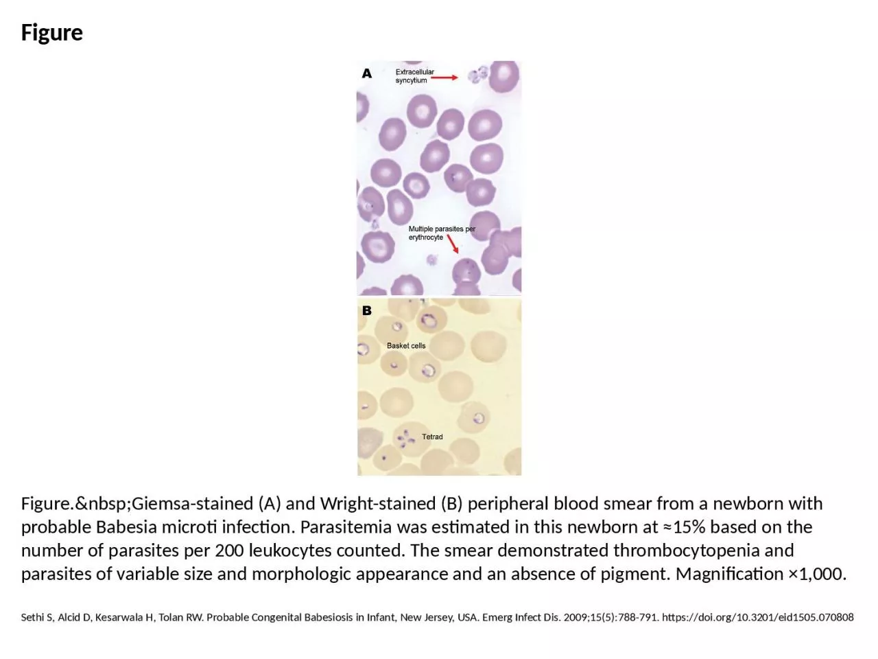
PPT-Figure Figure. Giemsa-stained (A) and Wright-stained (B) peripheral blood smear

Author : deena | Published Date : 2023-07-08

Sethi S Alcid D Kesarwala H Tolan RW Probable Congenital Babesiosis in Infant New Jersey USA Emerg Infect Dis 2009155788791 httpsdoiorg103201eid1505070808

Presentation Embed Code

Download Presentation

Download Presentation The PPT/PDF document "Figure Figure. Giemsa-stained (..." is the property of its rightful owner. Permission is granted to download and print the materials on this website for personal, non-commercial use only, and to display it on your personal computer provided you do not modify the materials and that you retain all copyright notices contained in the materials. By downloading content from our website, you accept the terms of this agreement.

Figure Figure. Giemsa-stained (A) and Wright-stained (B) peripheral blood smear: Transcript


Sethi S Alcid D Kesarwala H Tolan RW Probable Congenital Babesiosis in Infant New Jersey USA Emerg Infect Dis 2009155788791 httpsdoiorg103201eid1505070808. 1 4 WEEK Beginning Class The Vinery Stained Glass Studio 4317 W. Beltline Hwy., Madison, WI 53711 www.vineryglass.com 608 - 271 - 2490 2 Welcome! We hope you enjoy the next four weeks, meet new frien A well Made and well Stained Smear can provide: . Estimates of cell count. Proportions of the different types of WBC. Morphology. Peripheral Blood Smear. Objective. 1. Specimen Collection. 2. Peripheral Smear Preparation. By:Andrew. /6B. Church is a building which is used for religious activity. The church is a place where we held mass every week. In the church we have a priest which lead the mass and also we have the assistants and also there is the people who attend the mass.. Figure S3. MILI antibody specifically recognizes MILI.mili(+/+)mili(+/-mili(-/-) testes. Tubulin serveseIF3a, eIF4GII, and ribosomal protein S6 in MILI antibody co-precipitated MILI and eIF3a from 14 16 897 8 24 271 1504 903 790 1993 1998 Invited to screening Samples taken Nugent score graded slides Investigated all 4 times 1382 864 8 19 1494 928 771 2003 200 8 8 97 903 8 64 885 Samples ex A blood film is stained with Leishman’s stain and scanned under oil immersion, from one end to the other. . As each WBC is encountered, it is identified until 200 leukocytes have been examined. . The percentage distribution of each type of WBC is then calculated. . O'Rourke LG, Pitulle C, Hegarty BC, Kraycirik S, Killary KA, Grosenstein P, et al. Bartonella quintana in Cynomolgus Monkey (Macaca fascicularis). Emerg Infect Dis. 2005;11(12):1931-1934. https://doi.org/10.3201/eid1112.030045. Meissner EG, Bennett JE, Qvarnstrom Y, da Silva A, Chu EY, Tsokos M, et al. Disseminated Microsporidiosis in an Immunosuppressed Patient. Emerg Infect Dis. 2012;18(7):1155-1158. https://doi.org/10.3201/eid1807.120047. Shirwadkar CG, Samant R, Sankhe M, Deshpande R, Yagi S, Schuster FL, et al. Acanthamoeba Encephalitis in Patient with Systemic Lupus, India. Emerg Infect Dis. 2006;12(6):984-986. https://doi.org/10.3201/eid1206.060087. Aiki-Raji CO, Aguilar PV, Kwon Y, Goetz S, Suarez DL, Jethra AI, et al. Phylogenetics and Pathogenesis of Early Avian Influenza Viruses (H5N1), Nigeria. Emerg Infect Dis. 2008;14(11):1753-1755. https://doi.org/10.3201/eid1411.080557. Swayne DE, Suarez DL, Spackman E, Tumpey TM, Beck JR, Erdman DD, et al. Domestic Poultry and SARS Coronavirus, Southern China. Emerg Infect Dis. 2004;10(5):914-916. https://doi.org/10.3201/eid1005.030827. 10. Learning Objectives—Level l . At the end of this unit of study, the student should be able to:. List the assays typically included in the complete blood count (CBC).. Describe how to properly identify a patient prior to blood collection.. Safdar N, Love RB, Maki DG. Severe Ehrlichia chaffeensis Infection in a Lung Transplant Recipient: A Review of Ehrlichiosis in the Immunocompromised Patient. Emerg Infect Dis. 2002;8(3):320-323. https://doi.org/10.3201/eid0803.010249. Philbey AW, Kirkland PD, Ross AD, Davis RJ, Gleeson AB, Love RJ, et al. An Apparently New Virus (Family Paramyxoviridae) Infectious for Pigs, Humans, and Fruit Bats. Emerg Infect Dis. 1998;4(2):269-271. https://doi.org/10.3201/eid0402.980214.

Download Document

Here is the link to download the presentation.
"Figure Figure. Giemsa-stained (A) and Wright-stained (B) peripheral blood smear"The content belongs to its owner. You may download and print it for personal use, without modification, and keep all copyright notices. By downloading, you agree to these terms.

Related Documents