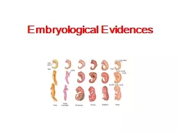
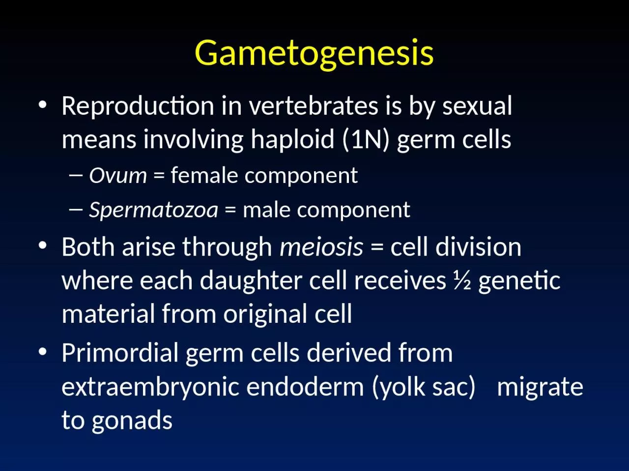
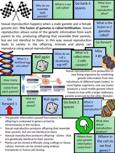
Ovum female component Spermatozoa male component Both arise through meiosis cell division where each daughter cell receives ½ genetic material from original cell Primordial germ cells derived from ID: 921369
Download Presentation The PPT/PDF document "Gametogenesis Reproduction in vertebrate..." is the property of its rightful owner. Permission is granted to download and print the materials on this web site for personal, non-commercial use only, and to display it on your personal computer provided you do not modify the materials and that you retain all copyright notices contained in the materials. By downloading content from our website, you accept the terms of this agreement.
Slide1
Gametogenesis
Reproduction in vertebrates is by sexual means involving haploid (1N) germ cells
Ovum
= female component
Spermatozoa
= male component
Both arise through
meiosis
= cell division where each daughter cell receives ½ genetic material from original cell
Primordial germ cells derived from
extraembryonic
endoderm (yolk sac)
migrate to gonads
Slide2Gametogenesis
Oogenesis occurs in Ovary within a follicle of epithelial cells
Spermatogenesis occurs in germinal epithelium lining seminiferous tubules of testis
Oogenesis begins with
oogonium
; Spermatogenesis begins with
spermatogonium
Both are normal 2N cells
Reduction in chromosome number accomplished via two meiotic divisions
Slide3Stages in Gametogenesis
Pairing and doubling of chromosomes in ____
gonia
, followed by growth as primary ____
ocyte
(2 X 2N)
1
st
meiotic division produces two 2 X 1N cells (= secondary ____
ocytes
)
2
nd
meiotic division produces four haploid cells (spermatids, ova)
Spermatids mature and differentiate to form functional spermatozoa
In spermatogenesis, all 4 sperm cells produced are viable
In oogenesis, only 1 of 4 cells produced is viable.
Others become abortive as polar bodies (only small amount of cytoplasm) that later degenerate
Slide4Fig 14.22
- Oogenesis
Slide5Fig 14.30
- Spermatogenesis
Slide6Egg Membranes and Structure
Cytoplasm enclosed within plasma membrane
Vitelline
membrane
= thin membrane closely attached to plasma membrane
Zona pellucida
= glycoprotein layer (mammals)
Corona
radiata
(mammals) = layer of follicle cells that become sloughed off after fertilization
Slide7Sperm Structure
Spermatozoa from different animals have a wide variety of forms
All have head and tail regions
Head region serves two functions:
Contains nucleus (genetic function)
Acrosomal cap = contains enzymes that allow sperm to break down membranes around egg and fertilize egg
Tail = flagellum that provides motility
Midpiece
between head and tail contains mitochondria that provide ATP to fuel swimming
Slide8Ovarian follicle
Zona pellucida
Primary follicle
Ovum
Spermatozoa
Slide9Fertilization
Several obstacles must be overcome for successful fertilization:
Sperm and egg must come into proximity
Cell to cell contact must occur
Sperm must penetrate egg cell
Slide10Mechanisms for Proximity
Transport occurs in liquid medium
EXTERNAL FERTILIZATION
Eggs and sperm simultaneously shed into water
Occurs in fishes (except
Chondrichthyes
) and most anurans
INTERNAL FERTILIZATION
Sperm introduced directly into female tract
Usually involves
copulatory
organs in males (none present in tuatara, birds, salamanders = copulation by
cloacal
“kiss”)
Occurs
in animals with shelled eggs or viviparous habits as sperm must reach egg before shell is added (
Chondrichthyes, most Amphibians, Amniotes)
Slide11Mechanisms for Contact
For internal fertilization, sperm travel within female tract by passive transport (dependent on muscular contractions
and
ciliary
currents provided by female tract).
Little active swimming by sperm for transport function
Contact in external fertilization accomplished by random swimming movements of sperm in water
Slide12Mechanisms for Egg Barrier Penetration
Once contact with egg has been established, the next step is to penetrate the egg so that nuclear materials can unite to form the diploid zygote.
Barrier penetration mechanisms are chemical in nature and involve
acrosomal reaction
Sperm
Lysins
= enzymes that locally dissolve egg membranes
Produced by acrosomal cap
Sperm
lysins
differ among animal groups as membranes surrounding eggs differ (e.g., jelly coat in amphibians, follicle cells of corona
radiata
in mammals)
Slide13Mechanisms for Egg Barrier Penetration
Acrosome Reaction
involves …
Release of sperm
lysins
Fusion of egg and sperm membranes
In some animals, acrosomal reaction involves exposure of binding sites on plasma membrane of sperm, via acrosomal tubule or filament, which bind to receptors on p.m. of egg in species-specific manner
This binding precedes fusion of sperm and egg plasma membranes
Slide14Acrosomal Reaction in Hemichordates
sperm
lysins
rupture
Binding sites exposed that bind to receptors on egg plasma membrane
Slide15Mechanisms for Egg Barrier Penetration
In mammals, there is no development of acrosomal filaments
Instead, fluids of female reproductive tract induce
capacitation
primes sperm for fertilization and includes removal of some components from sperm surface.
After capacitation,
hyaluronidase
on the sperm head is exposed and breaks down the hyaluronic acid cementing the follicle cells of corona
radiata
(which surround the egg) together
allows sperm passage through corona
radiata
to contact zona pellucida (a glycoprotein layer surrounding the egg)
Slide16Mechanisms for Egg Barrier Penetration
Zona pellucida has species-specific receptors for binding sperm
Binding causes rupture of acrosome, which releases contents that break down zona pellucida and allow contact with egg plasma membrane
Binding also exposes proteins on sperm surface that bind with receptors on egg plasma membrane to facilitate
fusion of sperm and egg
Fusion of plasma membranes releases sperm genetic material into egg as sperm
pronucleus
Male and female genetic material will soon combine forming a diploid
zygote
Slide17Slide18Post-fertilization Responses in Zygote
Formation of
Fertilization Cone
= outward bulge of egg cytoplasm that serves to engulf sperm
Occurs upon fusion of sperm and egg plasma membranes
Recession of cone brings sperm nucleus into egg cytoplasm
Egg Activation
Upon fusion (within 3 sec) get membrane depolarization or hyperpolarization (species-dependent)
blocks entrance of > 1 sperm (=
Fast block to
polyspermy
)
Slide19Post-fertilization Responses in Zygote
Next, get Ca
2+
release from internal stores within egg triggers cortical reaction
release of cortical granules to
perivitelline
space around egg
Cortical granule release causes development of fertilization membrane blocking further sperm entry (=
Slow block to
polyspermy
)
Slow block to
polyspermy
occurs about 25-30 sec post-fusion
Seems to occur only for
microlecithal
eggs (e.g., mammals); entrance of > 1 sperm into eggs of birds, reptiles and some amphibians common, but only 1 sperm contributes to zygote (others somehow inactivated)
Slide20Fast Block to
Polyspermy
Slow Block to
Polyspermy
Slide21Post-fertilization Responses in Zygote
Rearrangement of internal constituents within egg
Sets up gradients of certain substances and plane of bilateral symmetry within zygote for some animals
Fusion of Haploid Nuclei
In most vertebrates, meiosis within egg arrested after 1
st
meiotic division. Sperm entry stimulates 2
nd
meiotic division to produce female
pronucleus
(and 2
nd
polar body)
Once this 2
nd
division occurs, female
pronucleus
is ready for union with male pronucleus
Slide22Post-fertilization Responses in Zygote
Fusion of Haploid Nuclei (cont.)
Male and female
pronuclei
next approach each other (mechanism by which this movement occurs is not known with certainty)
Next get fusion of
pronuclei
In some animals (including most vertebrates),
pronucleus
membrane degenerates
free chromosomes arrange themselves at spindle (metaphase of mitosis)
completion of mitosis
dipolid
zygote
Slide23Parthenogenesis
Definition
= development of the egg in the absence of sperm
Occurrence suggests that:
(1) egg activation and nuclear fusion are separate developmental processes
(2) the ovum contains all the capacities necessary for embryo formation – all that is necessary is some triggering agent
Eggs can be activated by a number of chemical, thermal, electrical or mechanical means
Slide24Parthenogenesis
Parthenogenetic
individuals are expected to be haploid (and many are), but these embryos are often diploid.
Doubling of chromosomes accomplished in 3 ways:
Suppression of 2
nd
meiotic division – occurs only in eggs completing this division after fertilization
Refusion
with second polar body
Suppression of 1
st
mitotic division (= 1
st
cleavage division)
Slide25Parthenogenesis
Haploid embryos generally show premature developmental arrest
Parthenogenetic
diploid embryos also usually show premature developmental arrest
However, in several invertebrates parthenogenesis is normal (e.g., male drones of bee colony) and there are several species of naturally occurring
parthenogenetic
lizards (the entire population is female)
Artificial selection procedures have developed
parthenogenetic
strain of turkeys
Slide26The asexual, all-female whiptail
species
Cnemidophorus
neomexicanus
(
center), which reproduces via parthenogenesis, is shown flanked by two sexual species having males,
C.
inornaus
(
left)
and
C.
tigris
(right), which hybridized naturally to form the C. neomexicanus species.
Slide27Methods of Bearing Young
Oviparous
= egg laying
Primitive condition for vertebrates
Occurs in most fishes, amphibians, reptiles, all birds,
monotremes
Viviparous
= live-bearing
Advanced condition in vertebrates
Some live-bearers occur in all vertebrate classes except cyclostomes and birds
Evolved by retention of eggs within body to increase survival of young
Slide28“Placental Connections” in Viviparous Vertebrates
Anamniotes
with connection between yolk sac and maternal tissues through which exchange of metabolites occurs (e.g.,
Chondrichthyes
)
Reptiles use yolk sac,
chorion
, allantois (
extraembryonic
membranes) or some combination for connection
Mammals with a variety of connections
Slide29Early Development/Placentation in Mammals
After formation of zygote
cleavage
produces blastula
Blastula forms before embryo reaches uterus
Mammalian blastula consists of trophoblast and inner cell mass (ICM becomes embryo)
Upon reaching uterus, trophoblast overlying ICM makes contact with uterine endometrium trophoblast cells rapidly multiply and insert among epithelial cells lining endometrium and endometrial cells degenerate
implantation
Continued trophoblast cell division
placentation
; embryo becomes buried w/in endometrial lining
Slide30Fig 5.32
Slide31Mammalian Placenta Formation
Structure produced by apposition and fusion of
extraembryonic
membranes of embryo with uterine endometrium of mother
Extraembryonic
membranes
= tissues external to embryo not participating in embryo formation, but functioning in maintenance of the embryo
In Amniotes, four
extraembryonic
membranes exist
Slide32Extraembryonic
Membranes
Yolk Sac
= forms from
extraembryonic
hypomere
(
splanchnopleure
) that expands to enclose yolk
This is the only
extraembryonic
membrane present in
Anamniotes
, so it occurs in all vertebrates
Functions to derive nutrients from yolk in yolky eggs to nourish developing embryo
In Amniotes,
extraembryonic somatopleure grows over embryo by folding back on itself producing a double hood of somatopleureFrom this structure develop Amnion and Chorion
Slide33Extraembryonic
Membranes
Amnion
= forms from inner
somatopleure
+ ectoderm (inside)
Chorion
= forms from outer
somatopleure
+
ectoderm
(outside)
Amnion serves as fluid-filled sac for embryonic development
Replicates aquatic developmental environment of primitive vertebrates.
Allows complete conquest of terrestrial habitats
Chorion
functions in protection of embryo and in exchange of gases (and metabolites in placenta)
Slide34Extraembryonic
Membranes
Outgrowth of
splanchnopleure
from posterior region of gut in Amniotes eventually expands to fill
extraembryonic
coelom (= space between amnion and
chorion
)
This membrane is the
Allantois
= composed of splanchnic mesoderm (outside) + endoderm
Mesoderm fuses with mesoderm of
chorion
to form
Chorioallantoic
Membrane
= main gas exchange organ for Amniote embryosAllantois also serves waste storage function
Slide35Fig 5.29
–
Extraembryonic
membrane formation in a bird
Slide36Fig 5.30
Fig 5.30
–
Extraembryonic
membrane formation in a bird
Slide37Mammalian Placenta
From
chorion
(outermost
extraembryonic
membrane), finger-like processes grow outward to interlock with uterine endometrium
Blood streams of mother and fetus never mix – always separated by epithelial membrane, so exchange of gases and nutrients occurs by diffusion across this membrane
Chorion
is not in direct contact with embryo so some means of blood supply from embryo to placenta (and back) must occur
Slide38Mammalian Placenta
Blood Supply to developing embryo differs between marsupial and placental mammals
Marsupials = mostly
Choriovitelline
fetal placenta
Yolk sac associated with inner surface of
chorion
Blood vessels develop in mesoderm of yolk sac
This situation also occurs to some extent in several placental groups (e.g., rodents)
Placentals
=
Chorioallantoic
fetal placenta
Dominant connection to
chorion
provided by allantois, yolk sac usually degenerates
Allantoic
mesoderm forms blood vessels that function in gas & nutrient exchange, waste removal
Slide39Fig 5.33
– Fetal
extraembryonic
membranes in various Amniotes
Slide40Mammalian Placenta Types
Primitive Condition
= apposition without fusion (non-deciduous)
Advanced Condition
= fusion of maternal and fetal tissues (deciduous)
Four Types occur:
Epitheliochorial
= most primitive
Occurs in pig and some other mammals
Maternal and fetal blood separated by 6
layers: endothelium
, CT, epithelium, epithelium, CT,
endothelium
Slide41Mammalian Placenta Types
Syndesmochorial
= no uterine epithelium
Occurs in ruminant mammals (cattle, sheep, etc.)
Endotheliochorial
= no maternal epithelium or CT
Occurs in carnivores
Hemochorial
= advanced condition
Chorionic epithelium bathed in maternal blood
Occurs in primates and many rodents
Slide42