/
Journal of Surgery and Surgical Research Journal of Surgery and Surgical Research

Journal of Surgery and Surgical Research - PDF document

delcy
delcy . @delcy
Follow
342 views
Uploaded On 2022-08-21

Journal of Surgery and Surgical Research - PPT Presentation

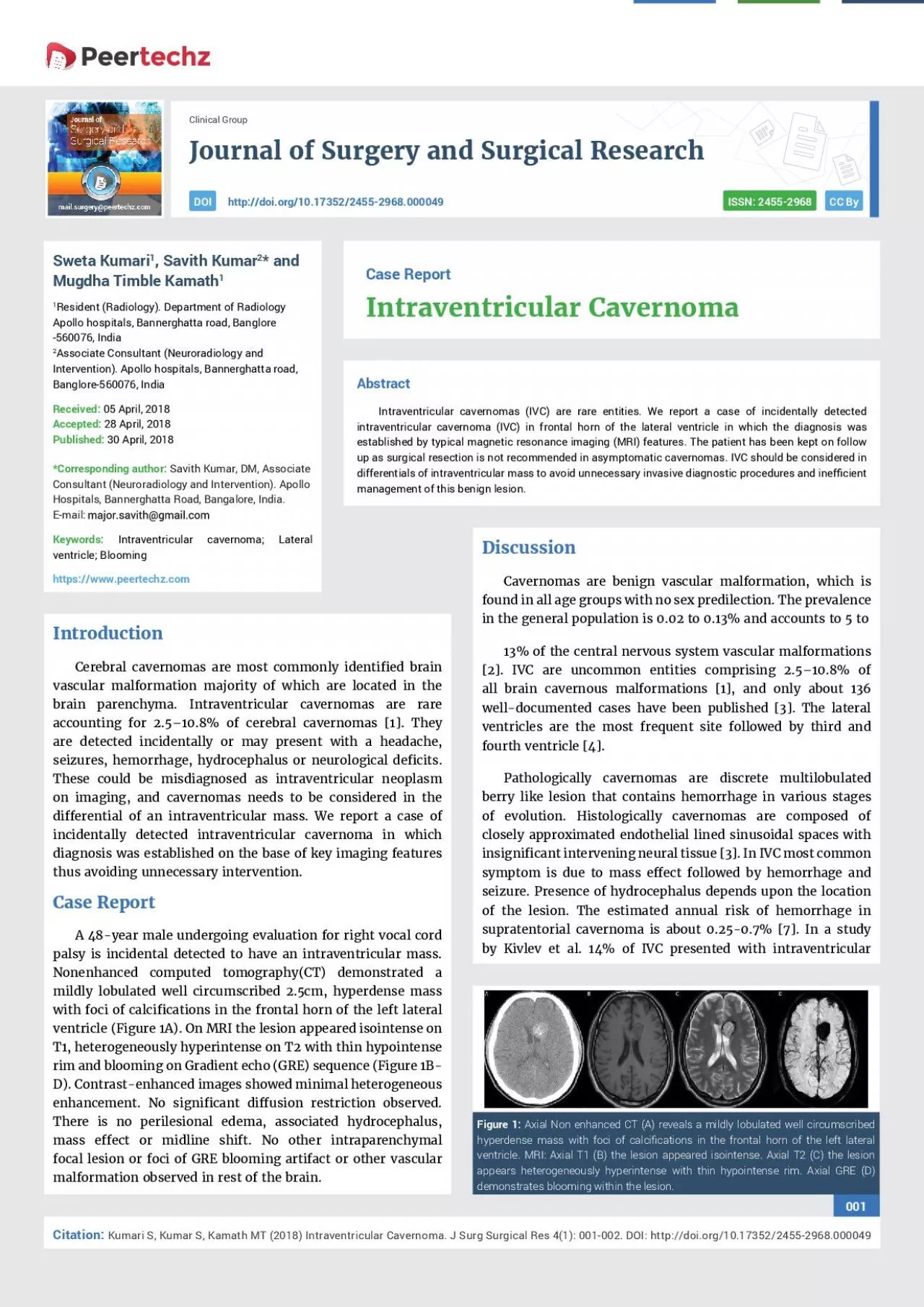
ISSN 24552968 CC By 001 Kumari S Kumar S Kamath MT 2018 Intraventricular Cavernoma J Surg Surgical Res 41 001002 DOI httpdoiorg1017352Clinical Group cient 1 Savith Kumar2 and M ID: 939416

lateral intraventricular common surgical intraventricular lateral surgical common cations 2018 imaging cavernomas doi cavernoma ivc 001 apollo signal 002

Share:

Link:

Embed:

Download Presentation from below link

Download Pdf The PPT/PDF document "Journal of Surgery and Surgical Research" is the property of its rightful owner. Permission is granted to download and print the materials on this web site for personal, non-commercial use only, and to display it on your personal computer provided you do not modify the materials and that you retain all copyright notices contained in the materials. By downloading content from our website, you accept the terms of this agreement.


Presentation Transcript

Journal of Surgery and Surgical Research ISSN: 2455-2968 CC By 001 Kumari S, Kumar S, Kamath MT (2018) Intraventricular Cavernoma. J Surg Surgical Res 4(1): 001-002. DOI: http://doi.org/10.17352Clinical Group cient 1, Savith Kumar2* and Mugdha Timble Kamath1 Resident (Radiology). Department of Radiology Apollo hospitals, Bannerghatta road, Banglore 2Associate Consultant (Neuroradiology and Intervention). Apollo hospitals, Bannerghatta road, Savith Kumar, DM, Associate Consultant (Neuroradiology and Intervention). Apollo Intraventricular cavernoma; Lateral ed brain brain are detected incidentally or may present with a headache, seizures, hemorrhage, hydrocephalus or neurological deÞ cits. cations in the frontal horn of the left lateral cant diffusion restriction observed. all brain cavernous malformations [1], and only about 136 well-documented cases have been published [3]. The lateral ventricles are the most frequent site followed by third and fourth ventricle [4].Pathologically cavernomas are discrete multilobulated berry like lesion that contains hemorrhage in various stages of evolution. Histologically cavernomas are composed of closely approximated endothelial lined sinusoidal spaces with insigniÞ cant intervening neural tissue [3]. In IVC most common Axial Non enhanced CT (A) reveals a mildly lobulated well circumscribed cations in the frontal horn of the left lateral 002 Kumari S, Kumar S, Kamath MT (2018) Intraventricular Cavernoma. J Surg Surgical Res 4(1): 001-002. DOI: http://doi.org/10.17352 IVC attain a larger size than their parenchymal counterpart as the intraventricular location allows for unrestricted growth and their increased tendency for intralesional rebleed [2].On imaging, IVC may appear similar to the intraparenchymal cavernoma. They are hyperdense on noncontrast CT and calciÞ cation is common. They show a core of mixed-signal cation is common. They show a core of mixed-signal Imaging differentials of intraventricular mass in adults includes central neurocytoma, subependymoma, men

ingioma, subependymal giant cell astrocytoma and metastasis [6].Central neurocytoma is typically lateral ventricular mass attached to the septum pellucidum. It is usually hypodense on nonenhanced CT. Punctate calciÞ cations are common. Cystic cations are common. Cystic Intraventricular meningiomas are located in trigon of the lateral ventricle in about 85% of cases. Sharply demarcated rounded or lobulated lesions with low T2 signal intensity, intense homogeneous enhancement, and moderate diffusion restriction are key features [6]. Subependymomas are well demarcated isodense partially calciÞ ed lesions on noncontrast © 2018 K This is an open-access article distributed under the terms of the Creative Commons Attribution License, which permits unrestri Complete surgical excision is the treatment of choice. Lateral ventricle caveromas are excised through transcortical or interhemispheric transcallosal approach. Surgical resection is not recommended for asymptomatic cavernomas especially if located in eloquent, deep and brainstem areas [7].In our case, as the lesion was incidentally detected, and the patient was asymptomatic he was considered to be kept on follow up. IVC are uncommon entities with wide differentials. Key imaging features are peripheral hyperintense rim on T2W, and blooming artifact on susceptibility weighted imaging favor the diagnosis of intraventricular cavernomas 1. Nigri F, Viana JDS, Pinto FPHDC, Simões EL, Ribeiro TCR (2018) 2. Faropoulos K, Panagiotopoulos V, Partheni M, Tzortzidis F, Konstantinou D Link: 3. Shirvani M, Hajimirzabeigi A (2017) Intraventricular Cavernous Malformation: Link: 4. Beechar VB, Srinivasan VM, Reznik OE, Sen A, Klisch TJ, et al. (2017) Link: 5. Kivelev J, Niemelä M, Kivisaari R, Hernesniemi J (2010) Intraventricular 6. Vandesteen L, Drier A, Galanaud D, Clarençon F, Leclercq D, et al. (2013) ndings of intraventricular and ependymal lesions. J Neuroradiol 7. Akers A, Dahlem K, Flemming K, Hart B, Kim H, et al. (2017) Synopsis of c Advisory Board Clinical Experts Panel