/
Some of the major advancements in Dentistry took place in the 1960s an Some of the major advancements in Dentistry took place in the 1960s an

Some of the major advancements in Dentistry took place in the 1960s an - PDF document

delcy
delcy . @delcy
Follow
343 views
Uploaded On 2021-09-15

Some of the major advancements in Dentistry took place in the 1960s an - PPT Presentation

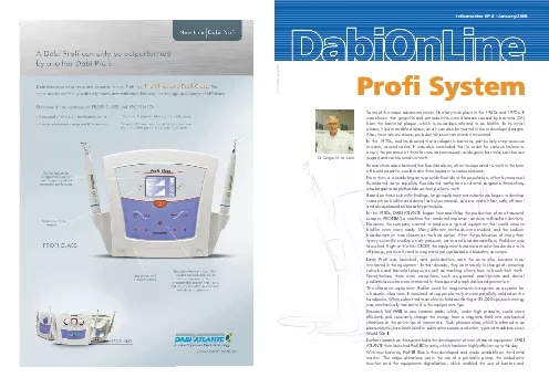
Dr Srgio N M LimaThe ltrasound is a piece of equipment that easily removes calculus nevertheless students begining to use it may make mistakes that will apparantely not compromise the procedure but wh ID: 881561

tip calculus profi ultrasound calculus tip ultrasound profi tooth removal biofilm patient tips equipment surface preparation dental irrigation solution

Share:

Link:

Embed:

Download Presentation from below link

Download Pdf The PPT/PDF document "Some of the major advancements in Dentis..." is the property of its rightful owner. Permission is granted to download and print the materials on this web site for personal, non-commercial use only, and to display it on your personal computer provided you do not modify the materials and that you retain all copyright notices contained in the materials. By downloading content from our website, you accept the terms of this agreement.


Presentation Transcript

1 Some of the major advancements in Dentis
Some of the major advancements in Dentistry took place in the 1960s and 1970s. It was shown that gingivitis and periodontitis were diseases caused by bacteria (SA) from the bacterial plaque, which is nowadays referred to as biofilm. In its initial phase, it is a reversible disease, and it can also be treated in more developed stages. Also, in certain situations, periodontal tissue can even be recovered. Dr. Sérgio N. M. Lima The ltrasound is a piece of equipment that easily removes calculus; nevertheless, students begining to use it may make mistakes that will apparantely not compromise the procedure, but which will delay work and sometimes make the procedure difficult and even inefficient.For educational purposes, the subject can be divided in three basic points: : it is usually whitish in smokers and dark in circumstances resulting from diet type. By using the correct tip indicated for supragingival calculus (Perio Supra Tip, for instance) and by applying the active tip on the limit between the calculus formation and the tooth surface, calculus will be removed without any difficulty. 2.Subgingival Calculus: it is usually dark and more adhered to the teeth. The subgingival tip (Perio Sub Tip, for instance.) is used, and it is also applied between the tooth and the mineralized deposit. In this circumstance, a novice professional may face some difficulty since the contour of such calculus formation is not always visible. Hence, the application of the active tip does not reach the limit area between the calculus and the tooth, which may hinder and even prevent the professional from effectively removing the calculus formation. 3.Pain when applying the ultrasound: If the patient feels pain during ultrasound application, which occurs particularly in the case of gingival retraction and subgingival calculus, the patient can be anesthetized. In order for a professional to develop good supra or subgingival calculus removal skills, it is essential that the patient remain comfortable and keep his/her head still. The difference between the points from which calculus is easily removed and those that are slowly worn out by the professional can be measured in millimeters.Hence, firstly training on an appropriate manikin is advised. Also, when performing on patients, calculus removal should always take place under direct view.In order to remove subgingival calculus without direct view and in a closed field, a lot of skill and experience is required so one can notice the limits of the tooth and the limits of calculus by touch. Under these circumstances, the most delicate tips in the collection supplied by the manufacturer must be used.Such sensitivity to touch would not be effective to any professionals if it were necessary to apply a lot of strength on the handpiece for calculus removal, that is, it is not the professional's strength that removes calculus, but rather the power of ultrasonic vibration.A simple test to confirm the abovementioned information is taking an empty can and applying the ultrasonic tip on its surface. In a few seconds, the tip will perforate the can.The sodium bicarbonate jet initially aims at removing and controlling non-mineralized supragingival biofilm in prophylaxis procedures. Application must be performed after coating bacterial biofilm, as for instance, using malachite green or another marker selected by the dental professional. The new Profi line provides a more concentrated bicarbonate flow, which makes it more efficient. The jet can be correctly directed to the area from where biofilm must be removed without discomfort to the patient's soft tissue during prophylaxis.The jet must directed at a distance ranging from 4mm to 1cm and at an angle of approximately 45 degrees in relation to the tooth's surface. Never position the jet at a larger distance than that, otherwise it will lose its efficiency and spread strong spray in the dental office, thus impairing suction by the auxiliary unit. SODIUM BICARBONATEJET APPLICATION ULTRASOUND APPLICATIONcontributed to biosafety in the dental office. At the beginning of the present century, all dental surgeons began to dedicate to and care about calculus removal and biofilm control in their patients. Peridontists were most concerned about such issue due to periodontal diseases and periodontal sacs that must be eliminated. Subgingival calculus is more difficult to be diagnosed and removed, but all dental professionals are alert since these bacterial deposits on the teeth interfere with the result of all types of treatment, regardless of their area of specialty.In 2008, DABI innovated again and presented its new Profi line.The same excellence level that has always been a feature of its equipment and that is a reference for quality on the dental market, but now with more resources, technology, precision and efficiency for use in all fields or modern Dentistry. The new line comprises models PROFI NEO and PROFI CLASS, which, in addition to the ultrasound and the bicarbonate jet, also have a peristaltic pump and a coupled irrigation solution reservoir, a resource that improves the post-operative period. Model PROFI CLASS features, as another differential item, a membrane-type digital keyboard with programmable power and irrigation solution outflow as well a

2 s the exclusive “Diamond-Function&#
s the exclusive “Diamond-Function” key for use of Dabi Restoration Tips (diamond tips) for maximum productivity of such tips and tooth preservation. Fig.AFig. A shows biofilm stained by malachite green in a patient who did not brush her teeth for two days during the The blue arrows show the correct directions for the bicarbonate jet to be used with the new Profi. Since the equipment features a very well directed jet, it is possible to remove biofilm (bacterial plaque) without discomfort for the patient.The red arrows indicate the non-desirable directions for the jet flow. The flow must be interrupted in the interdental spaces so as not to cause discomfort to the patient's soft tissues (mouth or tongue floor). Fig. B.shows teeth immediately after Profi prophylaxis. In this case, 0.12% chlorexidine in 25% solution was used, that is, 125ml of the product was placed into 375ml of drinkable water.As a routine and due to its great importance, it is always advised that 1.23% acidulated fluoride gel should be applied on all teeth after prophylaxis. Scientific studies show that, at this moment, fluoride incorporation by the tooth's enamel is maximum and virtually immediate.When applying the bicarbonate jet, the ideal procedure is to activate the footswitch intermittently. Such non-continuity causes the powder in the reservoir to whirl, which improves biofilm removal efficiency and, at the same time, saves time and powder. This procedure also makes the equipment's efficiency to remain independent of the amount of powder existing in the reservoir.Due to such benefits, it if is correctly used, the equipment will require a smaller amount of powder at each prophylaxis procedure.In order for the dental surgeon to take the best advantage from his/her equipment, a short, simple and fast training session out of the patient's mouth is advised.Important:The tips must never be applied on a straight angle in relation to the tooth's surface. The tip should always be inclined at approximately 45 degrees in relation to the tooth's axis. Didactically, a tip at an ideal angle would be on the alignment of the bisector formed between the tooth's surface and the calculus top area. By taking into account that the tooth's surface is a plane, the calculus limit makes such surface approximately a straight angle. The application of the ideal active tip would be a bisector in this micro space of action, with an angle of 45 degrees in relation to the plane of the surface from which calculus must be removed. As previously explained, the tooth's surface is not a horizontal plane; therefore, in order to keep the tips' efficiency, it is necessary to change their inclination according to the tooth's curvature. For greater efficiency, the limit between supra or subgingival calculus and the tooth must always be located. In case it is not very visible, a marker should be used and biofilm can be removed by a bicarbonate jet. Hence, it will be possible to correctly visualize such limit where the active tip of the ultrasound will be applied. With this procedure, a lot of time is saved and the patient is submitted to less stress. Fig.BFig.CFig.D ToothThis diagram better explains the position of a bisector in an ideal imaginary situation. PERIODONTOLOGYUltrasound has been used in periodontology since the 1960s. It all began when with the well-known Cavitron, but that equipment was criticized by many peridontists, who never stopped using their manual scrapers.In fact, DABI and its researchers did not launch Profilat in the 1980s with the purpose to replace manual scrapers, since the final finishing of a periodontal treatment and the normalization of the altered tissue on teeth require the use of scrapers such as curettes, which are essential irreplaceable.Today, the use of ultrasound in Periodontology is an unquestionable reality as it makes procedures faster and prevents cuts caused by curettes as they are not used on larger and more visible calculus that are easily removed by ultrasound. Also, according to studies by Lima et al. (1988), the use of ultrasound reduces microbiota on the gingival sulcus and on periodontal sacs in 99%, which makes its use very important in pre-surgical antisepsis in all dental fields. Even during absolute isolation such as in endodontics, this type of prophylaxis is indicated.A study by Lima et al (1988) also showed that risk patients require the use of preventive antiobiotic therapy prior to scraping since the use of ultrasonic and manual scrapers causes transient bacteriemia. This condition may be serious in risk patients with a history of cardiopathies, arthritis, rheumatism, immunodeficiencies, among others. Finally, with modern ultrasonic equipment and the use of appropriate techniques for its application, all periodontic equipment have become safer and more reliable in the practice of all dental specialitiesUltrasound power consists in the electromagnetic field power that is generated on the ceramic pads inside the handpiece. This effect, denominated as piezoelectric, moves the tip. The higher the selected power, the greater the vibration power; however, work must be performed within the biological limits of such canals, which are generally curved or atresic. The physical limits of files must be respected,

3 and remember that the vibrational effect
and remember that the vibrational effect of tips will produce heat within the canals. Therefore, in order to clean canals, power of approximately 30% of the equipment's capacity must be used with abundant irrigation so as not to heat their interior (possible injury to periapical tissue) and to prevent files from breaking. For lateral condensation in root canal filling, the equipment's power must be kept at approximately 30% or 40%, since slight heating is desirable, but not overheating, which would hinder filling. Ultrasound has been researched, used and discussed in endodontics since the 1950s, when the first ultrasound devices were launched. It has been discussed since last century because endodontists have always wanted to count on resources that can make clinical work easier and faster. Ultrasound is one of the technological solutions for such desire. These professionals use their valuable time to find openings to root canals, enlarge and widen root canal ducts and properly clean them, particularly those that are curved or atresic. Nowadays, time is a luxury and, therefore, a machine that can help them save time is of utmost importance.Dabi Atlante, which has always followed the desires and anxieties of endodontists, launched Profi Neo and Profi Class in 2008, two pieces of equipment that have brought encouragement to daily practice.Profi Neo and Profi Class feature a complete endodontics system which allows for time saving, ENDODONTICSOne of the greatest advantages of ultrasound is the cavitational effect that has already been mentioned by Dr. Sérgio Lima. The cavitational effect occurs when ultrasonic waves, in a liquid medium, generate such fast mechanical waves that micro bubbles are formed. These micro bubbles break so quickly that they lead cells, bactéria, DNA, hemoglobins, etc to break, thus strongly reducing the number of microorganisms. Adittionally, these waves exert a dissolving action on the surfaces with which they make contact. The cavitational effect increases the dissolving power of sodium hypochlorite fivefold. Profi Class and Profi Neo feature a coupled solution reservoir and functions for selecting the solution amount to be used (levels 1, 2, 3 or without irrigation). This allows the professional to choose which irrigation solution will be used and what its amount will be. The literature reports that the abundant irrigation obtained from the ultrasound is one of the major advantages of this technique. By associating irrigation abundance with the Biomechanical preparation by ultrasound is performed using the Endo L tip (insert picture) to which a file is adapted. This process is relatively simple and inexpensive. Conventional Flexofiles can be used by removing their cables and and adapting them to the tips during the procedure. It is fundamental to remember that the files used in ultrasound devices must always work freely inside the canals and that they do not produce the opening in depth, but always in laterality. During biomechanical preparation, ultrasonic files are only used after the initial widening and odontometry. A conventional number-15 file should be used, always with abundant irirgation.Some advantages of the biomechanical preparation cited in the book Endodontia: tratamento de canais radiculares: pricípios técnicos e biológicos (Mario Roberto Leonardo São Paulo: Artes Médicas, 2005) are: removing the pre-dentin layer and debris from inside the canal, allowing better cleaning of root canals; reducing working time, hence reducing operator fatigue; achieving greater wearing of dentin walls, thus allowing greater root conicity and facilitating filling. By using the Endo G tip (insert picture), a new resource for lateral condensation of gutta-percha cones is obtained. By interrupting irrigation, which is possible for Profi Class and Profi Neo, space is laterally created in order to place accessory cones with the advantage of creating heat through the tip, thus laterally condensating gutta-percha cones in a compact, homogeneous and radio-opaque fashion.By using the Endo G tip (picture), it is easy to remove gutta-percha cones. When this tip is applied with high power and is activated inside the canal, it creates space in the filling material for placing other instruments and dissolving . Work time is reduced and treatment quality is improved.Re-Treatment In order to remove posts or metallic cores, the Endo N tip, will work with medium high ultrasonic power and abundant irrigation. This will help the dental surgeon since the ultrasonic vibration will take cement to the fracture, thus facilitating post removal without heating it. For crown removal, the Endo C tip will work in a similar fashion and break cement adherence. Similarly to all other techniques, the use of ultrasound has advantages and disadvantages. The advantages are described above. The disadvantages are that training and investment are required. Opemindedness is necessary for acceptance of something new, as a challenge. After all, patience, tests and training are necessary to use a new technique. A few hours are required to learn how to handle the equipment, to test on extracted teeth, breaking files and tips and puzzling one's Finally, it is a feeling of having accomplished one's duty, the conscience of

4 doing something else for our patients a
doing something else for our patients and for ourselves. Patients realize it; they are satisfied. We work better and are satisfied too. Our patients come back again and recommned us, so we are even more satisfied! It is the compensation for our ergonomy and, above all, quality of work and life. These equipment items can be very easily used as their panels are simple and the functions are clear. Both of them enable the dentist to control work power as well as the presence and amount of irrigation solution. It is also possible to interchange the four tips (metal tips), each of which has a specific purpose abovementioned effects, it is concluded that excellent cleaning of the root canal system can be achieved by using the ultrasound. Drª. Carolina D. Lima0504 0706 IMPLANTOLOGY, SURGERY AND PROSTHESISThere are numerous situations in which implantologists and prosthesists have to perform procedures in a patient's mouth prior to its being submitted to appropriate antisepsis for such procedures.Various studies have shown that only mouthwashing with antiseptic solutions is not enough to achieve the higienization levels required by major procedures to be performed on surgery patients. Such studies have shown that not only the use of antiseptic silution is required, but so is the discerning removal of biofilm (bacterial plaque) and subgingival and supragingival calculus in order to accomplish ideal prophylaxis in the mouth and the safe performance of all surgical and prosthetic procedures.The new Profi line has been designed to give support to impantologists and surgeons in all phases of their work.Prior to beginning anethesia for surgery, it is necessary to coat the biofilm by using malachite green, for instance, since it would be fearful to expose bone tissue to a conaminated area during implantation.Therefore, in case biofilm exists, it is necessary to use the sodium bicarbonate jet. In the new Profi line, the flow is more concentrated, which prevents dispersion in the operating room. The use of antiseptic solution in the reservoir is always recommended so as to enhance the antibacterial action of the sodium bicarbonate jet, enhance the ultrasonic effect as a factor for calculus removal as well as its antibacterial effect and reduce the effects of contaminaning spray in the operating room. The presence of an antiseptic agent in the irrigation solution inactivates most of the bacterial flora in the biofilm.In general, after completely removing biofilm (bacterial plaque), there is always a calculus deposit that must be removed by using the ultrasound and the PERIO SUB or PERIO SUPRA tips, depending on calculus position.Once the operating field is completely clean, incisions for transplantation or other procedures required by patient's treatment can be safely performed.It must be clarified that having the surgeon's instruments, gloves and other essential items sterilized is not enough. Fundamentally, the operating field must also be submitted to rigorous prophylaxis.THE CASE OF PATIENT WITH IMPLANTS This is the case of a patient with implants who had no maintenance for 6 years. He attended our dental clinic due to a problem in another tooth on the upper arch.During clinical examination, a large amount of biofilm and calculus was found on the implants and pontics. However, prior to any other treatment, prophylaxis of the fixed prosthesis on the implants was begun.The existing biofilm and calculus can be seen in Fig. E. There is no doubt that the retractions occuring on the implants were caused by the presence of this biofilm on the prosthetic pieces and on the implants.Tip Perio E was used in order to adequately reach the prosthetic spaces and niches. The ultrasound power virtually “dissolves” the calculus deposits, which can be obserbed in Fig. F (red arrow). The professional must be very careful so as to never force the tip on the implant or prosthesis, since that could cause micro irregularities on those surfaces or even chip the porcelain. The ultrasound must always be applied on implants and prostheses by using the tip's ends and lateral parts, but never its top.Immediately after prophylaxis, the tissues already present a different aspect.As regard the spray, the use of IPE, that is, Individual Protection Equipment by the professional and his assistant is very important. They all must wear protection goggles, including the patient. Upon procedure completion, such goggles must be disinfected. All interventions in a patient's mouth must be preceeded by rigorous antisepsis, which is a simple and practical way to reduce the number of microorganisms present and spray contamination. By reducing the patient's oral microbiota, the professional will achieve more reliable results as well as greater safety for the therapeutic procedures to be performed. PERISTALTIC PUMP AND COUPLED IRRIGATION SOLUTION RESERVOIR It is very common for specialized dental surgeons to receive patients from colleagues for the performance of various types of interventions: extraction of embedded and semi-embedded third molars, interventions on tooth apexes, bone grafts and even periodontal treatments.Many specialists expect patients to come to their offices with their mouths ready to be submitted to such interventions. Nevertheless, tha

5 t, unfortunately, does not take place in
t, unfortunately, does not take place in most cases. The patients come with their mouths full of supra and subgingival calculus and a large amount of biofilm (bacterial Many studies have shown that there is no biosafety if intervention is performed under such circumstanes. Therefore, the removal of bacterial material adhered to the teeth and mouth tissues is essential as a measure to control postoperative In this case, the patient presented a large amount of calculus on all teeth. However, the lower right third molar was very painful and needed to be extracted since it was embedded and there were no feasible recovery conditions. Prior to extraction, comprehensive prophylaxis was performed in the patient's mouth. At first, the bicarbonate jet irrigated with an antiseptic solution was used, which was followed by calculus removal using the ultrasound.It is noteworhty that the new Profi line makes these procedures fast and safe and that such prophylaxis more objectively aims at the biosafety of the procedure rather than at the treatment of periodontitis presented by the patient. For fast and effective calculus removal by the ultrasound, its is important that the vibrating tip always be applied at an angle of approximately 45° in relation to the tooth's surface and on the limit between the tooth and the calculus deposit. The high-power aspirator, used in all surgeries, must be well positioned in order to maximally reduce spray dispersion in the A lot of time is wasted while trying to destroy the whole calcified mass by applying the active tip on distinct and random points of its surface. In the case of dark subgingival calculus, if the limit between the calculus and the tooth's surface is not localized, a lot of time is spent on the elimination of such mineralized biofilm, but when Profi is appropriately used, the whole calculus mass on the incisive mesial area comes off in a few seconds following the application of ultrasound. This technique, in combination with antiseptic irrigation, takes very little time.It must also be pointed out that it is not the strength applied to the ultrasound that will remove calculus. When used inappropriately, the equipment may lose its efficiency.Finally, the tooth (38) was removed and the tissues were saturated. The patient's postoperatice discomfort was minimal since no secondary contamination of the surgical wound occurred. Profi in action. The tip must be placed exactly between the tooth's surface and the calculus depositFig.E Fig.GFig.HFig.I Fig.J Fig.F 0908 Various BrandsTopical PVPI 50% - 250 ml 250 ml of drinkable waterof the product and50% - 250 ml and 250 ml of drinkable waterof the product Indicated Anti-SepticConcentrations that can be usedAccording to LIMA et al. (1988), the ultrasonic vibration itself can eliminate biofilm and break the bacterial wall. When its application is associated with an antiseptic solution, its effect is greatly enhanced. For this reason, all the equiment in the new Profi line feature a peristaltic pump and a coupled irrigation solution reservoir. Modern dentistry, with its preventive measures against caries and periodontal disease, refined surgical techniques which allow for transplants and implants, safe endodontic treatments and great reliability in routine treatment, has in antisepsis a relevant aspect in odontogenic infection control. Well performed mouth antisepsis can reduce microbiota on a tooth's cervical area in approximately 99.99%, thus decreasing the number of microorganisms in the spray formed during the use of rotating equipment, high-speed handpieces and others in 96% (LIMA et al. 1988).The sodium bicarbonate jet produces mechanical biofilm removal from the teeth. Nevertheless, once cleaning is completed the mechanisms of bacterial multiplication and adherence resume their activity and new biofilm will beging being formed on intra-oral surfaces. The use of irrigation allows for a residual antibacterial effect, which makes the prophylaxis performed by a professional more effective and longer lasting.Various antiseptic solutions can be used, namely chlorexidine, cetylpyridinium chloride, sodium hypochlorite, Listerine, topical PVPI and sodium hypochlorite. It is up to the dental surgeon to select the antiseptic solution and the concentration to be used.The conditions of the googles are observed after full prophylaxis.Operator’s googles. Picture taken with purple-light magnification in order to show deposits on the surface of the lens. Innovative solution for dental treatment, Dabi Aesthetic Tip features a unique diamond piece produced in accodance with Chemical Vapor Deposition technology, that covers the active molybdenum tip. This innovative technology was firstly conceived by researchers for use in air space applications at Instituto Nacional de Pesquisas Espaciais - INPE (National Institute for Air Space Research). The high adherence between the diamond end and the tip’s body allows for a new set of possibilities for profi application.The Dabi Aesthetic Tip Line is an alternative for cavity preparation according to the precepts of new dentistry. Its NEW USES OF PROFI WITH DABI ATLANTE DENTISTIC TIPSDentistic Tip: a single diamond stone with a rugged and well ordered surface. It does not wear out, hence it provi

6 des greater durability. Conventional dia
des greater durability. Conventional diamond tips: diamond grains aggregated on a metallic matrix which wears out due grain loss. * Reservoir capacity in the Profi line is of 500 m. high cutting precision and efficiency enables the association of maximum preservation and minimum restoration concepts which are necessary for less invasive dentistry.Major advantages: A painless procedure which eliminates the need for anesthesia in most cases. Minimum noise, providing comfort to the patient and the dentist.Precise and selective cutting that does not cut soft tissue, thus preventing bleeding and preserving dental structure. Much more durable. Better visibility and access. Finishing excellence. the tooth It produces less heat to hydrodynamic suction of odontoblasts. The vibration movement does not cause the prevents pain. The slight pressure that is applied Functioning of Dabi Dentistic TipsThe handling of Dentistic Tips is not like that of other tips or high-speed dvices. For this reason, attention is required so that the tips can provide optimal efficiency. The habits that must be changed are as follows:Replace pinching by continuous contact. When using high-speed devices, it is necessary to pinch in order to prevent teeth from overheating, which does not occur under the action of ultrasound. Moreover, continuous contact must be used in order to compensate for its cutting speed. Replace the extremely firm hand in order to allow small movements. The high cutting speed of high-speed devices makes the professional keep a firm hand so as to prevent unintentional movements that may damage or cut beyond the necessary point. With the ultrasound, cutting speed is much smaller and the risk of cutting beyond the necessary point is almost inexistent. Additionally, speed reduction helps the dental surgeon make small movements that increase cutting precision. Reduce pressure on the tip while cutting. The pressure necessary for ultrasound cutting is much smaller than that when using high-speed devices. On average less than one third of that pressure is necesssary. Since the cutting speed of ultrasound is smaller, professionals tend to apply more pressure when they first use the equipment as if they were using a blind drill, and this inhibits the vibration movement. Cutting is efficient only when very slight pressure is applied and when the tip vibrates freely. Avoid cutting with the lateral part of the tip. The best cutting position is achieved by using the top of the cosmetic tips on the tooth's surface. The lateral areas cut less and must be used for finishing work. Dabi Dentistic Tips have a characteristic fold that forms a 60º angle in relation to the handpiece's axis. Such fold provides better access to the area to be treated, thus offering a better ergonomic condition for most operations required by cavity preparation.Additionally, such fold provides a tip vibration condition that characterizes the operation of the Dentistic Tips. Under the ultrasound action and due to the fold, the active region vibrates forwards and backwards, that is, in the antero-posterior direction, on the same plane defined by the tip.This antero-posterior movement allows the lateral surfaces of the active tip to have distinct areas.The front part and the back part cause an impact on the surface to be cut and, for this reason, are good for cutting. Due to the angle formed, the back part is more efficient.No impact occurs on the two lateral surfaces as they are on a paralel plane in relation to the antero-posterior movment. This movement is characteristic of scraping or smoothing, with ideal cutting power for finishing work.Vibration of the Dabi Dentistic Tips Fig.LFig.MFig.NFig.O 1110 Indications: pediatric dentistry, compound resin finishing; removal of excessive restorative material, Interpapillary, supra- and sub-gingival; and preparation of grooves with incipient lesions. Indications: removal of tartar and stains, supra- and sub-gingival, removal of old restorations, finishing, beveling, occlusal adjustment, preparation in grooves with more advanced lesions, gingival scraping (2nd phase of gingival peeling). Preparations that require parallelism, angle preparation, beveling, more extensive preparations, posterior. Papillary contour, roof and floor preparation in proximal cavities, tunnel preparation, access to anterior teeth, decayed tissue removal. Removal of alloy and temporary cement, angle preparation, mechanical retentions, deeper gingival penetration to form micro channels, preparation of more extensive cavities. Indications: Paring of large areas, with flat faces, interproximal access, apical surgery, cavity enlargement. Indications: Pediatric dentistry (baby-bottle caries), removal of old resin and alloy restorations, removal of temporary cement, removal of decayed tissue, mechanical micro-retentions, preparation in regions with demineralized enamel. Lines PERIODONTICS / ENDODONTICS / RETROSURGERY of hard tartar, in EndodonticsRemo CIndicated for removal of prosthetic and from root canal interior.root canal filing, guta- RetrosurgeryRetro A3Indicated for retro cavity preparation in Indicated for retro cavity Retro A5Indicated for retro cavity preparation in Retro R5Indicated for retro cavity preparation in Retro R3 Periodontic