PDF-Dorsal Sacral Rami Block 02282014rdswteachingsheet REVIEWED iewed 3
Author : della | Published Date : 2021-05-15
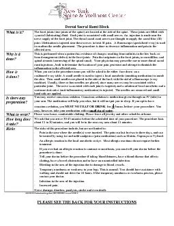
The facet joints true joints of the spine are located at the side of thsynovial lubricating fluid Each joint is associated with small nerves An injection is made
Presentation Embed Code
Download Presentation
Download Presentation The PPT/PDF document "Dorsal Sacral Rami Block 02282014rdswtea..." is the property of its rightful owner. Permission is granted to download and print the materials on this website for personal, non-commercial use only, and to display it on your personal computer provided you do not modify the materials and that you retain all copyright notices contained in the materials. By downloading content from our website, you accept the terms of this agreement.
Dorsal Sacral Rami Block 02282014rdswteachingsheet REVIEWED iewed 3: Transcript
Download Rules Of Document
"Dorsal Sacral Rami Block 02282014rdswteachingsheet REVIEWED iewed 3"The content belongs to its owner. You may download and print it for personal use, without modification, and keep all copyright notices. By downloading, you agree to these terms.
Related Documents