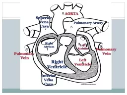
Why do you think that ventricles walls are thicker What makes a heart beat Todays Agenda Science Starter Beating Heart Heart Conduction System ECG amp Arrhythmia Exit Quiz https youtubeNEuSCPifKhA ID: 778765
Download The PPT/PDF document "Science Starter Which ventricle of the h..." is the property of its rightful owner. Permission is granted to download and print the materials on this web site for personal, non-commercial use only, and to display it on your personal computer provided you do not modify the materials and that you retain all copyright notices contained in the materials. By downloading content from our website, you accept the terms of this agreement.
Slide1
Science Starter
Which ventricle of the heart has thicker walls, left or right?
Why do you think that ventricle’s walls are thicker
?
What makes a heart beat?
Slide2Today’s Agenda
Science Starter
Beating Heart
Heart Conduction System
ECG & Arrhythmia
Exit Quiz
Slide3https
://youtu.be/NEuSCPifKhA
Slide4Slide5Think, Write, Pair, Share
Take a minute and think about why the heart could beat outside the body
Write down your thoughts
Share with your neighbor
A few volunteers will share their neighbors’ thoughts
Slide6Unit 5: Cardiovascular System
5.3: Heart
Contractions
Slide7A
.
The
Heart: Conduction System
The heart is not just an inert bundle of muscle
It contains
control centers (nodes) and conductive pathways that control its contractions
These pathways can function without input from the nervous system
Slide8B
. Heart
Contractions
Contraction is initiated by the
sinoatrial
node
Sequential stimulation occurs at other
autorhythmic
cells
“Auto-”: self
Slide9C. Heart
Contraction Sequence
Sinoatrial
(SA) node initiates contraction (L & R Atria contract)
Impulse from SA node reaches
a
trioventricular
(AV) node, a “delay switch”
AV node sends signal down atrioventricular bundle (AV bundle/Bundle of His
)
Slide10C. Heart
Contraction Sequence
Signal from AV bundle splits at bundle branches
Signal travels through Purkinje Fibers, which cause ventricles to contract
Chambers contract as one unit because cardiac muscle cells are electrically linked
Slide11How do we measure this?
With a device called an EKG or ECG (electrocardiogram)
This is the beep-beep thing in medical
tv
shows
Slide12D. ECG
– electrocardiogram
–
a recording of the electrical events (changes) during a cardiac cycle
P Wave
–
depolarization
(contraction)
of the atria
(atrial contraction – systole)
QRS Complex
– depolarization of the ventricles (ventricular contraction, systole)T Wave – Repolarization of the ventricles
Slide13E. What
happens if this goes wrong?
We get an abnormal heartbeat
A.K.A. an
arrhythmia
This can be diagnosed by examining specific portions of an ECG trace
A few examples:
http://www.practicalclinicalskills.com/ekg-reference-guide.aspx
Slide14Arrhythmia Dance!
http://youtu.be/r9yUIDNmCsw
Slide15Cardiac Conduction Practice
Use the link on my website to practice with cardiac conduction
Mrhoutzscience.weebly.com
Match the heart condition with the EKG strip
Make sure you complete the final question
Slide16Exit Quiz