/
RADIOLOGY  BONE   DISEASE RADIOLOGY  BONE   DISEASE

RADIOLOGY BONE DISEASE - PowerPoint Presentation

dorothy
dorothy . @dorothy
Follow
342 views
Uploaded On 2022-06-08

RADIOLOGY BONE DISEASE - PPT Presentation

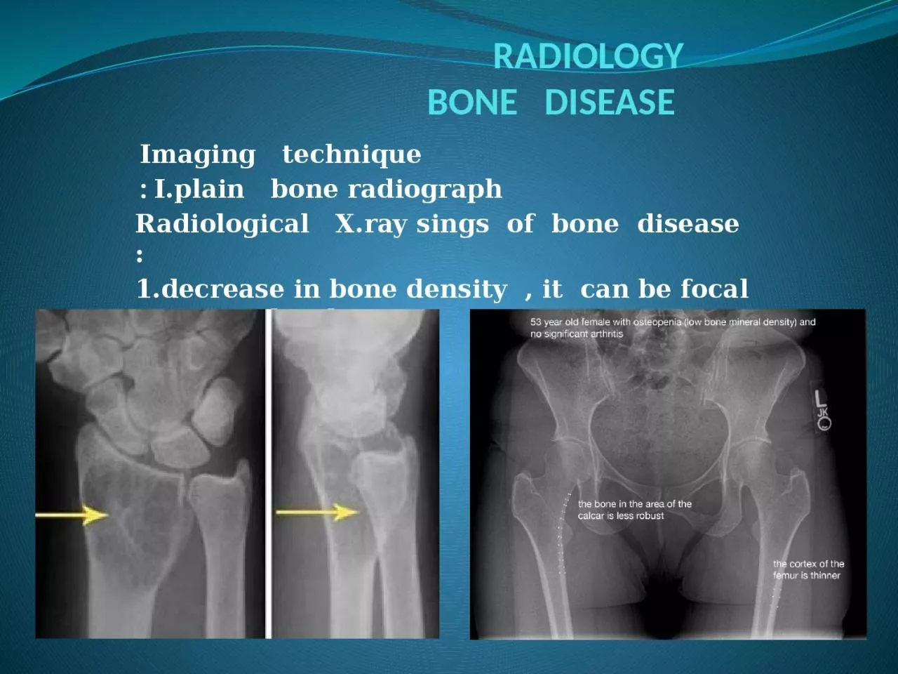
Imaging technique Iplain bone radiograph Radiological Xray sings of bone disease 1decrease in bone density it can be focal or generalized 2increase bone density sclerosis can also be focal or generalized ID: 915708

amp bone disease tumour bone amp tumour disease lesion malignant alteration diagnose benign tissue soft primary sarcoma tumours langerhans

Share:

Link:

Embed:

Download Presentation from below link

Download Presentation The PPT/PDF document "RADIOLOGY BONE DISEASE" is the property of its rightful owner. Permission is granted to download and print the materials on this web site for personal, non-commercial use only, and to display it on your personal computer provided you do not modify the materials and that you retain all copyright notices contained in the materials. By downloading content from our website, you accept the terms of this agreement.


Presentation Transcript

Slide1

RADIOLOGY BONE DISEASE

Imaging techniqueI.plain bone radiograph :Radiological X.ray sings of bone disease :1.decrease in bone density , it can be focal or generalized

Slide2

2-increase bone density ( sclerosis ) can also be focal or generalized

Slide3

3-periosteal

reaction

Slide4

4- cortical

thickening

Slide5

5. alteration in

trabecular pattern

Slide6

6- alteration in the shape of bone

e.g acromegaly 7- alteration in bone age II. U/S in musculoskeletal disease III. radionuclide bone scanning

Slide7

IV

. computed tomography in bone disease (CT scan ) :1.Demonestrating abnormality in the pelvis and spine 2.Demonstrating the extent & characterization of bone tumour in selected cases to complement MRI 3.As gide of bone biopsy

Slide8

V.MRI (magnetic resonance imaging in bone disease

):.demonstrate disc herniation and spinal cord or nerve root compression . to diagnose bone metastasis .show extent of primary bone tumor & demonstrate myloma & lymphoma . image soft tissue mass . to diagnose osteomylitis & show any soft tissue abnormality

. to diagnose a vascular necrosis & other joint pathology .

Slide9

Slide10

Bone diseases

Solitary bone lesion are usually one of the following .bone tumor: malignant ( primary or secondary ) , benign . osteomyelitis . bone cyst, fibrous dysplasia or other non –neoplastic defects of bone . condition of uncertain nature such as langerhans histiocystosis

the initial radiological decision is usually to try &decide whether the solitary lesion is benign or its aggressive by looking for the following features on plain radiographs & CT

:

1.Zone

of

transition

2.The adjacent cortex

Slide11

Slide12

3.Expansion

Slide13

4.Periosteal

reaction

Slide14

the causes of localized

peiosteal reactions adjacent to a lytic or sclerotic lesions are :.Osteomyelitis .Malignant bone tumour , particularly Ewing sarcoma & osteosarcoma.Occasionally metastasis , particularly neuroblastoma .

Langerhans histiocytosis

.Trauma

5.

Calcific

densities within the

lesion

Slide15

6. Soft tissue

swelling7.Site:The site of a lesion is most important as certain lesions tend to occur at certain sites.

Slide16

Bone

tumours Primary malignant tumours Osteosarcoma ( osteogenic sarcoma )

Slide17

Chondrosarcoma

:

Slide18

Slide19

Ewing

sarcoma

Slide20

Giant cell

tumour

Slide21

Benign

tumour & tumour like condition Enchondromas :

Slide22

Fibrous cortical defects ( non ossifying

fibromas )

Slide23

A simple bone cyst

:

Slide24

Aneurysmal

bone cysts :

Slide25

Osteoid

osteoma :

Slide26

Eosinophilic

granuloma :