/
wwwthelancetcomVol 383   May 31 2014 wwwthelancetcomVol 383   May 31 2014

wwwthelancetcomVol 383 May 31 2014 - PDF document

elise
elise . @elise
Follow
342 views
Uploaded On 2022-08-16

wwwthelancetcomVol 383 May 31 2014 - PPT Presentation

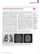
Seminar Atrial septal defectsTal Geva Jose D Martins Rachel M WaldAtrial septal defects are the third most common type of congenital heart disease Included in this group of pulmonary circulations ID: 936876

septal atrial closure defect atrial septal defect closure defects heart left pulmonary secundum 147 patients 133 device septum disease

Share:

Link:

Embed:

Download Presentation from below link

Download Pdf The PPT/PDF document "wwwthelancetcomVol 383 May 31 2014" is the property of its rightful owner. Permission is granted to download and print the materials on this web site for personal, non-commercial use only, and to display it on your personal computer provided you do not modify the materials and that you retain all copyright notices contained in the materials. By downloading content from our website, you accept the terms of this agreement.


Presentation Transcript

Seminar www.thelancet.comVol 383 May 31, 2014 Atrial septal defectsTal Geva, Jose D Martins, Rachel M WaldAtrial septal defects are the third most common type of congenital heart disease. Included in this group of pulmonary circulations. Most children with isolated atrial septal defects are free of symptoms, but the rates of exercise defects. Surgical closure is safe and e ective and when done before age 25 years is associated with normal life Search strategy and selection criteria Seminar www.thelancet.comVol 383 May 31, 2014 gure 2). The tissue that tissue that separates the coronary sinus from the left Patent foramen ovalePatent foramen ovale is the space between a well developed (valve-competent) septum primum and a normally formed septum secundum (“ gure 3). It is a normal interatrial communication during fetal life, characterised by streaming of oxygen-rich ” ow from the ductus venosus and, to a lesser extent, from the inferior vena cava, through the foramen ovale to the left atrium. After birth, left atrial pressure normally exceeds right atrial pressure and, septum primum apposes septum secundum, and the foramen ovale narrows. A patent foramen ovale is seen in almost all newborn babies, but its frequency decreases Complete anatomical closure of Secundum atrial septal defectSecundum atrial septal defect is a defect within the fossa gure 2B). Septum secundum is well-formed in most patients. Most secundum defects are not uent with the vena cavae, right pulmonary veins, coronary sinus, or the atrioventricular valves. With the ciency, or even complete Primum atrial septal defectPrimum atrial septal defect is one of several variants of common atrioventricular canal defects (also termed atrioventricular septal defect) with an interatrial communication located between the anterior-inferior margin of the fossa ovalis and the atrioventricular valves. The defect is characterised by a common atrioventricular ori“ ce with two distinct atrioventricular valve annuli completed by valve tissue adhering to the crest of the ventricular septum. The atrioventricular tissue occludes the space that accounts for the ventricular septal defect component in the complete form of the malformation (“ gure 2B). In addition to the septal defect, the atrioventricular valves in this anomaly are almost always abnormal, including a cleft in the anterior mitral lea” et. Unlike other types of atrial septal defects, the position and course of the conduction axis Sinus venosus defectThis defect is a communication between one or more of the right pulmonary veins and the cardiac end of the superior vena cava (superior vena cava type) or the posterior-inferior atrial wall just above the inferior vena cava-right atrial junction (inferior sinus venosus defect; gure 2B). About 4…11% of atrial septal defects are sinus The most common location of the Development of the atrial septum(A) At 28 days after gestation, septum primum (Sep 1°)„the “ rst septum to appear in the developing atria„develops as a crescent-shaped structure. Its leading edge is covered by a layer of mesenchymal cells called mesenchymal cap (shown in red). The space between the developing septum primum and the developing endocardial cushions is called foramen primum or ostium primum. Septum secundum (Sep 2°; limbus of the fossa ovalis) is a crescent-shaped muscular infolding of the atria wall that appears shortly thereafter to the right of septum primum. (B) At 35 days, both septum primum and septum secundum continue to develop. The openings within septum primum are called foramina secundi. (C) At 60 days, the atrial septum is nearly fully formed and the foramina secundi close by way of coalescing fenestrations within septum primum. The foramen ovale remains patent throughout pregnancy. Modi“ ed from Geva, with permission from Wiley-Blackwell Publishing. CPV=common pulmonary vein. IVC=inferior vena cava. LA=left atrium. LV=left ventricle. LVV=left venous valve. IVCSVC LVVRALARVLV Anatomy of the

atrial septum and neighbouring structures(A) Right atrial aspect of the normal atrial septum. (B) Types of interatrial communications. Modi“ ed from Geva,with permission from Wiley-Blackwell Publishing. ASD=atrial septal defect. Ao=aorta. CS=coronary sinus. CT=crista terminalis. EV=Eustachian valve. FO=foramen ovale. IVC=inferior vena cava. RA=right atrial. RLPV=right lower pulmonary vein. RMPV=right middle pulmonary vein. RUPV=right upper pulmonary vein. SLB=superior limbic band (septum secundum). SVC=superior vena cava. TBV=Thebesian valve. RMPV RUPVSVC CTRA appendage SLB RLPVFOIVCEVTBVCS SinusvenosusASD 1°ASD 2°AB Ao Seminar www.thelancet.comVol 383 May 31, 2014 ) is between the right upper pulmonary vein and the superior vena cava, resulting from de“ ciency of the tissue that separates these two The left atrial ori“ ce of the right upper pulmonary vein allows for communication between the left atrium and the cardiac end of the superior vena cava. Less frequently, the defect involves the posterior or inferior aspects of the right atrium with or without involvement of gure 2B).Coronary sinus defect ng of the tissue separating the coronary sinus from the left atrium, allowing a shunt ce. The left superior vena cava is termed Raghib syndrome.Common atrium is present when septum primum, septum secundum, and the atrioventricular canal septum are absent, which is often seen in patients with heterotaxy syndrome. Remnants of atrial septal tissue can sometimes PathophysiologyIn most patients an atrial septal defect results in left-to-which relate to the compliances of the left and right At birth, ventricular compliance is low, changing gradually to a ndings in secundum atrial septal defect include left-to-right shunt mostly during late atrial contraction and expiration. Most defects smaller ow ratio can exceed 1·5 and triggers a cascade chamber enlargement with diastolic septal shift towards the left ventricle and adverse interventricular interaction resulting in decreased left ventricular compliance and a shift from a circular to a D-shape short-axis geometry.These changes result in decreased left ventricular diastolic “ lling, increased pulmonary-to-systemic ”output. Left ventricular systolic dysfunction can develop Factors that decrease left-to-right ” ow include anatomical (eg, andcellular injury manifesting as increased serum leading to arteriolar narrowing and pulmonary hyper-tension. Mild increase in pulmonary artery pressure is The natural history of isolated atrial communications c factors. Sinus venosus and primum defects are cant shunt, do not decrease in size, and usually need surgical closure. By contrast, the natural history of secundum defects vary widely. Spontaneous closure occurs frequently in young patients with small defects. Hanslik in a study of 200 consecutive patients up 4·5 years), reported spontaneous closure in 56% of patients with an initial defect size of 4…5 mm, 30% in Atrial septal components and patent foramen ovaleArrow indicates patent foramen ovale (PFO). Modi“ ed from Geva, with permission from Wiley-Blackwell Publishing. AVS=atrioventricular septum. FO=fossa ovalis. ILB=inferior limbic band. LA=left atrium. LV=left ventricle. RARVSLBILB AVSFO PFOSep 1°LALV Seminar www.thelancet.comVol 383 May 31, 20146…7 mm defects, 12% in 8…10 mm defects, and in none of those with a larger defect. In that study, 39% of patients diagnosed at younger than 1 year had spontaneous closure by contrast with only 19% of those diagnosed later.In patients whose secundum defect does not close spontaneously, defect size can increase or decrease with In general, 70% of initially small (4 mm) defects decrease in size, 12% remain unchanged, and 18% increase. By contrast, in those with an initial defect size greater than 8…12 mm, only 9% decreased in size, 15% remain unchanged, and 76% increased. Notably, most reports of change in defect size have reported absolute values. Whether changes in defect size are

proportional to increase in heart size or to somatic growth is unclear.Most patients remain asymptomatic throughout most of childhood. Even those with a large left-to-right shunt might not have overt symptoms until adulthood. Incidental diagnosis by an echocardiogram obtained because of a heart murmur or an abnormal “ nding on a chest radiogram or an electrocardiogram is common. Rarely an isolated atrial septal defect is found in an infant with tachypnoea, slow weight gain, or recurrent respiratory In such cases, a careful search for associated non-cardiac anomalies and pulmonary hypertension In the second decade of life, none or subtle symptoms of shortness of breath with exertion or palpitations are common. By contrast, most adult patients with a large defect present with symptoms, including fatigue, exercise intolerance, palpitations, syncope, shortness of breath, peripheral oedema, manifestations of Exercise capacityExercise intolerance is uncommon in young children with Nonetheless, pulmonary function is often impaired in this age group. The frequency of exercise intolerance increases insidiously with age. Exercise capacity and peak oxygen consumption are decreased in most adults with unrepaired secundum defect, often at 50…60% of predicted values in healthy controls.Major arrhythmias are uncommon in children with atrial septal defects. The most common arrhythmias are atrial utter and “ brillation, incidences of which increase with age. Among the 211 adult patients with atrial septal defects only one patient younger than 40 years had atrial ” utter compared with 15% of the patients aged 40…60 years; 16% and 19% had atrial utter and “ brillation, respectively, in those older than 60 years. In addition to tachyarrhythmias, atrioventricular ) has Pulmonary hypertension is uncommon in children with Pulmonary vascular obstructive disease with or without right-to-left atrial-level shunting Patients with atrial septal defect and Echocardiographic imaging of atrial defects(A) Secundum atrial septal defect in the centre of the fossa ovalis (*). Note the left-to-right ” ow imaged by colour Doppler (arrow). (B) Superior vena cava (SVC)-type sinus venosus defect located above the fossa ovalis between the SVC and the right upper pulmonary vein as it enters the left atrium (LA). RA=right atrium. RPA=right pulmonary artery. A BLARA*LARPASVCRA* Seminar www.thelancet.comVol 383 May 31, 2014 anorexigenic agents (eg, fen” uramine) should be The availability of surgical closure of atrial septal defects since 1952 hampers the evaluation of its natural history in the modern era. Nonetheless, evidence clearly suggests that untreated large secundum defects are associated with a reduced lifespan. Campbell reported a low annualised mortality rate in the “ rst two decades of life (0·6% and 0·7% per year, respectively), increasing to 4·5% per year in the fourth decade and 7·5% per year in the sixth This “ nding is supported by a subsequent study can have few or no symptoms. The precordium is often hypertension. A soft systolic ejection murmur is usually heard over the pulmonary area in the left upper sternal border. A diastolic rumble over the left lower sternum ow through the tricuspid valve. A holosystolic ” ow murmur over the apex Signs of right heart failure are rare, but can be utter or brillation. Right ventricular hypertrophy is evident in patients with pulmonary hypertension. Left axis deviation cant atrial septal defects. Right atrial and anterior-posterior projection whereas right ventricular Similarly, left atrial dilatation (associated with mitral Transthoracic echocardiography is the primary diagnostic Two-dimensional imaging with colour Doppler ” ow (“ gure 4). Spectral Doppler further ow and allows determination ow velocity. Three-dimensional imaging allows en-face views of the defect from the right and left atrial perspectives, allowing appreciation of the defects shape gure 5).ventricular, and pulmonary

artery size. Right ventricular erence between erences between manifests as diastolic septal ” attening (deviation towards the left ventricle) and pressure overload manifests as septal ” attening during systole. Three-dimensional imaging of a secundum atrial septal defect imaged by a transoesophageal echocardiogram before (A) and after (B) device closure(A) En-face view of the defect as seen from the right atrium. (B) En-face view of the occluding device as seen from the left atrium. Images are from di erent patients. Ao=ascending aorta. ASD=atrial septal defect. IVC=inferior vena cava. SVC=superior vena cava. TV=tricuspid valve. A BSVCAoASDIVCTV SA Seminar www.thelancet.comVol 383 May 31, 2014those with restricted acoustic windows due to obesity, large body habitus, and previous thoracic surgery. Transoesophageal echocardiography provides an Contrast echocardiography with injection of agitated left superior vena cava, injection of agitated saline in a left arm venous cannula can diagnose a coronary sinus septal defect with appearance of the contrast in the left MRI and CTAdvances in cardiac MRI techniques allow anatomical delineation of atrial septal defects and quantitative cally, cine steady-state free precession imaging is currently the gold standard technique for measurements of ventricular volumes and function whereas phase velocity ow mapping allows accurate quanti“ cation of the ow ratio. Magnetic resonance angiography allows imaging of associated pulmonary and systemic venous anomalies. In patients with isolated secundum or primum defects, cardiac MRI is seldom necessary. Exceptions include those in whom the location of the defect or its haemodynamic burden is in question. In contrast, cardiac MRI is an important diagnostic gure 6). The posterior location of these defects and the frequently associated anomalous pulmonary venous drainage hamper the ability of transthoracic echocardiography to adequately evaluate these defects as patients body size increases.High-resolution contrast CT is capable of anatomical delineation of atrial septal defects. However, the risk of Cardiac catheterisation is seldom done solely for diagnostic purposes. Most catheterisations are done with the intention to close the defect percutaneously. A diagnostic procedure typically precedes device placement, including determination of pressures and pulmonary and systemic ow ratios. In selected patients, angiography is done to delineate associated anomalies not shown by non-invasive imaging. In adult patients at risk of coronary artery disease and in those with pulmonary hypertension, diagnostic TreatmentIndications and contraindication for defect closure cant shunt that cant shuntwas classically de“ in the absence of other causes (class IIa, level of up expectantly, keeping in mind the possibility of Pulmonary hypertension is not an absolute contra-baseline or after pulmonary vasodilator acute challenge a pulmonary-to-systemic ” ow ratio greater than 1·5 vascular resistance greater than 8 Woods units generally left shunt such as Eisenmenger syndrome. Inferior sinus venosus defect imaged by cardiac MRI(A) Cine steady-state free precession image in an oblique sagittal plane showing the defect in the posterior-inferior atrial junction (arrow), just above the entrance of the inferior vena cava (IVC) to the right atrium (RA). Note that the fossa ovalis is intact (arrow head). (B) Phase velocity mapping in the same orientation showing ” ow from the left atrium (LA) to the right atrium through the defect. SVC=superior vena cava. A BLARAIVCSVC Seminar www.thelancet.comVol 383 May 31, 2014 valve), including severe obstructive or restrictive right or left heart lesions. Patients with pulmonary Timing of defect closure cant atrial septal defect rmed. Although there is no lower limit of age for 3…5 years. At the other end of the age spectrum, evidence ective in Treatment strategies for defect closureSinus venosus, primum, and coronary sinus septal defects need sur

gical closure. Secundum defects can be using an occluding device delivered by a catheter. Transcatheter closure might not be feasible in some Since the pioneering reports of Murray who “ rst closed an atrial septal defect without direct visualisation in 1948 and Lewis and Tau“ c who used hypothermia and in” ow occlusion to close an atrial septal defect under direct vision more than 50 years of experience have resulted in a safe and e ective operation with almost no mortality and little morbidity. The defect is closed under direct vision using cardiopulmonary bypass, either by direct suture or with a pericardial or a synthetic patch. Access to the heart is accomplished through median sternotomy, sub-mammary incision, lateral thoracotomy, transxiphoid, and other approaches. A small skin incision improves the cosmetic results and video-assisted thoracoscopic Results of secundum defect closure in the modern era are excellent with near-zero mortality for isolated defects. Morbidities such as arrhythmias, bleeding, usions are usually transient. Arrhythmia and prolonged stay in �the intensive care unit (3 days) are more common in The long-term results of surgical closure of secundum defects are excellent for patients operated on when younger than 25 years with an actuarial survival curve that is indistinguishable from that of the general population„98% versus 99% in those operated on when younger than 12 years and 93% versus 97% in those operated on between the ages of 12 and 24 years. By contrast, those operated on at age 25…41 years 59%) had Transcatheter closureSince King and colleagues rst transcatheter eld has evolved substantially with a range of occluding devices and delivery systems now available (table). Improvements in device design and ease of use coupled with avoidance of cardiac surgery have led many centres to adopt rst Secundum defects larger than 36…40 mm in maximum diameter, inadequate margins to anchor the device, and interference of the device with atrioventricular valve function or with systemic or pulmonary venous The device is typically introduced through a sheath in the femoral vein and its deployment is guided by a combination of ” uoroscopy and echo cardiography. Echocardiographic guidance can be accomplished through a transoesophageal approach (real-time two-dimensional and three-dimensional), intracardiac ultrasound, or Most practitioners prescribe antiplatelet drugs after device implantation, but data to Current results of device closure of secundum defects cacy pro“ le. A retrospective multicentre review of 478 patients undergoing implantation of an Amplatzer septal occluding device (St Jude Medical, Plymouth, MN, USA) at a median age of 6 years showed technical success in 96% with an occlusion rate at 24 h of A study in 650 adult patients at a mean age of 45years showed similarly good early results. Results of a prospective multicentre cohort evaluating the Helex device (W L Gore & Associates, Flagsta , AZ, USA) showed a Complications occur in inverse relation to institutional respectively. Meta-analysis of 142 case series showed (0·1%). Cardiac erosion by the device was the focus of and a review by the US Food and Drug Administration. New-onset or worsening Although several studies have compared costs, clinical outcomes, e cacy, and rates of complications between transcatheter and surgical closure of secundum Seminar www.thelancet.comVol 383 May 31, 2014 no prospective randomised trial has been published at the time of writing. In general, these studies have shown no major advantage of one approach compared with the other with each having speci“ c advantages and The aforementioned discussion emphasises the importance of long-term follow-up after Clinical and haemodynamic results of defect closurePatients commonly report subjective improvement in symptoms after closure of atrial septal defects. Some adults after defect closure, Conversely, cant improvement after The haemodynamic res

ponse to defect closure includes Most of the decrease occurs immediately with some further remodelling 1…2 years later. A younger age at closure and a lesser degree of chamber enlargement before repair are associated with a higher likelihood of Persistent enlargement of the right heart has been reported in up to a third of patients, mostly in adults with severely dilated Echo cardiographic indices of right ventricular function before and after defect closure have shown mixed results with some measures showing an improvement from before to after closure, whereas others show either no Left ventricular “ lling improves However, in adults with pre-existing decreased left ventricular compliance, the acute increase in preload associated with defect closure can lead to worsening left atrial and pulmonary venous Pulmonary artery pressure decreases to normal except in some patients with moderate to severe pulmonary A salutary e ect of defect closure on atrial arrhythmias has been shown in meta-analysis of 26 studies spanning four decades, including 1841 surgical closures and Considering all studies, the incidence of atrial arrhythmias decreased in the short term (odds ratio 0·66, 95% CI 0·57…0·77). However, when only studies with at least 5 years of follow-up were considered, cial e ect was lost. Other studies have identi“ ed persistent rhythm and conduction abnormalities These observations have led the American Heart Association and the European Society of Cardiology to consider a Maze antiarrhythmic Adults with atrial septal defectsAn isolated atrial septal defect can occasionally go undiagnosed for decades. It accounts for 25…30% of Although many young adults have no subjective symptoms, exercise testing usually unveils subnormal exercise capacity. With advancing age, however, overt symptoms of fatigue, exercise intolerance, shortness of breath, palpitations, and manifestations of heart failure Onset of ischaemic heart disease and other comorbidities associated CompanyDesignMaximum Regulatory Nitinol mesh of self-centring double disc with 40 mmEasy to use; versatile; easily withdrawn and redeployable; small delivery sheath (8/9 Fr for 18 mm device); largest experience worldwideAvoid in nickel allergy; late erosions (rare); sti er delivery cable (distorts anatomy before release) FDA and CE W L Gore & , AZ, covered with ePTFE 18 mmFlexible, compliant (no erosion, conforms to le; metal almost completely covered by ePTFE; Not self-centred; slightly more challenging deployment; care with de“ cient rims; more device embolisation and residual shunt; needs larger sheath (12 Fr for closure of 18 mm defect)FDA and CE Nitinol mesh of self-40 mmSame as Amplatzer device, but left atrial component ” at and latest generation has tilting of attachment with delivery cable Same as Amplatzer device, but needs slightly larger sheaths (10 Fr for 18 mm device); less Nitinol mesh of self-42 mmSame as Amplatzer device, but has a ” at left atrial component and latest generation has tilting with delivery cable (allows better Same as Amplatzer device, but needs slightly larger sheaths (10 Fr for 18 mm device); scarce Nitinol double disk self-34 mmLow pro“ le, ” exible (conforms to atrial anatomy), retrievable and re-deployable, less metal, which is coveredRequires only slightly larger delivery sheath Amplatzer device (9 Fr for 18 mm device); no published experience with newest design, See appendix for images of devices. FDA=US Food and Drug Administration. CE=Conformité Européenne. ePTFE=expanded polytetra” Table: Examples of devices for transcatheter closure of atrial septal defect See Online Seminar www.thelancet.comVol 383 May 31, 2014 with decreased left ventricular compliance (eg, essential hypertension, aortic valve stenosis, ageing) lead to increased left-to-right shunting across the defect, which further aggravate the symptoms and lead to clinical deterioration. Atrial ” utter and “ brillation are important causes of mor

bidity, seen in 21% of adults older than Repaired defectsafter age 40 years confers morbidity and mortality ts compared with medical therapy alone.exercise capacity, decrease in right atrial and right ts are less pronounced after age In view of the risk of unmasking left utter and “ brillation, remains high after defect closure Maternal complications are uncommon in isolated atrial septal defects not complicated by pulmonary Yap and colleagues found similarly low rates of maternal complications in women with repaired and unrepaired defects, including arrhythmias (4%) and transient ischaemic attack (1%). None of the 98 women with 188 pregnancies had a stroke, heart failure symptoms, or endocarditis. Pre-pregnancy history of arrhythmia and maternal age older than 30 years were risk factors for maternal cardiac complications. By comparison with the general population, women with unrepaired atrial septal defects had an increased risk of pre-eclampsia, fetal loss, and low birthweight. By contrast, the outcome for o spring of women with a repaired defect was similar to that of the Pregnancy should be avoided in women with an atrial septal defect and severe pulmonary hypertension. In a contemporary study maternal mortality was prohibitively high (28%) in women with congenital heart disease and pulmonary hypertension, despite use of pulmonary Maternal deaths tended to occur shortly after delivery and were often caused by heart failure, thromboembolism, All authors took part in the review of the literature, drafting the Seminar, nal version.Declaration of interestsWe declare that we have no competing interests.References1 Ho man JI, Kaplan S. The incidence of congenital heart disease. J Am Coll Cardiol2 Botto LD, Correa A, Erickson JD. Racial and temporal variations Pediatrics3 Caputo S, Capozzi G, Russo MG, et al. Familial recurrence of congenital heart disease in patients with ostium secundum atrial Eur Heart J4 Chen Y, Han ZQ, Yan WD, et al. A novel mutation in GATA4 gene associated with dominant inherited familial atrial septal defect. J Thorac Cardiovasc Surg 684…87.5 Maitra M, Schluterman MK, Nichols HA, et al. Interaction of Gata4 and Gata6 with Tbx5 is critical for normal cardiac development. 368…77.6 Schott JJ, Benson DW, Basson CT, et al. Congenital heart disease 1998; 7 Benson DW, Silberbach GM, Kavanaugh-McHugh A, et al. Mutations in the cardiac transcription factor NKX2.5 a ect diverse cardiac 8 McElhinney DB, Geiger E, Blinder J, Benson DW, Goldmuntz E. NKX2.5 mutations in patients with congenital heart disease. J Am Coll Cardiol9 Lee SA, Lee SG, Moon HS, Lavulo L, Cho KO, Hyun C. GATA4gene in a family of Doberman Pinschers with an atrial septal defect. J Genet 241…47.10 Hirayama-Yamada K, Kamisago M, Akimoto K, et al. Phenotypes with GATA4 or NKX2.5 mutations in familial atrial septal defect. Am J Med Genet A11 DAmato E, Giacopelli F, Giannattasio A, et al. Genetic investigation in an Italian child with an unusual association of atrial septal defect, GATA4 gene mutation, and neonatal Diabet Med12 Ching YH, Ghosh TK, Cross SJ, et al. Mutation in myosin heavy Nat Genet13 Okubo A, Miyoshi O, Baba K, et al. A novel GATA4 mutation completely segregated with atrial septal defect in a large Japanese family. J Med Genet e97.14 Ikeda Y, Hiroi Y, Hosoda T, et al. Novel point mutation in the cardiac transcription factor CSX/NKX2.5 associated with congenital heart 15 Hosoda T, Komuro I, Shiojima I, et al. Familial atrial septal defect and atrioventricular conduction disturbance associated with a point CSX/NKX2-5 in a Japanese jørnstad PG, Leren TP. Familial atrial septal defect in the oval fossa with progressive prolongation of the atrioventricular conduction Cardiol Young 2009; 17 Rifai L, Maazouzi W, Se“ ani A. Novel point mutation in the gene in a Moroccan family with atrioventricular conduction Cardiol Young18 Gelernter-Yaniv L, Lorber A. The familial form of atrial septal defect. Acta Paediatr19 Elliott DA, Kirk EP, Yeoh T, et al. Cardiac ho

meobox gene mutations and congenital heart disease: associations with atrial septal defect and hypoplastic left heart syndrome. J Am Coll Cardiol20 Strauss RM, Ferguson AD, Rittey CD, Cork MJ. Microcephaly-lymphoedema-chorioretinal-dysplasia syndrome Pediatr Dermatol21 Yoshihara K, Ozawa T, Sakuragawa H, et al. [Noonan syndrome associated with atrial septal defect, pulmonary stenosis, and completely unroofed coronary sinus without LSVC: a case report]. Kyobu Geka 134…37 (in Japanese).22 Cunningham ET Jr, Eliott D, Miller NR, Maumenee IH, Green WR. Familial Axenfeld-Rieger anomaly, atrial septal defect, and sensorineural hearing loss: a possible new genetic syndrome. Seminar www.thelancet.comVol 383 May 31, 201423 Diegeler A, Van Son JA, Mohr FW. Budd-Chiari syndrome as late complication of secundum atrial septal defect closure. Eur J Cardiothorac Surg24 Shimizu K, Arai H, Sakamoto T, Sunamori M, Suzuki A. Jarcho-Levin syndrome associated with atrial septal defect and partial anomalous J Card Surg25 Aynaci FM, Ozdemir M, Iik Y. Atrial septal defect in Hallermann syndrome. Genet Couns26 Holt M, Oram S. Familial heart disease with skeletal malformations. Br Heart J27 Ganie MA, Laway BA, Ahmed S, Alai MS, Lone GN. Mayer-Rokintansky-Kuster-Hauser syndrome associated with atrialseptal defect, partial anomalous pulmonary venous connectionand unilateral kidney…an unusual triad of anomalies. J Pediatr Endocrinol Metab28 Freeman SB, Bean LH, Allen EG, et al. Ethnicity, sex, and the incidence of congenital heart defects: a report from the national Genet Med29 Burd L, Deal E, Rios R, Adickes E, Wynne J, Klug MG. Congenitalheart defects and fetal alcohol spectrum disorders. Congenit Heart Dis30 Alverson CJ, Strickland MJ, Gilboa SM, Correa A. Maternal smoking and congenital heart defects in the Baltimore-Washington Infant Study. Pediatrics31 Lee LJ, Lupo PJ. Maternal smoking during pregnancy and the risk ofcongenital heart defects in o spring: a systematic review and Pediatr Cardiol 398…407.32 Louik C, Lin AE, Werler MM, Hernández-Díaz S, Mitchell AA. First-trimester use of selective serotonin-reuptake inhibitors and N Engl J Med33 Polen KN, Rasmussen SA, Riehle-Colarusso T, Reefhuis J, and the National Birth Defects Prevention Study. Association between reported venlafaxine use in early pregnancy and birth defects, national birth defects prevention study, 1997…2007. Birth Defects Res A Clin Mol Teratol34 Bakker MK, Kerstjens-Frederikse WS, Buys CH, de Walle HE, deJong-van den Berg LT. First-trimester use of paroxetine and congenital heart defects: a population-based case-control study. Birth Defects Res A Clin Mol Teratol35 Correa A, Gilboa SM, Besser LM, et al. Diabetes mellitus and birth Am J Obstet Gynecol36 Parker SE, Werler MM, Shaw GM, Anderka M, Yazdy MM, and theNational Birth Defects Prevention Study. Dietary glycemic index 37 Miller A, Riehle-Colarusso T, Si el C, Frías JL, Correa A. Maternalage and prevalence of isolated congenital heart defects Am J Med Genet A 2011; 38 Reefhuis J, Honein MA, Schieve LA, Correa A, Hobbs CA, Rasmussen SA, and the National Birth Defects Prevention Study. Assisted reproductive technology and major structural birth defects Hum Reprod39 Geva T. Anomalies of the atrial septum. In: Lai WW, Mertens LL, Cohen MS, Geva T, eds. Echocardiography in pediatric and congenital heart disease: from fetus to adult. Oxford: Wiley-Blackwell Publishing, 2009; 158…74.40 Anderson RH, Brown NA, Webb S. Development and structure of Heart42 Mommersteeg MT, Soufan AT, de Lange FJ, et al. Two distinct pools of mesenchyme contribute to the development of the atrial septum. 43 Wessels A, Anderson RH, Markwald RR, et al. Atrial development inthe human heart: an immunohistochemical study with emphasis 44 Van Praagh S, Carrera ME, Sanders SP, Mayer JE, VanPraagh R.Sinus venosus defects: unroo“ ng of the right ndings Am Heart J45 Blom NA, Gittenberger-de Groot AC, Jongeneel TH, DeRuiter MC, Poelmann RE,

Ottenkamp J. Normal development of the pulmonary veins in human embryos and formulation of a morphogenetic Am J Cardiol46 Banka P, Bacha E, Powell AJ, Benavidez OJ, Geva T. Outcomes of inferior sinus venosus defect repair. J Thorac Cardiovasc Surg 2011; 47 Chin AJ, Murphy JD. Identi“ cation of coronary sinus septal defect (unroofed coronary sinus) by color Doppler echocardiography. Am Heart J 1655…57.48 Hagen PT, Scholz DG, Edwards WD. Incidence and size of patent foramen ovale during the “ rst 10 decades of life: an autopsy study Mayo Clin Proc49 Connuck D, Sun JP, Super DM, et al. Incidence of patent ductus Am J Cardiol 244…47.50 Fisher DC, Fisher EA, Budd JH, Rosen SE, Goldman ME. Theincidence of patent foramen ovale in 1,000 consecutive patients. A contrast transesophageal echocardiography study. 1995; 51 Schneider B, Zienkiewicz T, Jansen V, Hofmann T, Noltenius H, Meinertz T. Diagnosis of patent foramen ovale by transesophageal ndings. Am J Cardiol52 Attenhofer Jost CH, Connolly HM, Danielson GK, et al. Sinus venosus atrial septal defect: long-term postoperative 53 Raghib G, Ruttenberg HD, Anderson RC, Amplatz K, Adams P Jr, Edwards JE. Termination of left superior vena cava in left atrium, atrial septal defect, and absence of coronary sinus; a developmental 54 Fuse S, Tomita H, Hatakeyama K, Kubo N, Abe N. E ect of size of a Am J Cardiol 2001; 55 Walker RE, Moran AM, Gauvreau K, Colan SD. Evidence of adverse ventricular interdependence in patients with atrial septal defects. Am J Cardiol 1374…77.56 Masutani S, Senzaki H. Left ventricular function in adult patients with atrial septal defect: implication for development of heart failure after transcatheter closure. J Card Failalvo G, Drago M, Pacileo G, et al. Atrial function after surgical and percutaneous closure of atrial septal defect: a strain rate imaging study. 58 Sugimoto M, Ota K, Kajihama A, Nakau K, Manabe H, Kajino H. Volume overload and pressure overload due to left-to-right shunt-induced myocardial injury. Evaluation using a highly sensitive cardiac Troponin-I assay in children with congenital heart disease. 59 Steele PM, Fuster V, Cohen M, Ritter DG, McGoon DC. Isolatedatrial septal defect with pulmonary vascular obstructive disease„long-term follow-up and prediction of outcome after 60 Sachweh JS, Daebritz SH, Hermanns B, et al. Hypertensive pulmonary vascular disease in adults with secundum or sinus Ann Thorac Surg61 Hanslik A, Pospisil U, Salzer-Muhar U, Greber-Platzer S, Mal-e C. defect in children: a longitudinal study. Pediatrics 2006; 62 Helgason H, Jonsdottir G. Spontaneous closure of atrial septal Pediatr Cardiol63 McMahon CJ, Feltes TF, Fraley JK, et al. Natural history of growth ofsecundum atrial septal defects and implications for transcatheter Heart64 Saxena A, Divekar A, Soni NR. Natural history of secundum atrial septal defect revisited in the era of transcatheter closure. Indian Heart J65 Andrews R, Tulloh R, Magee A, Anderson D. Atrial septal defect withfailure to thrive in infancy: hidden pulmonary vascular disease? Pediatr Cardiol66 Lammers A, Hager A, Eicken A, Lange R, Hauser M, Hess J. Need for closure of secundum atrial septal defect in infancy. J Thorac Cardiovasc Surg 1353…57.67 Goetschmann S, Dibernardo S, Steinmann H, Pavlovic M, Sekarski N, Pfammatter JP. Frequency of severe pulmonary hypertension complicating isolatedŽ atrial septal defect in infancy. Am J Cardiol Seminar www.thelancet.comVol 383 May 31, 2014 68 Rhodes J, Patel H, Hijazi ZM. E ect of transcatheter closure of atrialseptal defect on the cardiopulmonary response to exercise. Am J Cardiol69 Lee YS, Jeng MJ, Tsao PC, et al. Pulmonary function changes inchildren after transcatheter closure of atrial septal defect. Pediatr Pulmonol70 Van De Bruaene A, Buys R, Vanhees L, Delcroix M, Moons P, Budts W. Cardiopulmonary exercise testing and SF-36 in patients with atrial J Cardiopulm Rehabil Prev71 Berger F, Vogel M, Kramer A, et al. Incidence of atrial ”

; brillation in adults with atrial septal defect before and after surgery. Ann Thorac Surg72 Nouira S, Kamoun I, Ouragini H, et al. Clinical and genetic investigation of atrial septal defect with atrioventricular conduction defect in a large consanguineous Tunisian family. Arch Med Res 2008; 73 Khoury GH, Hawes CR. Atrial septal defect associated with J Pediatr74 Humenberger M, Rosenhek R, Gabriel H, et al. Bene“ t of atrial Eur Heart J 2011; 75 Yalonetsky S, Lorber A. Comparative changes of pulmonary arterypressure values and tricuspid valve regurgitation following transcatheter atrial septal defect closure in adults and the elderly. Congenit Heart Dis76 Engelfriet P, Meijboom F, Boersma E, Tijssen J, Mulder B. Repaired and open atrial septal defects type II in adulthood: an epidemiological Int J Cardiol77 Campbell M. Natural history of atrial septal defect. Br Heart J 1970; 78 Murphy JG, Gersh BJ, McGoon MD, et al. Long-term outcome after surgical repair of isolated atrial septal defect. Follow-up at 27 to N Engl J Med79 van den Bosch AE, Ten Harkel DJ, McGhie JS, et al. Characterization of atrial septal defect assessed by real-time 3-dimensional echocardiography. 80 Rosenzweig BP, Nayar AC, Varkey MP, Kronzon I. Echo contrast-enhanced diagnosis of atrial septal defect. 155…57.81 Valverde I, Simpson J, Schae ter T, Beerbaum P. 4D phase-contrast ow cardiovascular magnetic resonance: comprehensive quanti“ cation and visualization of ” ow dynamics in atrial septal defect and partial Pediatr Cardiol82 Teo KS, Disney PJ, Dundon BK, et al. Assessment of atrial septal transoesophageal echocardiography. J Cardiovasc Magn Reson83 Powell AJ, Tsai-Goodman B, Prakash A, Greil GF, Geva T. Comparison between phase-velocity cine magnetic resonance imaging and invasive oximetry for quanti“ cation of atrial shunts. Am J Cardiolalente AM, Sena L, Powell AJ, Del Nido PJ, Geva T. Cardiacmagnetic resonance imaging evaluation of sinus venosus defects: comparison to Pediatr Cardiol85 Mathews JD, Forsythe AV, Brady Z, et al. Cancer risk in 680,000 people exposed to computed tomography scans in childhood oradolescence: data linkage study of 11 million Australians. 86 Warnes CA, Williams RG, Bashore TM, et al, and the American College of Cardiology, and the American Heart Association Task Force on Practice Guidelines (Writing Committee to Develop Guidelines on the Management of Adults With Congenital Heart Disease), and the American Society of Echocardiography, and the Heart Rhythm Society, and the International Society for Adult Congenital Heart Disease, and the Society for Cardiovascular Angiography and Interventions, and the Society of Thoracic Surgeons. ACC/AHA 2008 guidelines for the management of adults with congenital heart disease: a report of the American College of Cardiology/American Heart Association Task Force on Practice Guidelines (Writing Committee to Develop Guidelines on the Management of Adults with Congenital Heart Disease). Developed in collaboration with the American Society of Echocardiography, Heart Rhythm Society, International Society for Adult Congenital Heart Disease, Society for Cardiovascular Angiography and Interventions, and Society of Thoracic Surgeons. J Am Coll Cardiol87 Baumgartner H, Bonhoe er P, De Groot NM, et al, and the Task Force on the Management of Grown-up Congenital Heart Disease of the European Society of Cardiology (ESC), and the Association for European Paediatric Cardiology (AEPC), and the ESC Committee for Practice Guidelines (CPG). ESC Guidelines for the Eur Heart J 2915…57.88 Kim YH, Yu JJ, Yun TJ, et al. Repair of atrial septal defect with Eisenmenger syndrome after long-term sildena“ l therapy. Ann Thorac Surg89 Hanninen M, Kmet A, Taylor DA, Ross DB, Rebeyka I, VonderMuhll IF. Atrial septal defect closure in the elderly is Can J Cardiol90 Nakagawa K, Akagi T, Taniguchi M, et al. Transcatheter closure Catheter Cardiovasc Interv91 Nyboe C, Fenger-Grøn M, Nielsen-Kudsk JE, Hjortdal V. Eur J Cardiothora

c Surg 752…57.92 Murray G. Closure of defects in cardiac septa. Ann Surg93 Lewis FJ, Tau“ c M. Closure of atrial septal defects with the aid Surgery94 Suematsu Y, Kiaii B, Bainbridge DT, del Nido PJ, Novick RJ. Robotic-assisted closure of atrial septal defect under real-time three-dimensional echo guide: in vitro study. Eur J Cardiothorac Surg95 King TD, Thompson SL, Steiner C, Mills NL. Secundum atrial septal defect. Nonoperative closure during cardiac catheterization. 96 Opotowsky AR, Landzberg MJ, Kimmel SE, Webb GD. Trends inthe 97 Meier B. Percutaneous atrial septal defect closure: pushing the envelope but pushing it gently. Catheter Cardiovasc Interv98 Valente A, Rhodes JF. Current indications and contraindications Am Heart J99 Kleinman CS. Echocardiographic guidance of catheter-based Pediatr Cardiol100 Balzer J, van Hall S, Rassaf T, et al. Feasibility, safety, and everett AD, Jennings J, Sibinga E, et al. Community use of the amplatzer atrial septal defect occluder: results of the multicenter MAGIC atrial septal defect study. Pediatr Cardiol 240…47.102 Majunke N, Bialkowski J, Wilson N, et al. Closure of atrial septal Am J Cardiol103 Jones TK, Latson LA, Zahn E, et al, and the Multicenter Pivotal Study of the HELEX Septal Occluder Investigators. Results of the U.S. multicenter pivotal study of the HELEX septal occluder for percutaneous closure of secundum atrial septal defects. J Am Coll Cardiol104 Opotowsky AR, Landzberg MJ, Kimmel SE, Webb GD. Percutaneousclosure of patent foramen ovale and atrial septal defect in adults: the impact of clinical variables and hospital procedure Am Heart J105 Sadiq M, Kazmi T, Rehman AU, Latif F, Hyder N, Qureshi SA. Device closure of atrial septal defect: medium-term outcome with Cardiol Young106 Abaci A, Unlu S, Alsancak Y, Kaya U, Sezenoz B. Short and long term complications of device closure of atrial septal defect and patent foramen ovale: meta-analysis of 28,142 patients from 203 studies. Catheter Cardiovasc Interv107 Divekar A, Gaamangwe T, Shaikh N, Raabe M, Ducas J. Cardiacperforation after device closure of atrial septal defects with the Amplatzer septal occluder. J Am Coll Cardiol Seminar www.thelancet.comVol 383 May 31, 2014108 Amin Z, Hijazi ZM, Bass JL, Cheatham JP, Hellenbrand WE, Kleinman CS. Erosion of Amplatzer septal occluder device after closure of secundum atrial septal defects: review of registry of complications and recommendations to minimize future risk. Catheter Cardiovasc Interv109 Taggart NW, Dearani JA, Hagler DJ. Late erosion of an Amplatzer septal occluder device 6 years after placement. J Thorac Cardiovasc Surg110 Gaithersburg H. FDA executive summary memorandum. Transcatheter ASD occluders: clinical update and review of events. 2012. http://www.fda.gov/downloads/AdvisoryCommittees/CommitteesMeetingMaterials/MedicalDevices/MedicalDevices AdvisoryCommittee/CirculatorySystemDevicesPanel/UCM304924.pdf (accessed Feb 7, 2014).111 Wertman B, Azarbal B, Riedl M, Tobis J. Adverse events associated with nickel allergy in patients undergoing percutaneous atrial septal J Am Coll Cardiol 2006; 1226…27.112 Kutty S, Hazeem AA, Brown K, et al. Long-term (5- to 20-year) outcomes after transcatheter or surgical treatment of hemodynamically signi“ cant isolated secundum atrial septal defect. Am J Cardiol113 Suchon E, Pieculewicz M, Tracz W, Przewlocki T, Sadowski J, Podolec P. Transcatheter closure as an alternative and equivalent method to the surgical treatment of atrial septal defect in adults: Med Sci Monit 2009; CR612…17.114 Butera G, Carminati M, Chessa M, et al. Percutaneous versus surgical closure of secundum atrial septal defect: comparison Am Heart J115 Rhee EK, Evangelista JK, Nigrin DJ, Erickson LC. Impact of Am J Cardiol116 Takaya Y, Taniguchi M, Akagi T, et al. Long-term e ects of transcatheter closure of atrial septal defect on cardiac remodeling and exercise capacity in patients older than 40 years with a reduction J Interv Cardiol117 Zaqout M, De Baets F, Schelstraet

e P, et al. Pulmonary function inchildren after surgical and percutaneous closure of atrial septal Pediatr Cardiol118 Thomas VC, Vincent R, Raviele A, Diehl H, Qian H, Kim D. Transcatheter closure of secundum atrial septal defect in infants less than 12 months of age improves symptoms of chronic lung disease. Congenit Heart Dis119 Du ZD, Cao QL, Koenig P, Heitschmidt M, Hijazi ZM. Speedofnormalization of right ventricular volume overload after transcatheter closure of atrial septal defect in children and adults. Am J Cardiol120 Monfredi O, Luckie M, Mirjafari H, et al. Percutaneous device closure of atrial septal defect results in very early and sustained changes of right and left heart function. Int J Cardiol 2013; 121 Veldtman GR, Razack V, Siu S, et al. Right ventricular form and function after percutaneous atrial septal defect device closure. J Am Coll Cardiol122 Schoen SP, Kittner T, Bohl S, et al. Transcatheter closure of atrial septal defects improves right ventricular volume, mass, function, pulmonary pressure, and functional class: a magnetic resonance imaging study. Heart123 Vitarelli A, Sardella G, Roma AD, et al. Assessment of right ventricular function by three-dimensional echocardiography and myocardial strain imaging in adult atrial septal defect before and afterpercutaneous closure. Int J Cardiovasc Imaging124 Salehian O, Horlick E, Schwerzmann M, et al. Improvements in cardiac form and function after transcatheter closure of secundum J Am Coll Cardiol125 Masutani S, Taketazu M, Ishido H, et al. E ects of age on hemodynamic changes after transcatheter closure of atrial septal Heart Vessels 2012; amman A, Haberer K, et al. Outcomes in patients with pulmonary hypertension undergoing percutaneous atrial septal defect Heart127 Vecht JA, Saso S, Rao C, et al. Atrial septal defect closure is associated with a reduced prevalence of atrial tachyarrhythmia in the short to Heart 2010; 1789…97.128 Cottens D, Van De Bruaene A, Troost E, Willems R, Moons P, Budts W. In” uence of percutaneous transcatheter closure of an atrial Acta Cardiol 2011; 129 Thilén U, Carlson J, Platonov PG, Olsson SB. Atrial myocardial pathoelectrophysiology in adults with a secundum atrial septal defect is una ected by closure of the defect. A study using high resolution Int J Cardiol 2009; 130 Lindsey JB, Hillis LD. Clinical update: atrial septal defect in adults. 131 Diller GP, Dimopoulos K, Okonko D, et al. Exercise intolerance inadult congenital heart disease: comparative severity, correlates, 132 Attie F, Rosas M, Granados N, Zabal C, Buendía A, Calderón J. Surgical treatment for secundum atrial septal defects in patients J Am Coll Cardiol 2001; 133 Konstantinides S, Geibel A, Olschewski M, et al. A comparison ofsurgical and medical therapy for atrial septal defect in adults. N Engl J Med134 Giardini A, Donti A, Formigari R, et al. Determinants of cardiopulmonary functional improvement after transcatheter atrial J Am Coll Cardiol 2004; 135 Schussler JM, Anwar A, Phillips SD, Roberts BJ, Vallabhan RC, Grayburn PA. E ect on right ventricular volume of percutaneous Am J Cardiol 2005; 136 Khan AA, Tan JL, Li W, et al. The impact of transcatheter atrial septaldefect closure in the older population: a prospective study. JACC Cardiovasc Interv137 Silversides CK, Haberer K, Siu SC, et al. Predictors of atrial arrhythmias after device closure of secundum type atrial septal Am J Cardiol 683…87.138 Gatzoulis MA, Freeman MA, Siu SC, Webb GD, Harris L. Atrialarrhythmia after surgical closure of atrial septal defects N Engl J Med139 Drenthen W, Pieper PG, Roos-Hesselink JW, et al, and the ZAHARAInvestigators. Outcome of pregnancy in women with congenital heart disease: a literature review. J Am Coll Cardiol 2007; 140 Yap SC, Drenthen W, Meijboom FJ, et al, and the ZAHARA investigators. Comparison of pregnancy outcomes in women 2009; 141 Bédard E, Dimopoulos K, Gatzoulis MA. Has there been any progressmade on pregnancy outcomes among women with Eur He