/
wwwthelancetcomVol 386   October 3 2015 wwwthelancetcomVol 386   October 3 2015

wwwthelancetcomVol 386 October 3 2015 - PDF document

samantha
samantha . @samantha
Follow
342 views
Uploaded On 2022-09-21

wwwthelancetcomVol 386 October 3 2015 - PPT Presentation

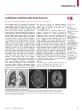
After a forklift accident a 44yearold man was admitted to our hospital in July 2014 with fractures of the right bula and tibia the left femur and the pelvis A proximal femoral nail antirotati ID: 954527

embolism usion left fat usion embolism fat left mri bilateral days opacities kpa pulmonary 148 weighted femur developed

Share:

Link:

Embed:

Download Presentation from below link

Download Pdf The PPT/PDF document "wwwthelancetcomVol 386 October 3 2015" is the property of its rightful owner. Permission is granted to download and print the materials on this web site for personal, non-commercial use only, and to display it on your personal computer provided you do not modify the materials and that you retain all copyright notices contained in the materials. By downloading content from our website, you accept the terms of this agreement.


Presentation Transcript

www.thelancet.comVol 386 October 3, 2015 After a forklift accident, a 44-year-old man was admitted to our hospital in July, 2014, with fractures of the right bula and tibia, the left femur, and the pelvis. A proximal femoral nail antirotation was done to stabilise the left femur. 1 h after the operation, he developed acute gure), without pulmonary embolism oxygen 6ยท7 kPa; normal range 11…13 kPa). We started 4 days later, we stopped sedative medication to allow Glasgow Coma Scale score of 3. He developed axillar and inguinal petechiae, so 10 days after surgery we did MRI A C B Figure: Radiographical assessment of a cerebral fat embolism(A) CT angiography; coronal multiplanar reconstruction of the thorax shows thickened interlobular septa, bilateral ground-glass opacities, and distinct nodular opacities consistent with “ ndings in pulmonary fat embolism. Axial ” air (B) and di usion-weighted (C) MRI images show bilateral con” uent white matter abnormalities in the periventricular and subcortical regions, the posterior limb of the capsula interna, and splenium of the corpus callosum, with corresponding usion restriction on di usion-weighted imaging.