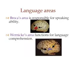
/
   Broca’s area  is responsible for speaking ability.    Broca’s area  is responsible for speaking ability.

Broca’s area is responsible for speaking ability. - PowerPoint Presentation

ellena-manuel
ellena-manuel . @ellena-manuel
Follow
344 views
Uploaded On 2020-04-03

Broca’s area is responsible for speaking ability. - PPT Presentation

Wernickes area functions for language comprehension Language areas Lateralization of hemispheres corpus callosum Cerebellum compares motor cortex output with what is happening in body Important for acquiring physical skills procedural memory ID: 774997

memory cord spinal cortex memory cord spinal cortex nerves brain nerve disease eeg limbic neurons term area system short

Share:

Link:

Embed:

Download Presentation from below link

Download Presentation The PPT/PDF document " Broca’s area is responsible for sp..." is the property of its rightful owner. Permission is granted to download and print the materials on this web site for personal, non-commercial use only, and to display it on your personal computer provided you do not modify the materials and that you retain all copyright notices contained in the materials. By downloading content from our website, you accept the terms of this agreement.


Presentation Transcript

Slide1

Broca’s area is responsible for speaking ability. Wernicke’s area functions for language comprehension.

Language areas

Slide2

Slide3

Lateralization of hemispheres

Slide4

corpus callosum

Slide5

Cerebellum – compares motor cortex output with what is happening in body. Important for acquiring physical skills (procedural memory) Basal nuclei – inhibits unwanted movements. Associated with Parkinson’s disease, Huntington’s disease

Coordination and smooth movements require additional input

Slide6

Basal nuclei

inhibits muscle tone

selects and maintains purposeful muscle activity while inhibiting useless movement

monitors and controls slow, sustained contractions (posture)

Slide7

Hypotonia

- low muscle tone

normal

Slide8

EEG records synchronous firing of pyramidal neurons in cortex (many combined dipoles). EEG measures combined activity of ~10 million neurons

Basic EEG (‘brainwaves’)

Slide9

Basic EEG (‘brainwaves’)

Slide10

A set of interconnected brain areas that fxn in motivation, emotion, and memory

Limbic association cortex

Slide11

Involves both cortical and subcortical regions “Feeling and reacting brain” vs. thinking brain of frontal cortex. Fear, anger, pleasure and sexual drive Reward and punishment centers exist

Limbic system

Slide12

Receives signals right from thalamus when experiencing fearful stimuli (emotional stimuli)Amygdala is associated with hippocampus, learning.

Limbic system: amygdala

Slide13

Short-term memory – stored for a few minutes and can contain only a few items or concepts (like a phone number). Items can get ‘bumped’ from STM. Long-term memory – lasts years or more – generally stored in cortex. Includes factual info about the world, and personal events.

Memory

Slide14

Hippocampus

Short-term memory is converted to long-term memory using signals involving the

hippocampusHippocampus is an area where new neurons can be produced.

Slide15

Newly acquired

information

Usuallypermanently lost

Rapid retrieval

Inability toretrieve

“Forgetting”

“Remembering”

Searching andreadout

Short-termmemory stores

(Practice)

Consolidation

Long-termmemory stores

Slower retrieval, except forthoroughly ingrained memories,which are rapidly retrieved

Usually onlytransiently unableto access stores

Storage in temporal lobes, limbic system, cerebellum

How we remember

Slide16

In Alzheimer’s disease, abnormal proteins cause damage to neurons (amyloid beta, tau)The hippocampus is the first to be affected

Alzheimer’s disease

Slide17

Idea that areas of the brain distinctly focus on one sense has been discardedThere is a lot of ‘cross-talk’ between the senses (i.e. what you see influences what you hear and so on)

Our brain is not really a ‘swiss army knife’

Then Now

Slide18

Figure 5.28Page 173

Cervical

cord

Thoracic cord

Lumbar cord

Sacral cord

Cervical nerves

Thoracic nerves

Lumbar nerves

Sacral nerves

Coccygealnerve

Cauda

equina

Slide19

Dermatome - a patch of skin innervated by the same spinal nerve

Slide20

Spinal

cord

Spinal

nerve

Vertebra

Meninges

layers

Slide21

Nerve- a bundle of peripheral axons. Spinal nerves contain afferent and efferent fibers.

Slide22

Cell body of

efferent neuron

Cell body ofafferent neuron

From receptors

To effectors

Interneuron

Spinal nerve

Slide23

Stimulus

Biceps

(flexor)contracts

Handwithdrawn

Triceps(extensor)relaxes

Ascending pathwayto brain

Response

Reflex arcReceptorAfferent pathwayIntegrating centerEfferent pathwayEffector organs

Integrating center(spinal cord)

Thermalpain receptorin finger

Efferent Pathway

Effectororgans

Afferent

Pathway

Withdrawal reflex