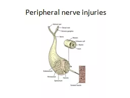
NERVE STRUCTURE AND FUNCTION Peripheral nerves are bundles of axons conducting efferent motor impulses from cells in the anterior horn of the spinal cord to the muscles and afferent sensory impulses from peripheral receptors via cells ID: 550876
Download Presentation The PPT/PDF document "Peripheral nerve injuries" is the property of its rightful owner. Permission is granted to download and print the materials on this web site for personal, non-commercial use only, and to display it on your personal computer provided you do not modify the materials and that you retain all copyright notices contained in the materials. By downloading content from our website, you accept the terms of this agreement.
Slide1
Peripheral nerve injuriesSlide2
NERVE STRUCTURE AND FUNCTION
Peripheral
nerves are bundles of axons conducting
efferent (motor) impulses from cells in the anterior
horn of the spinal cord to the muscles, and afferent
(sensory) impulses from peripheral receptors via cells
in the posterior root ganglia to the cord. They also
convey
sudomotor
and vasomotor
fibres
from ganglion
cells in the sympathetic chain. Some nerves are
predominantly motor, some predominantly sensory;
the larger trunks are mixed, with motor and sensory
axons running in separate bundles.Slide3
Pathological classification
Nerves
can be injured by
ischaemia
,
compression,traction
, laceration or burning. Damage varies
in
severity
from transient and quickly recoverable loss
of
function
to complete interruption and degeneration
.
SEDDON CLASSIFICATION OF NERVE INJURIES:
1-Transient
ischaemia
:
Acute
nerve compression causes numbness and tingling within 15 minutes, loss of pain sensibility
after30
minutes and muscle weakness after 45 minutes.
Relief
of compression is followed by intense
paraesthesiae
lasting
up to 5 minutes (the familiar ‘pins
and
needles
’ after a limb ‘goes to sleep’); feeling
is
restored
within 30 seconds and full muscle
power
after
about 10 minutes.
These
changes are due
to
transient
endoneurial
anoxia and they leave no
trace of
nerve damage.Slide4
2-Neurapraxia
Seddon
(1942) coined the term ‘
neurapraxia
’ to
describe a reversible physiological nerve
conduction
block
in which there is loss of some types of
sensation and
muscle power followed by spontaneous
recovery
after
a few days or weeks. It is due to mechanical
pressure
causing
segmental
demyelination
and is seen
typically
in
‘crutch palsy’, pressure paralysis in states
of
drunkenness
(‘Saturday night palsy’
) and the
milder
types
of tourniquet palsy
.Slide5
3-AXONOTEMESIS
This is a more severe form of nerve injury, seen
typically
after
closed fractures and dislocations. The term
means,
literally
, axonal interruption. There is loss of
conduction
but
the nerve is in continuity and the neural
tubes
are
intact. Distal to the lesion, and for a few
millimetres
retrograde
, axons disintegrate and are
resorbed
by
phagocytes
.
This
wallerian
degeneration
(named
after
the
physiologist, Augustus Waller, who described
the
process
in 1851) takes only a few days and is
accompanied
by
marked proliferation of Schwann cells
and
fibroblasts
lining the
endoneurial
tubes.
The
denervated
target
organs (motor end-plates and
sensoryreceptors
) gradually atrophy, and if they are not
reinnervated
within
2 years they will never recover.
Axonal
regeneration
starts within hours of
nerve
damage
, probably encouraged by
neurotropic
factors
produced
by Schwann cells distal to the injury.
From
the
proximal stumps grow numerous fine
unmyelinated
tendrils
, many of which find their way into
the
cell-clogged
endoneurial
tubes. These
axonal
processes
grow at a speed of 1–2 mm per day,
the
larger
fibres
slowly acquiring a new myelin
coat
.
Eventually
they
join to end-organs, which enlarge and
start
functioning again.Slide6
Wallerian
degenerationSlide7
4-Neurotmesis
In
Seddon’s
original classification,
neurotmesis
mean division
of the nerve trunk, such as may occur in
an
open
wound. It is now recognized that severe
degrees
of damage may be inflicted without actually dividing the nerve. If the injury is more severe, whether
the
nerve
is in continuity or not, recovery will not occur.
As in
axonotmesis
, there is rapid
wallerian
degeneration,
but
here the
endoneurial
tubes are
destroyed
over
a variable segment and scarring thwarts any
hope
of
regenerating axons entering the distal segment
and
regaining
their target organs. Instead,
regenerating
fibres
mingle with proliferating Schwann cells
and
fibroblasts
in a jumbled knot, or ‘
neuroma
’, at the
site
of
injury. Even after surgical repair, many new
axons
fail
to reach the distal segment, and those that do
may
not
find suitable Schwann tubes, or may not reach
the
correct
end-organs in time, or may remain
incompletely
myelinated
. Function may be adequate but
is
never
normal.Slide8
The ‘double crush’ phenomenon
There
is convincing evidence that proximal
compression
of
a peripheral nerve renders it more
susceptible
to
the effects of a second, more peripheral injury.
This
may
explain why peripheral entrapment syndromes are often associated with cervical or lumbar spondylosis
.
A similar type of ‘sensitization’ is seen in
patients
with
peripheral neuropathy due to diabetes or alcoholismSlide9
Clinical features
Acute nerve injuries are easily missed, especially
ifassociated
with fractures or dislocations, the
symptoms of
which may overshadow those of the
nervelesion
.
Always
test for nerve injuries following any
significanttrauma
.
If a nerve injury is present, it is crucial also to look for an accompanying vascular
injury.
Ask
the patient if there is numbness,
paraesthesia
ormuscle
weakness in the related area.
Then examine
the
injured limb systematically for signs of
abnormal
posture
(e.g. a wrist drop in radial nerve palsy), weakness
in specific muscle groups and changes in
sensibility.Areas
of altered sensation should be
accuratelymapped
. Each spinal nerve root serves a specific
dermatome
the skin feels
dry
due
to lack of sweating. If this is not obvious,
the‘plastic
pen test’ may help. The smooth barrel of
the
pen
is brushed across the
palmar
skin: normally
there
is
a sense of slight stickiness, due to the thin layer
ofsurface
sweat, but in
denervated
skin the pen
slips
along
smoothly with no sense of stickiness in
the
affected
area.
The neurological examination must be repeated
at
intervals
so as not to miss signs which appear
hours
after
the original injury, or following manipulation
or
operation
.
In
chronic nerve injuries
there are other
characteristic
signs
. The
anaesthetic
skin may be smooth
and
shiny
, with evidence of diminished sensibility such
as
cigarette
burns of the thumb in median nerve palsy
or
foot
ulcers with sciatic nerve palsy. Muscle groups
will
be
wasted and postural deformities may become fixed.Slide10
Assessment of nerve recovery
The
presence or absence of distal nerve function can
be
revealed
by simple clinical tests of muscle power
and
sensitivity
to light touch and pin-prick. Remember
that
after nerve injury motor recovery is slower than sensory recovery.
More
specific assessment is required to
answer
two
questions: How severe was the lesion? How well
is
the
nerve functioning now?
THE DEGREE OF INJURY
The
history
is most helpful. A low energy injury
is
likely
to have caused a
neurapraxia
; the patient
should
be
observed and recovery anticipated. A high
energy
injury
is more likely to have caused axonal
and
endoneurial
disruption
and
so recovery is less predictable. An
open
injury
, or a very high energy closed injury, will
probably
have
divided the nerve and early exploration
is
called
for.
Tinel’s
sign
– peripheral tingling or
dysaesthesia
provoked
by
percussing
the nerve – is important.
In
neurapraxia
,
Tinel’s
sign is negative. In
axonotmesis
,
it
is positive at the site of injury because of
sensitivity
of
the regenerating axon sprouts. After a delay of
a
few
days or weeks, the
Tinel
sign will then advance
at
a
rate of about 1 mm each day as the
regenerating
axons
progress along the Schwann-cell tube.
Motor
activity
also should progress down the limb. Failure of
If the
Tinel
sign proceeds very slowly, or if muscle
groups
do
not sequentially recover as expected, then a
good
recovery
is unlikely and here again exploration
must
be
considered.Slide11Slide12
Nerve conduction study and
Electromyography
(EMG) studies
can be helpful. If
a
muscle
loses its nerve supply, the EMG will
show
denervation
potentials by the third week excludes neurapraxia but of course it does not
distinguish between axonotmesis and neurotmesis;
this
remains
a clinical distinction, but if one waits too
long
to
decide then the target muscle may have
failed
irrecoverably
and the answer hardly mattersSlide13
PRINCIPLES OF TREATMENT
1-Nerve exploration
:
Closed
low energy injuries usually recover
spontaneously
and
it is worth waiting until the most
proximally
supplied muscle should have regained function.
Exploration
is indicated:
(
1) if the nerve was seen
to
be
divided and needs to be repaired; (2) if the type
of
injury
(e.g. a knife wound or a high energy
injury)
suggests
that the nerve has been divided or
severely
damaged
; (3) if recovery is inappropriately
delayed
and
the diagnosis is in doubt.
Vascular injuries, unstable fractures,
contaminated
soft
tissues and tendon divisions should be dealt
with
before
the nerve lesion.
The
incision will be long,
as
the
nerve must be widely exposed above and
below
the
lesion before the lesion itself is repaired.
Thenerve
must be handled gently with suitable instruments.
Bipolar diathermy and magnification are
essential.
An
operating microscope is ideal but
magnifying
loupes
are better than nothing. A nerve stimulator
is
essential
if scarring makes recognition uncertain.
If
microsurgical
equipment and expertise are not
available,
the
nerve lesion should be identified
and
the
wound closed pending
transferral
to an
appropriate facility
.Slide14
2-Primary
repair
A divided nerve is best repaired as soon as this can
be
done
safely. Primary suture at the time of wound
toilet has considerable advantages: the nerve
ends
have
not retracted much; their relative rotation is
usually
undisturbed; and there is no fibrosis.A clean cut nerve is sutured without further
preparation;
a
ragged cut may need paring of the
stumps
with
a sharp blade, but this must be kept to a
minimum.
The
stumps are anatomically orientated
and
fine
(10/0) sutures are inserted in the
epineurium
.
There
should be no tension on the suture line.
Opinions
are
divided on the value of fascicular repair
with
perineurial
sutures.
Sufficient relaxation of the tissues to permit
tension-
free
repair can usually be obtained by
positioning
the
nearby joints or by mobilizing and re-routing
the
nerve
. If this does not solve the problem then a
primary
nerve
graft must be considered. A traction
lesion
especially of the brachial plexus – may leave a
gap
too
wide to close. These injuries are best dealt with in
specialized
centres
, where primary grafting or
nerve
transfer
can be carried out.
If a tourniquet is used it should be a
pneumatic
one
; it must be released and bleeding stopped
before
the
wound is
closed.
The
limb is splinted in a position to ensure
minimal
tension
on the nerve; if flexion needs to be
excessive,
a
graft is required. The splint is retained for 3
weeks
and
thereafter physiotherapy is encouraged.Slide15
3-Delayed
repair
Late repair, i.e. weeks or months after the injury,
may
be
indicated because: (1) a closed injury was left
alone
but
shows no sign of recovery at the expected time
;
(2) the diagnosis was missed and the patient present late; or (3) primary repair has failed. The options
must be
carefully weighed: if the patient has adapted to
the
functional
loss, if it is a high lesion and
re-
innervation
.
is
unlikely within the critical 2-year period, or if
there
is
a pure motor loss which can be treated by
tendon
transfers
, it may be best to leave well alone.
Excessive
scarring
and intractable joint stiffness may,
likewise,
make
nerve repair questionable; yet in the hand it
is
still
worthwhile simply to regain protective sensation.
The lesion is exposed, working from normal
tissue
above
and below towards the scarred area. When
the
nerve
is in continuity it is difficult to know
whether
resection
is necessary or not. If the nerve is
only
slightly
thickened and feels soft, or if there is
conduction
across
the lesion, resection is not advised; if
the
‘
neuroma
’ is hard and there is no conduction
on
nerve
stimulation, it should be
resected
, paring
back
the
stumps until healthy fascicles are exposed.
How to deal with the gap? The nerve must
be
sutured
without tension. The stumps may be
brought
together
by gently mobilizing the proximal and
distal
segments
, by flexing nearby joints to relax the soft
tissues,
or
(in the case of the
ulnar
nerve) by
transposing
the
nerve trunk to the flexor aspect of the elbow.
In this
way
, gaps of 2 cm in the median nerve, 4–5 cm in
the
ulnar
nerve and 6–8 cm in the sciatic nerve can
usually
be
closed, the limb being splinted in the ‘relaxing’
position
for
4–6 weeks after the operation. Elsewhere, gaps
of more than 1–2 cm usually require grafting.Slide16
4-Nerve
grafting
Free
autogenous
nerve grafts can be used to
bridge
gaps
too large for direct suture. The
sural
nerve
ismost
commonly used; up to 40 cm can be obtainedfrom each leg. Because the nerve diameter is small, several strips may be used (cable graft
).
The
graft
should
be long enough to lie without any tension,
and
it
should be routed through a well-
vascularized
bed.
The
graft is attached at each end either by fine sutures
or with fibrin
glue.
It
is crucial that the motor and sensory fascicles
are
appropriately
connected by the graft.
There
are
various
techniques
which can
help. If
the
ulnar
and median nerves are both damaged (
e.g.
in
Volkmann’s
ischaemia
) a pedicle graft from
the
ulnar
nerve may be used to bridge the gap in
the
median
. It is also possible to use free
vascularized
grafts
for certain brachial plexus lesions.Slide17
5-PRINCIPLES
OF TENDON
TRANSFER
Assess the problem
Which muscles are missing?
Which muscles are available?
The donor muscle should
be:expendablepowerful
enough
an
agonist or
synergist
,
The
recipient site
should:be
stable
have
mobile joints and supple tissues
The transferred tendon should be:
routed
subcutaneouslyplaced
in a straight line of
pull
,
capable of
firm fixation
The patient should be:
motivated able
to comprehend and attend hand
therapy
intercostal
nerves or the spinal accessory nerve to
the
stump
of the original nerve supplying that muscle.
Care of
paralysed
parts
While
recovery is awaited the skin must be
protected
from
friction damage and burns. The joints should
be
moved
through their full range twice daily to
prevent
stiffness
and minimize the work required of muscles
when
they recover. ‘Dynamic’ splints may be
helpful.
Tendon
transfers
Motor
recovery may not occur if the axons,
regenerating
at
about 1 mm per day, do not reach the
muscle
within
18–24 months of injury.
This
is most
likelywhen
there is a proximal injury in a nerve
supplying
distal
muscles. In such circumstances, tendon
transfers
should
be considered. The principles can be
summarized
in
the Box on the previous
page.
Slide18
PROGNOSIS
1-Type
of lesion
Neurapraxia
always recovers
fully;
axonotmesis
may or may not;
neurotmesis
will not
unless
the nerve is repaired.2- Level of lesion The higher the lesion, the worse theprognosis
.
3- Type
of nerve
Purely motor or purely sensory
nerves
recover
better than mixed nerves, because there is
less
likelihood
of axonal confusion.
4- Size
of gap
Above the critical resection length,
suture
is
not successful.
5- Age
Children do better than adults. Old people
do
poorly
.
6- Delay
in suture
This is a most important adverse
factor.
The
best results are obtained with early
nerve
repair
. After a few months, recovery following suture
becomes
progressively less likely.
7- Associated
lesions
Damage to vessels, tendons
and
other
structures makes it more difficult to obtain
recovery
of
a useful limb even if the nerve itself
recovers.
8-
surgical
techniques
Skill, experience and suitable
facilities
are
needed to treat nerve injuries. If these
arelacking
, it is wiser to perform the essential wound
toiletand
then transfer the patient to a specialized
centre
.