PPT-Development of MRI-defined Structural Tissue Damage after Anterior Cruciate Ligament Injury
Author : eloise | Published Date : 2022-02-15
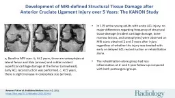
Roemer F W et al Published Online March 9 2021 httpsdoiorg101148radiol2021202954 In 119 active young adults with acute ACL injury no major differences regarding
Presentation Embed Code
Download Presentation
Download Presentation The PPT/PDF document "Development of MRI-defined Structural Ti..." is the property of its rightful owner. Permission is granted to download and print the materials on this website for personal, non-commercial use only, and to display it on your personal computer provided you do not modify the materials and that you retain all copyright notices contained in the materials. By downloading content from our website, you accept the terms of this agreement.
Development of MRI-defined Structural Tissue Damage after Anterior Cruciate Ligament Injury: Transcript
Download Rules Of Document
"Development of MRI-defined Structural Tissue Damage after Anterior Cruciate Ligament Injury"The content belongs to its owner. You may download and print it for personal use, without modification, and keep all copyright notices. By downloading, you agree to these terms.
Related Documents