/
Polycystic Ovarian Disease and Endometriosis Polycystic Ovarian Disease and Endometriosis

Polycystic Ovarian Disease and Endometriosis - PowerPoint Presentation

elyana
elyana . @elyana
Follow
0 views
Uploaded On 2024-03-13

Polycystic Ovarian Disease and Endometriosis - PPT Presentation

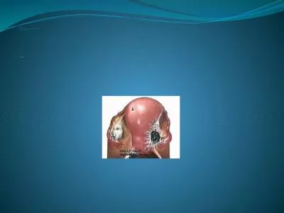
SUFIA HUSAIN MBBS MD Pathology FRCPath Polycystic Ovarian Disease PCOD Polycystic ovaries are characterized by bilaterally enlarged polycystic ovaries chronic anovulation and clinical manifestations secondary to excessive production of estrogens and androgens mainly androgens ID: 1047065

endometriosis polycystic endometrial ovarian polycystic endometriosis ovarian endometrial cysts lesions menstrual clinical anovulation syndrome disease ovary chocolate ovulation brown

Share:

Link:

Embed:

Download Presentation from below link

Download Presentation The PPT/PDF document "Polycystic Ovarian Disease and Endometri..." is the property of its rightful owner. Permission is granted to download and print the materials on this web site for personal, non-commercial use only, and to display it on your personal computer provided you do not modify the materials and that you retain all copyright notices contained in the materials. By downloading content from our website, you accept the terms of this agreement.


Presentation Transcript

1. Polycystic Ovarian Disease and EndometriosisSUFIA HUSAIN, MBBS, MD Pathology, FRCPath.

2. Polycystic Ovarian Disease (PCOD)Polycystic ovaries are characterized by bilaterally enlarged polycystic ovaries, chronic anovulation and clinical manifestations secondary to excessive production of estrogens and androgens, mainly androgens.The initial abnormality resulting in the syndrome is not known but is believed to be related to hypothalamus-pituitary dysfunction leading to oversecretion of luteinizing hormone (LH). LH in turn stimulates the ovary to produce excess androgens. Secretion of follicle stimulating hormone (FSH) is inhibited resulting in repression of ovulation with follicle cyst formation.High level of LH and low FSH

3. Polycystic Ovarian DiseaseOther names for this syndrome include polycystic ovarian syndrome and Stein-Leventhal syndrome

4. Polycystic Ovarian DiseaseClinical appearances.The usual clinical presentation is a young woman (between 15 and 30 years) with - secondary amenorrhea with anovulation - oligomenorrhea or irregular menses - infertility - hirsutism - virilism due to excessive amounts or effects of androgenic (masculinizing) hormones - obesity - acne

5. Polycystic Ovarian DiseaseGrosslyBoth ovaries are markedly enlarged usually twice normal in size and have a thickened, fibrotic gray white capsule with smooth pearl-white outer surface, studded with sub cortical cysts 0.5 to 1.5 cm in diameter.

6. PCOD

7. PCOD7

8. Polycystic Ovarian DiseaseHistologyMicroscopically, the outer portion of the cortex is thickened and fibrotic many follicle cysts lined by granulosa cells are present in the subcapsular cortex. The cysts have prominent outer theca interna layer, which is often luteinized. Corpora lutea are frequently absent due to the anovulation and occasional focal stromal luteinization is present. Cortical stromal fibrosisThe chronic anovulation results in unopposed estrogenic stimulation of the endometrium leading to a variety of appearances ranging from mild atypical hyperplasia to well-differentiated endometrial adenocarcinoma

9. PCOD

10. Polycystic Ovarian DiseaseClinical behaviorTreatment with drugs that either induce ovulation (clomiphene or hCG) or regulate the menstrual cycle restores fertility. Reduction of ovarian volume by wedge resection is also successful in initiating ovulation and restoring fertility. The endometrial changes usually regress once ovulation is achieved.

11. Polycystic OvariesStein-Leventhal SyndromeWomen with PCOS are at risk for the following:Endometrial hyperplasia and endometrial cancerInsulin resistance/Type II diabetes High blood pressure Depression/Anxiety DyslipidemiaCardiovascular disease Strokes Weight gain Miscarriage Acanthosis nigricans (patches of darkened skin under the arms, in the groin area, on the back of the neck) Autoimmune thyroiditis 11

12.

13. Endometriosis This is the presence of ectopic endometrial glands and stroma outside the uterus. The lesions are usually found on the peritoneal surfaces of the reproductive organs and adjacent pelvic organs. The most frequent location is the ovary (approx. 50%) followed by the pouch of Douglas, uterine ligaments. Occasional sites include the cervix, vagina, perineum, bladder, large bowel and the umbilicus. Rare lesions are seen as far as small bowel, kidneys, lungs and brain.It has been reported in men. The sites involved have been the bladder, scrotum and prostate

14. Endometriosis Non-neoplastic.Like the uterine endometrium it is responsive to the hormonal variations of the menstrual cycle.It is characterized by menstrual type bleeding at the site of the ectopic endometrium, resulting in blood filled areas (e.g.chocolate cysts).

15. Endometriosis :Clinical appearancesClinical presentation depends on the site of endometriosis. Dysmenorrhea, cyclic abdominal pain and dyspareunia are common symptoms. Usually there is severe menstrual-related pain.Often results in infertility.Endometriosis usually appears as multiple red or brown (due to hemosiderin) 1mm to 5mm nodules (some may form larger masses or cysts). Dense fibrous adhesions may surround the foci.

16. Endometriosis Repeated hemorrhage into foci in the ovary with each menstrual cycle produces cysts, which contain inspissated, chocolate-brown material, called "chocolate cystsā€¯ in which the ovaries turn into large cystic masses filled with brown fluid Clinical behaviorBenign with no malignant potential. May recur after surgical excision but the risk is low.

17. Endometriosis

18. Chocolate cyst of ovary (endometriotic cyst)

19. EndometriosisHistologyMicroscopic diagnosis of endometriosis is made by the presence of ectopic endometrial glands and stroma. Macrophages containing hemosiderin (siderophages) may be present in lesions with previous hemorrhage. When endometriosis develops in a muscular viscus, the smooth muscle around it is often hyperplastic.

20. Endometriosis

21. Adenomyosis This is defined as the presence of endometrial glands and stroma in the myometrium. The condition involves the posterior wall more often than the anterior wall but it may affect both walls in the same uterus. The disease is primarily a disorder of parous women and occurs infrequently in the nullipara. It is associated with menorrhagia and severe dysmenorrhea. In about a third of patients there are no symptoms and the lesions are discovered accidentally.

22. AdenomyosisWhen extensive the lesions produce myometrial thickening with small yellow or brown cystic spaces containing fluid or blood.Occasionally, a proliferation of smooth muscle around a focus of adenomyosis produces a tumor called adenomyoma, which resembles uterine leiomyoma.Clinical behavior.This is a benign condition with no known malignant potential that regresses after the menopause

23. Adenomyosis

24.