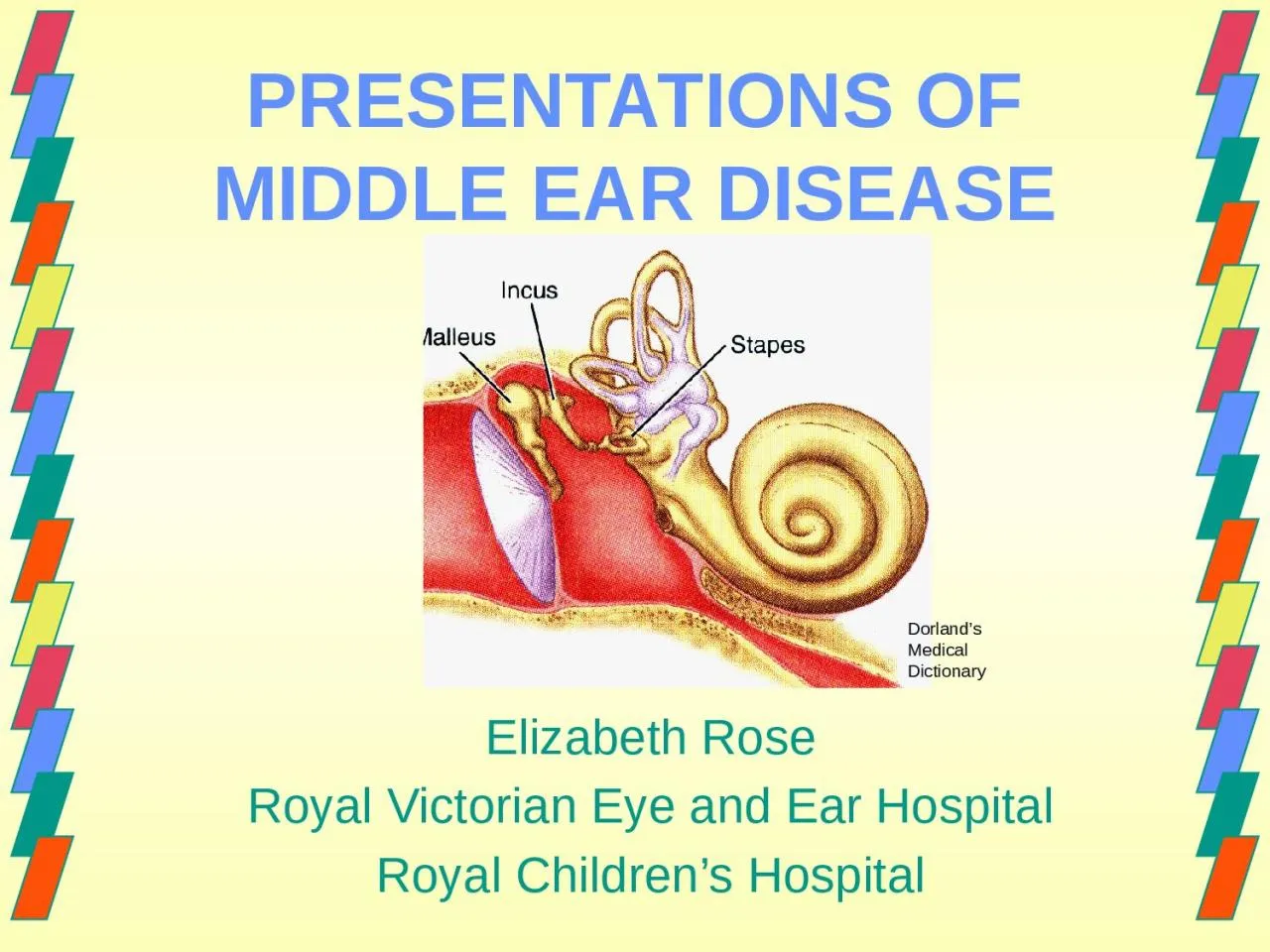
/
PRESENTATIONS OF MIDDLE EAR DISEASE PRESENTATIONS OF MIDDLE EAR DISEASE

PRESENTATIONS OF MIDDLE EAR DISEASE - PowerPoint Presentation

emily
emily . @emily
Follow
342 views
Uploaded On 2022-06-07

PRESENTATIONS OF MIDDLE EAR DISEASE - PPT Presentation

Elizabeth Rose Royal Victorian Eye and Ear Hospital Royal Childrens Hospital Dorlands Medical Dictionary A look and learn lecture Middleear conditions Management of otitis media ID: 914082

https ear middle otitis ear https otitis middle http media hawkelibrary hearing php www itemid main tympanic effusion perforation

Share:

Link:

Embed:

Download Presentation from below link

Download Presentation The PPT/PDF document "PRESENTATIONS OF MIDDLE EAR DISEASE" is the property of its rightful owner. Permission is granted to download and print the materials on this web site for personal, non-commercial use only, and to display it on your personal computer provided you do not modify the materials and that you retain all copyright notices contained in the materials. By downloading content from our website, you accept the terms of this agreement.


Presentation Transcript

Slide1

PRESENTATIONS OF MIDDLE EAR DISEASE

Elizabeth RoseRoyal Victorian Eye and Ear HospitalRoyal Children’s Hospital

Dorland’s Medical Dictionary

Slide2

A “look and learn” lecture

Middle-ear conditionsManagement of otitis media Differential diagnosis of ear painClinical cases

Slide3

Normal TM

http://me.hawkelibrary.com/new/main.php?g2_itemId=315

Slide4

Translucent

(can see through it)

Handle of the malleus

Incus

Stapedius tendon

Round window niche

Normal TM

http://me.hawkelibrary.com/new/main.php?g2_itemId=315

Slide5

OTITIS MEDIAA

SPECTRUM OF DISEASEacute otitis media

chronic otitis media with effusionatelectasis of the tympanic membranechronic adhesive otitis mediachronic suppurative otitis mediatubotympanic (“safe”)atticoantral (“unsafe”)

and may be a

continuum

of disease

Slide6

Acute otitis media

Otitis media with effusion

Normal

Usual

sequence after

AOM

http://me.hawkelibrary.com/new/main.php?g2_itemId=187

http://me.hawkelibrary.com/new/main.php?g2_itemId=315

(all accessed 12-03-13)

http://me.hawkelibrary.com/new/main.php?g2_itemId=524

Slide7

Acute otitis media

Perforation

with discharge (

otorrhoea

)

Normal (perforation heals)

Persistent perforation

But

AOM may also lead to a

perforation

of the TM

http://me.hawkelibrary.com/new/main.php?g2_itemId=369

http://me.hawkelibrary.com/new/main.php?g2_itemId=524

http://otitismedia.hawkelibrary.com/aom/3_2_CSOM_Otorrhea

http://otitismedia.hawkelibrary.com/normal/tm_2

Slide8

Acute otitis media

Otitis media with effusion

Cholesteatoma

Atelectasis and adhesions

with continuing Eustachian tube dysfunction

… and more severe problems

http://hawkelibrary.com

http://me.hawkelibrary.com/new/main.php?g2_itemId=524

http://me.hawkelibrary.com/new/main.php?g2_itemId=187

http://me.hawkelibrary.com/new/main.php?g2_itemId=1225

http://me.hawkelibrary.com/new/main.php?g2_itemId=1486

Slide9

http://www.dr-li.net/Hearing%20Health.html

Slide10

Diagnosis of acute otitis media

(Therapeutic Guidelines)

A history of acute onset of symptoms and signsotalgia, ear pulling, otorrhoea, fever, anorexia, vomitingAND

2. Middle ear effusion (otorrhoea if a TP perforation)

AND

3. signs and symptoms of

middle ear inflammation

,

characterised

by redness of the tympanic membrane.

Slide11

Management of AOM

Pain reliefDecongestants (oral/topical) and antihistamines

do not make the eustachian tube function better Not recommended

Slide12

Management of AOM

Who should be treated with antibiotics?

Slide13

Antibiotic therapy

if severe symptomspain/fever

perforationbilateral diseasepossible suppurative complicationmastoiditisMeningitis or other intracranial complication≤ 2 years oldIndigenous (ATSI , Maori and Pacific Islanders)

immune deficiency

cochlear implant

only hearing ear

Slide14

Antibiotic therapy

Recommended treatment is: amoxicillin 45mg/kg/day in 3 doses

Can give up to 100mg/kg/dayContinue for 10 daysIf no improvement in 2 days change to amoxicillin/clavulanate

Slide15

If an

allergy to penicillin

cefuroxime child 3 months to 2 years: 10 mg/kg up to 125 mg; 2 years or more: 15 mg/kg up to 500 mg orally, 12-hourly for 5 days

Slide16

Antibiotic therapy

older children who can be accurate about their symptoms should be treated symptomaticallyif no improvement after 2 days consider treatment with antibiotics

Slide17

CHRONIC OTITIS MEDIA WITH EFFUSION(COME)

the presence of a middle ear effusionasymptomatic apart from some hearing loss

Slide18

CHRONIC OTITIS MEDIA WITH EFFUSION

Normal OME

Slide19

AOM COME

http://me.hawkelibrary.com/new/main.php?g2_itemId=524

Slide20

AOM COME

TM bulging

Cannot see lateral process

of the

malleus

Opaque

White from the pus in the ME

Inflammation/prominent

vessels

TM is retracted

Malleus handle is more prominent

Opaque

Yellow from effusion

https://sydneyentclinic.com/sean-flanagan/patient-resources-2/patient-resources/hearing-loss/glue-ear-middle-ear-infections-and-grommets/

https://www.drrahmatorlummc.com/serousotitismedia.htm

Slide21

MIDDLE-EAR EFFUSION

https://www.emaze.com/@AOOROWLL

https://www.drrahmatorlummc.com/serousotitismedia.htm

Middle ear full of fluid

Air-fluid level

Slide22

CHRONIC ADHESIVE OTITIS MEDIA

TM is thin and adheres to the

ossicles

, often with erosion

https://www.medical.theclinics.com/article/S0025-7125(18)30068-3/fulltext

Slide23

TYMPANIC MEMBRANE PERFORATIONS

https://journals.sagepub.com/doi/abs/10.1093/innovait/inp007?journalCode=inoa

https://dontforgetthebubbles.com/otitis-media/

Likely to have hearing impairment

Hearing may be normal

Slide24

CHRONIC SUPPURATIVE OTITIS MEDIA

(CSOM)“deafness and discharge”

persistent diseaseinsidious onsetsevere destructionirreversible sequelae

Slide25

1.

tubotympanic disease (“safe”)central perforation2. atticoantral disease

(“unsafe”)cholesteatomathe presence of keratinising squamous epithelium in the middle ear

Slide26

TUBOTYMPANIC

(central perforation)

https://dontforgetthebubbles.com/otitis-media/

Central perforation with active discharge (

otorrhoea

)

Slide27

ATTICOANTRAL DISEASE

CHOLESTEATOMA(marginal or attic perforation)

https://www.southlakeent.com/services-procedures/ears-hearing/ear-concerns/cholesteatomas

Erosion in the attic with keratin

Can see cholesteatoma in the

mesotympanum

as well

Likely to surround the

ossicles

(incus)

Slide28

Cholesteatoma

A non-neoplastic accumulation of keratinising stratified squamous epithelium in the tympanic cavity (middle ear) or mastoid air cells

Resorbs (damages) underlying bonePotentially dangerous to the patient

Slide29

CHOLESTEATOMA

Complications

Hearing loss Intracranial

.tympanic membrane .

ossicles

.inner ear

Vertigo

Tinnitus

Facial paralysis

https://www.vectorstock.com/royalty-free-vector/ear-anatomy-vector-23904762

Slide30

MANAGEMENT OF CHRONIC

OTITIS MEDIA WITH EFFUSION(and also retraction/atelectasis of the tympanic membrane)

Slide31

What are grommets?

Slide32

https://jaydeeautocables.com.au/products/rubber-grommet-assortment-kit

Any tube with flanges

https://twen.rs-online.com/web/p/rubber-grommets/1879453/

https://venuspack.com.au/product-category/poly-bags-netting/tarpaulins/

Slide33

?Young surfers

https://kialoa.com/blogs/talkstory/endearing-photos-of-young-surfers

https://diannewolfer.com/books/picture-books/granny-grommet-and-me/

Slide34

Wallace and gromit

https://www.radiotimes.com/movies/is-wallace-and-gromit-on-netflix/

Slide35

MIDDLE EARVENTILATION TUBES

https://slideplayer.com/slide/14431309/

Slide36

AKA

grommetstubes pressure equalisation tubes

middle ear ventilation tubestympanostomy tubesNOT a drainage tube(it’s role in life is to ventilate!)

Slide37

https://www.slideshare.net/bethfernandezaud/unit-3-review-30629514

Mucoid middle ear effusion

Slide38

Ventilates

the middle ear

https://www.healthinfo.org.nz/index.htm?Ventilation-tubes-grommets.htm

Slide39

COME

Who should have middle ear ventilation tubes?

(for OME)

Slide40

Natural history

of OME after AOM

Slide41

1. COME for

4 months at least, with hearing loss

2. COME in a child ‘at risk’ regardless of the hearing3. COME and structural damage to the tympanic membrane

Slide42

1. Hearing loss

median hearing loss is mild but there is a wide range

no data on the criteria for what is a significant hearing loss

Slide43

O

]

O

O

O

O

O

X

X

X

X

X

[

[

[

[

[

]

]

]

]

X

50%

of children with persistent OME have hearing thresholds at

20 dB

Slide44

O

]

O

O

O

O

O

X

X

X

X

X

[

[

[

[

[

]

]

]

]

X

20%

of children with persistent OME have hearing thresholds at

>35 dB

Slide45

2. An

‘at risk’ child has an increased risk of developmental difficulties due to factors not related to the OME:

physicalsensory, e.g. blindnesscognitive, e.g. developmental delaybehavioural

Slide46

https://www.pinterest.com.au/pin/350999364682083551/

Slide47

3. Structural damage to the tympanic membrane

https://app1.unmc.edu/medicine/heywood/otology/unit4-middle-ear-disease-diagnosis.cfm

Retraction of the tympanic membrane can get worse and damage the

ossicles

or become a cholesteatoma

Slide48

Children with persistent OME

who:Do not have other risk factorsDo

not have significant hearing lossDo not have damage to the tympanic membrane or middle earCan be evaluated every three months

Slide49

Take it to Telehealth - 1

remember referred otalgia

If the ear is normal seek the cause of the pain

Slide50

Take it to Telehealth – 2

More is missed in medicine by not looking than by not knowing

https://www.thesun.co.uk/living/2712715/can-you-spot-the-dog-on-the-rug-hilarious-photos-show-animals-blend-into-the-background-when-it-suits-them/