/
Nerve Injuries of the  U Nerve Injuries of the  U

Nerve Injuries of the U - PowerPoint Presentation

evans
evans . @evans
Follow
66 views
Uploaded On 2023-05-23

Nerve Injuries of the U - PPT Presentation

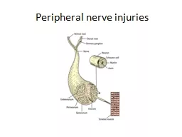
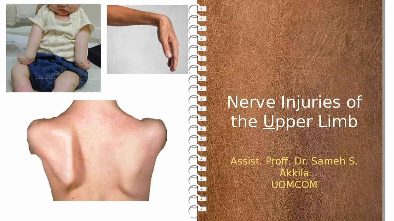
pper Limb Assist Proff Dr Sameh S Akkila UOMCOM Objectives Why How 1 Understand why nerve injuries of the upper limb are common amp important 2 Classify the types of nerve injuries amp the different outcomes of each ID: 999326

nerve amp ulnar wrist amp nerve wrist ulnar shoulder fingers plexus commonly forearm lat paralyzed muscles compressed injuries cut

Share:

Link:

Embed:

Download Presentation from below link

Download Presentation The PPT/PDF document "Nerve Injuries of the U" is the property of its rightful owner. Permission is granted to download and print the materials on this web site for personal, non-commercial use only, and to display it on your personal computer provided you do not modify the materials and that you retain all copyright notices contained in the materials. By downloading content from our website, you accept the terms of this agreement.


Presentation Transcript

1. Nerve Injuries of the Upper LimbAssist. Proff. Dr. Sameh S. AkkilaUOMCOM

2. ObjectivesWhy? How?1. Understand why nerve injuries of the upper limb are common & important.2. Classify the types of nerve injuries & the different outcomes of each.3. Review the course of the major nerves of the upper limb & how / where they can be injured with the resulting outcome.

3. Why? How?SummaryTypes of nerve injuriesA nerve can be injured completely or partially. It can be compressed (e.g., by a tumor or an enlarged lymph node) or severed (e.g., by a stab wound, a bullet or a fractured bone). A compressed nerve will have its fibers short-circuiting causing unreal sensation (paresthesia) and irregular muscle movement or weakness.A severed (cut) nerve will not transmit any impulsesleading to complete loss of sensation & complete lossof muscle function. Overtime, paralyzed muscles ATROPHY (shrink in size).In Division: The nerve fibers (axons) shrink back and 'rest' for about a month; then they begin to grow again. Axons will regenerate about 1mm per day. The extent to which a nerve recovers is variable, and it will always be incomplete.

4. RadiculopathyWhere?Brachial plexus injuriesBrachial plexus injuries = Radiculopathy (myotomes& dermatomes) affecting multiple nerves that share the root.C5+C6= Upper trunk (UT)C7 = Middle trunk (MT)C8+T1= Lower trunk (LT)Each trunk divides into ant. & post. divisions.Ant. divisions of UT & MT form lateral cord (LC)Ant. division of LT forms medial cord (MC)All 3 post. divisions form posterior cord (PC)The plexus is usually injured at the peripheries:Upper = C5, C6orLower = C8, T1

5. How?Like thisUpper Brachial plexus injuriesTraction injuries to the neck affecting the upper roots: (C5 & C6)  Erb-Duchenne palsy.- In Infants (Congenital): hasty head delivery.- In adults (Acquired): stretching the head to one side & the shoulder to the opposite side (e.g., falling from a height).It affects all nerves with C5, C6 root value.

6. Main nerves affectedResultUpper Brachial plexus injuriesAffected nervesParalyzed ResultSuprascapular (C5, C6)SupraspinatusInfraspinatus-No Shoulder Abduction-No Shoulder Lat. rotationAxillary (C5, C6)DeltoidTeres minor-Weak all shoulder movements except adduction-No Shoulder Lat. rotationThe shoulder is ADDUCTED & MEDIALLY ROTATED with the elbow EXTENDED and the palm facing backwards due to a PRONATED FOREARM (Supinator is paralyzed & biceps CANNOT supinate an extended elbow!).Affected nerveParalyzed ResultLong thoracic (C5, C6, C7)- Most of Serratus anterior- Winging of scapulaMusculocutaneous (C5,C6,C7)- Elbow flexors myotome- Greatly weakened elbow flexionRadial branch to supinator C5- Supinator- Forearm pronatedWrist flexors (C6 myotome)- Wrist flexion-Weak wrist flexion

7. How?Like thisLower Brachial plexus injuries = Klumpke’s PalsyAffect the C8 & T1 root distributed to the small muscles of the HAND via fibers that travel through the ULNAR & MEDIAN nerves.An abducted arm is pulled away from the body.- In Infants (Congenital): hasty arm delivery.- In adults (Acquired): commonly associated withshoulder dislocation (i.e., abduction under weight).

8. Causes & Affected nervesResultNerveParalyzed Median- Wrist pronators- Wrist flexors- Thenar mm.Ulnar- Small muscles of the handForearm supinated + Wrist extendedwith Thumb paralyzed & Fingers clawed

9. ResultNerve reviewLong thoracic nerve injuryThe nerve arises from C5, C6, C7 roots & descends behind the 1st part of the axillary artery along the lateral thoracic wall running along & supplying serratus anterior muscle. It may be cut during radical mastectomy or by a stab wound along its course leading to paralysis of serratus anterior.In addition to weakness in protraction, the scapula is no longer held against the thorax and when the patient pushes forwards, this leads to WINGING of the scapula = the medial border is pushed backwards,

10. ResultNerve reviewAxillary nerve injuryThe axillary nerve (C5, C6) arises from the post. cord of the brachial plexus & passes through the quadrangular space to run deep to deltoid around the surgical neck of the humerus. It supplies DELTOID, TERES MINOR & the skin over the lower ½ of deltoid (upper lateral cutaneous nerve of the arm).It’s commonly injured by inferior shoulder dislocation, fracture of the surgical neck of the humerus or penetrating injuries to the axilla.With deltoid paralyzed, shoulder abduction can only be initiated by SUPRASPINATUS but cannot be continued. Overtime, deltoid atrophies leading to loss of the shoulder round and prominent coracoid & acromion. There’s also small area of anesthesia of the skin over the lower ½ of deltoid.

11. How? Where?Nerve reviewRadial nerve injuryThe radial nerve (C5-T1) is the continuation of the pos. cord of the brachial plexus. In the axilla it gives muscular branches to triceps & the post. Cut. N. of the arm &then passes through the triangular interval to run in the radial groove where it gives muscular branches to triceps & anconeus &the lower lat. Cut. N. of the arm & post. Cut. N. of the forearm. After which, the radial groove leads it back to the anterior compartment of the distal arm.Here it lies between & supplies ECRL, brachioradialis, & part of brachialis and then divides in deep & superficial branches. The deep branch passes to the muscles of the posterior compartment of the forearm between the two heads of supinator. The superficial branch runs the lateral side of the forearm and wrist to the skin of the lateral 2/3 of the dorsum of the hand.The nerve may be injured in the AXILLA, RADIAL GROOVE or its deep branch may be compressed by supinator.

12. Wrist DropFinger DropSupinator syndrome= Finger dropOveruse of supinator can impinge the deep branch of the radial nerve leading to weakness of all posterior forearm extensor muscles EXCEPT ECRL. Wrist extension is still possible but always combined with abduction. Digital extension is greatly weakened leading to FINGER DROP. THERE IS NO SENSORY LOSS.Radial groove injuryWith wrist drop the patient CANNOT MAKE A FIST. The sensory loss is limited to the back of the forearm & hand.

13. ResultMechanismRadial nerve injury in the axillaMore commonly a compression than a division.Extensive damageThe elbow, wrist & fingers are all unopposedly flexed. Paresthesia is extensive.

14. Where? How?Nerve reviewMedian nerve injuryThe nerve is most commonly COMPRESSED at the carpal tunnel  CARPAL TUNNEL SYNDROME.Less commonly it can be divided at the wrist (Distal injury)  MEDIAN CLAWIt may be less commonly compressed by pronator teres  PRONATOR SYNDROMEIt can also be divided by a fractured medial epicondyle.

15. ResultCausesMotor: great weakness in thumb opposition and less weakness in other movements EXCEPT ADDUCTION. If not treated, thenar muscles atrophy EXCEPT for ADDUCTOR POLLICIS.Carpal tunnel syndromeSensory: paresthesia in the lat. 3 ½ fingers but NOT THE PALM.

16. Distal injuryDistal injuryMedian ClawAll the intrinsic thumb muscles (EXCEPT ADDUCTOR POLLICIS) are paralyzed & then atrophy. The thenar eminence is lost, the thumb is kept in adduction with slight lateral rotation due to pull from the abductor pollicis longus.Paralysis of the lat. 2 lumbricals with preserved function of extrinsic flexors. This imbalance leads to permanent flexion of the index & middle fingers phalanges (aggravated when trying to extend the fingers).Recurrent branch damage

17. Proximal divisionProximal CompressionPronator teres syndromeWith lumbricals I & II paralyzed, trying to make a fist leaves the index & middle fingers extended. The thumb is kept in an extended/adducted position. (MOTOR > SENSORY).Repeated pronation compresses the nerve between the heads of the pronator teres. Patients complains of pain and tenderness in the proximal aspect of the anterior forearm aggravated by resisted pronation, and paresthesia of palmar aspects of the lat. 3 ½ fingers & lat. 2/3 of the palm. SENSORY > MOTORProximal cutBenediction Sign

18. Where? How?Nerve reviewUlnar nerve injuryThe nerve is most commonly cut by a fracture of the medial epicondyle.It may be less commonly compressed as it passes anterior to the flexor retinaculum.Rarely, it is compressed at the axilla or by FCU.

19. Cut >> CompressionGuyon’s (ulnar) canalDistal ulnar nerve injuryGuyon’s (ulnar) tunnel houses the ulnar nerve & artery as they pass between the wrist ligaments, pisiform, hook of the hamate & abductor digiti minimi superficial to the flexor retinaculum.>

20. Ulnar paradoxProximal cutProximal ulnar nerve injuryFCU & The medial 2 tendons of FDP are also paralyzed. So, flexing the wrsit is combined with abduction. When flexing the fingers the ring & little fingers remain extended fiving the OK SIGN.Ulnar PRADOX = distal injury is more devastating than proximal injury. Related to the function of FDP.

21. The End