/
Seminar topic:  “Venous Seminar topic:  “Venous

Seminar topic: “Venous - PowerPoint Presentation

everfashion
everfashion . @everfashion
Follow
342 views
Uploaded On 2020-06-18

Seminar topic: “Venous - PPT Presentation

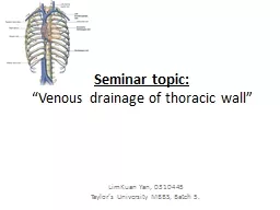
drainage of thoracic wall LimKuan Yan 0310445 Taylors University MBBS Batch 5 Veins that we going to discuss today Internal thoracic vein Intercostal vein Ant amp Post Azygos ID: 781172

intercostal vein thoracic left vein intercostal left thoracic azygos post artery upper lumbar subcostal part internal side spaces veins

Share:

Link:

Embed:

Download Presentation from below link

Download The PPT/PDF document "Seminar topic: “Venous" is the property of its rightful owner. Permission is granted to download and print the materials on this web site for personal, non-commercial use only, and to display it on your personal computer provided you do not modify the materials and that you retain all copyright notices contained in the materials. By downloading content from our website, you accept the terms of this agreement.


Presentation Transcript

Slide1

Seminar topic: “Venous drainage of thoracic wall”

LimKuan Yan, 0310445Taylor’s University MBBS, Batch 5.

Slide2

Veins that we going to discuss todayInternal thoracic vein

Intercostal vein (Ant & Post.)Azygos VeinHemiazygos veinAccessory vein

Slide3

Internal Thoracic Vein the (Internal Mammary

Vein)

I

s a vessel that drains the

chest wall

and breasts.

Bilaterally, it arises from the superior epigastric vein, Accompanies the internal thoracic artery along its course and terminates in the brachiocephalic vein.

Slide4

Intercostal Vein

Slide5

Intercostal Vein

Anterior intercostal Vein

In each of the upper nine spaces

Two anterior intercostal veins

They accompany the corresponding arteries

Termination of Ant.

I

ntercostal.V

In upper 3 spaces, the veins end in the internal thoracic vein

In upper 4-6 spaces, the veins end in vena

comitantes

accompanying internal thoracic artery.

In lower 7-9 spaces, the

viens

end in vena

comitantes

accompanying

muculophrenic artery

Slide6

Musculophrenic artery & Internal Thoracic artery

Slide7

Intercostal Vein

Posterior intercostal Vein

In each of upper nine spaces

One posterior intercostal vein

One collateral vein

Each vein accompany the corresponding artery and lies superiorly to the artery

Collateral branch of the posterior intercostal vein

Posterior intercostal Vein

V.A.N

Slide8

Termination of Post. Intercostal Vein

Postrior

intersotal

Veins (P.I.V)

P.I.V

on the right drain into

P.I.V

on the left drain into

1st

Right

brachiocephalic.V

Left

Brachiocephalic.V

2-4

th

(Right

side =

Joined

together and form right sup. Intercostal vein)

(Left side = Joined together and form left sup. intercostal vein)

Azygous.V

Left

Brachiocephalic.V

5-8

th

Azygous.V

Accesory

hemiazygos

.V9-11th + subcostal V (12th)Azygous.VHemiazygos.V

Right sup. Int. costal vein

Left sup. Int. costal vein

Right subcostal vein

Left subcostal vein

Accesory

hemizygous V.

Hemizygous

V.

Slide9

Azygos (Unpaired) Vein

General infoAzygos vein drain the thoracic wall and the upper lumbar region.It forms and important channel connecting the sup. & inf.

Vanae

cavae

The vein occupies the upper part of post. Abdominal wall and post. Mediastinum

It connects portal venous system to caval venous system.FormationThe

azygos.V

is formed by union of

Lumbar

azygos.V (Abdominal part of azygos.V)

Lies to the right of lumbar vetebraeIt’s lower end communicates with IVC Ascending lumbar.V (Formed by verticle anastomoses that connect the lumbar vein) Right subcostal.V

(Accompany by right subcostal artery)

Slide10

Azygos (Unpaired) Vein

Course Ascends upPass through the aortic opening of the diaphragmEnter throax

Continue ascend up to T4

Form arches forward over the root of the right lung

Joined the post aspect of

sup.vena

cava (just before piercing into the pericardium) THE ENDRelation Ant = Oesophagus

Post = Lower 8

throcic

vetebraes ( T5-T12) = Right post. Intercostal arteriesRight = Right lung

= PleuraLeft = (lower part) => Thoracic duct + aorta = (upper part) =>Oesophagus + trachea + vagus nerve

Slide11

Hemiazygos (Inferior hemiazygous) Vein

General infoIt is mirror immage of lower part of the

azgos

vein.

Formation

Hemiazyos

vein is formed by union of Left lumbar azygos.VLeft ascending

lumbar.V

Left

subcostal.V

Slide12

Hemiazygos (Inferior hemiazygous) Vein

CourseAscend upwardPierce the left

crura

of the diaphragm

Go upward along with the left side of the

vetebrae

Overlapped by aortaAt T9 go right side of the vetebrae Passes behind oesophagus

Joins the

azygos.V

THE END

Tributaries9th

to 11th left post. Intercostal.VOesophagus.V

Slide13

Accessory (Superior hemiazygos) Vein

General infoIt is the mirror image of upper part of azygos vein.

Course

Begin with medial end of 4

th

or 5

th intercostal spaceDescends downward on the left side of the vertebral columnAt the level of T8,it turns to the right side of vetebrae

P

asses behind the aorta & thoracic duct

Join with azygos.VTHE ENDTributaries

5th to 8th left post. Intercostal.V

Slide14

References

Slide15

Thank you !!!