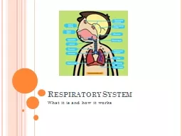
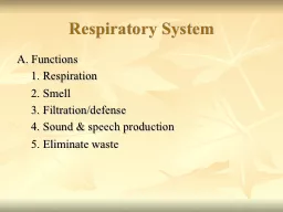
Dr Pallav Shekhar Assitant Professor Unit 2 Objectives To study the method of recording of respiration rate in animals Procedure Examination of respiration is important in ID: 920489
Download Presentation The PPT/PDF document "Practical No.3 : Special examination of..." is the property of its rightful owner. Permission is granted to download and print the materials on this web site for personal, non-commercial use only, and to display it on your personal computer provided you do not modify the materials and that you retain all copyright notices contained in the materials. By downloading content from our website, you accept the terms of this agreement.
Slide1
Practical No.3 : Special examination of Respiratory system
Dr. Pallav ShekharAssitant Professor
Unit 2
Slide2Objectives: To study the method of recording of respiration rate in animals
Procedure: Examination of respiration is important in diagnosing respiratory system diseases as well as other systemic disorders. Rate, rhythm, type and depth of respiration should be noted when assessing the animal’s respiratory movements. Rate is the number of respiratory cycles (inhalation, exhalation and rest) in a minute. The rate is counted by noting either the inhalations or exhalations.
Slide3RESPIRATORY CYCLE
Slide4Caudal Boundary of Pulmonary Percussion Area (Intercostal Space).
Sl. No.
Animal
No. of ribs
At level of external angle of ileum
Middle Thorax
Ventral Border
1.
Horse
18
16
11
6
2.
Cow
13
11
9
5
3.
Sheep & goats
13
11
7
6
4.
Pig
14-15
11-12
8-9
4-5
5.
Dog & Cat
13
11
9
6
6.
Rabbit
12
10
-
7
Slide5Manifestation of Respiratory Insufficiency (Terminology for normal and abnormal lung and thoracic sounds).
Sound Heard
Term Replaced
Example
Present finding in patients (To be filled by the students)
i. Normal Lung Sound
Normal breath sound
Alveolar vesicular bronchial sound/tone
ii.Abnormal lung sound
i.Increased breath sound
-
Fever, exercise, high environmental temperature
ii.Decreased/absence of breath sound
-
Pneumothorax,
iii. Crackels
Moist rales
Interstitial pneumonia
Wheezes
Dry rales, Ronchi
CODP, Chronic stages of pneumonia
Pleuritic friction
-
Pleurisy
Stridors
-
DRT disease
ANATOMY OF NORMAL LUNGS
Slide7CHEST X-RAY
Slide8Slide9Respiratory rates in different animals (Respiration/ Min.):
Animal
Normal Respiration Rate/Min.
Present Respiration Rate/Min. (To
be filled by the student)
Animal
Normal Respiration Rate/Min.
Present Respiration
Rate/Min.
(To
be filled by the student).
Horse
10-14
Dogs
15-30
Ox
10-30
Cats
20-30
Yearling
15-40
Rabbit
30-45
Sheep & Goat
20-30
Camel
5-12
Pigs
8-18
Note: Exercise and exposure to high atmospheric temperatures increase the respiratory rate as the animal’s body is trying to get rid of excessive heat. Pathological conditions that increase the respiratory rates are Fever, Various pulmonary diseases, Upper respiratory tract diseases, Cardiac diseases, Pleurisy or peritonitis, Anaemia etc.
Slide11Types of Respiration: On the basis of involvement of muscle in respiration
The muscles involved in respiration are abdominal muscles, intercostal muscles and the diaphragm. 1. Abdominal Respiration- In abdominal respiration, the abdominal muscles predominate in creating the respiratory movement. This type of respiration is evident in cattle.
Slide122. Costo
-abdominal Respiration- When both abdominal and costal muscles are similarly sharing in respiratory movements the type of respiration is termed costo-abdominal. This type of respiration is seen in horses.
Slide133. Costal respirations-
The costal muscles are predominant in movement in costal respirations. This type of respiration is evident in dogs and cats.
Slide14The rhythm of respiration is altered in some diseased conditions (advanced renal and cardiac disease, severe toxaemia, menengitis). The respiratory depth is also altered in some respiratory disorders (hydrothorax, pneumothorax).
On the basis of rhythm and depth it can be classified in following steps:-
Slide151. Hyperpnoea
Hyperpnoea is the increased pulmonary ventilation (increased respiratory rate with or without increase in the amplitude or width of the movement.
Slide162. Polypnoea
-Polypnoea is increased respiratory rate with reduction in the depth of movements.
Slide173. Oligopnoea
Oligopnoea is reduced respiratory rate. This occasionally occurs in animals suffering from space-occupying lesions of the brain and uraemia. The above mentioned condition comes under dyspnoeaDyspnoea: Difficulty in breathing due to diseased conditions is termed dyspnoea. Acceleration of the rate of respiration without change in type or depth defines the simple
dyspnoea. It is often seen in fever, pneumonia, and pulmonary tuberculosis and in some cardiac affections.
Slide18OLIGOPNOEA/HYPOPNEA
Slide19Laboured breathing is called aggravated
dyspnoea. Animals suffering from this type of dyspnoea show the following signs; Widely distended nostrils, Open mouth, Extension of the head and neckAdduction of the forelimbs.
Laboured
breathing or aggravated dyspnoea
- categorized into inspiratory, expiratory and mixed
dyspnoea
Slide20Inspiratory dyspnoea- Observed in severe unilateral paralysis of the pharynx, paralysis or rupture of the diaphragm, advanced cases of pneumonia and bronchitis, stenosis or obstruction of the nasal passages.
Slide21Expiratory
dyspnoea- seen when exhalation is made difficult. Exaggerated movement of the abdominal muscles- Evident in this type of dyspnoea, as animals need more force to expel air from the lungs. This occurs in vesicular and interstitial emphysema and chronic bronchitis.
Slide22Mixed dyspnoea
- Apparent when the respiratory rate is accelerated as well as difficulty encountered during inspiration and expiration. This occurs in all severe diseases of the respiratory tract (Pneumothorax and hydrothorax) as well as some infectious diseases.PNEUMOTHORAX
Slide23PNEUMOTHORAX
Slide24PULSE OXIMETRY POSITION
Slide25Pulse Oximeter