/
Darkfield Darkfield

Darkfield - PowerPoint Presentation

faustina-dinatale
faustina-dinatale . @faustina-dinatale
Follow
366 views
Uploaded On 2017-08-19

Darkfield - PPT Presentation

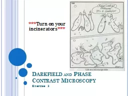
and Phase Contrast Microscopy Exercise 3 Turn on your incinerators Darkfield Microscopy Last week we took a look at brightfield microscopy Bright background dark specimen ID: 580135

motility movement phase light movement motility light phase microscopy darkfield bacteria contrast water focus needle fine background agar organisms

Share:

Link:

Embed:

Download Presentation from below link

Download Presentation The PPT/PDF document "Darkfield" is the property of its rightful owner. Permission is granted to download and print the materials on this web site for personal, non-commercial use only, and to display it on your personal computer provided you do not modify the materials and that you retain all copyright notices contained in the materials. By downloading content from our website, you accept the terms of this agreement.


Presentation Transcript

Slide1

Darkfield and Phase Contrast Microscopy

Exercise 3

***

Turn on your incinerators

***

Slide2

Darkfield Microscopy

Last week, we took a look at brightfield microscopyBright background, dark specimenDarkfield

MicroscopyDark background, specimen is lit upUse of special filterBlocks out central light rays Only scattered light hit the specimen at different anglesSlide3

Darkfield Microscopy

Advantages:Viewing live organisms

More detailed view of external featuresAdding a fluorescent dye increase the ability to see your specimen Disadvantages:Better when the room is completely dark Needs an intense amount of light from the microscope to workCan easily mistake dust for an organismSlide4

How to use Darkfield microscopy

Using a wet mount of a pond water sample. Focus your microscope normally up to the 40x/High Dry Objective.Condenser all the way up, Diaphragm all the way open, Light Control turned all the way up

Remove the blue filter attached to the condenser (put this in a spot where it wont go missing)Replace the blue filter with your darkfield adapter Use the fine focus to fine tune your image.https://www.youtube.com/watch?v=qVylp5rAsTASlide5

Phase Contrast Microscopy

Another type of microscopyUses a special adapter that slows down the wavelength of light by ¼ (phase shift)Developed by Frits Zernike

The phase shift results in the cell having a different refractive index then its surroundingsThe light passes through the organisms vs the background differently Organisms have a halo, bluish background Slide6

How to use phase contrastUsing a wet mount of a pond water sample. Focus your microscope normally up to the 40x/High Dry Objective

.Condenser all the way up, Diaphragm all the way open, Light Control turned all the way up

Make sure your blue filter is in place Push in the phase contrast adapter (PH knob under the stage)Fine tune the image with fine focus https://www.youtube.com/watch?v=7pR7TNzJ_pA&list=PLJYr_JA9Jjd4hW1V-8PIb5Ot97Jj1-K5rSlide7

Make a wet mount of the pond water View under

Brightfield, Phase Contrast and Darkfield

Make notes of your observations Check out the guides provided to see if you can identify any of the organisms you find Slide8

Motility Exercise 4Slide9

Motility Movement of bacteria Allows bacteria to move towards favorable environments or away from unfavorable conditions

Chemotaxis- chemicalsPhototaxis- light

Aerotaxis- oxygen Organelle for motility: flagella Monotrichous- single flagellum Amphitrichous : flagella at each end of the cellPeritrichous : flagellum on all sidesLophotrichous : several flagella on one side Slide10

Motility Brownian movement

Random movement due to the bombardment of molecules of the solventNot true movement Shake or jiggle Water movement

Movement due to movement of water under slideNot a true movement“Goes with the flow”True motilityRapid swimming movement, abrupt changes in directions, swimming against the current Slide11

Motility Another way to determine motility is the use of Sulfide Indole Motility (SIM) Agar

Semi-soft agar that allows for the movement of bacteria through the medium Non-motile bacteria will only grow along the inoculation line Motile bacteria will grow along and away the inoculation line Slide12

Inoculation needle

2 test tubes of SIM Agar/group

1 tube/student

What do we need?

Cultures

Pseudomonas aeruginosa

Staphylococcus aureus

LABEL YOUR TUBES

Name/Initials

Class Section

Name of Organism used Slide13

Slightly vortex your bacterial cultures

Flame your needle to sterilize

Dip your sterile needle into your assigned organism

Stab the SIM agar (2/3 the way) as straight as you can, one time.

Flame your loop to sterilize again before putting your needle away

Place your inoculated tube in the class rack

Incubates at 37°C for 24 hours

We will check results next week Slide14

Expected results

Related Contents


Next Show more