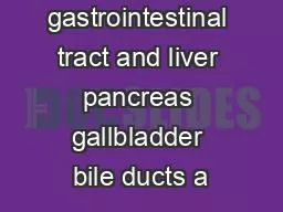
October 2 nd 2014 David Tang MD Faculty Manreet Kaur MD Jason Hou MD Case 64 year old White man who presented for colonoscopy 2007 Liver transplant for primary sclerosing ID: 679122
Download Presentation The PPT/PDF document "Gastroenterology Grand Rounds" is the property of its rightful owner. Permission is granted to download and print the materials on this web site for personal, non-commercial use only, and to display it on your personal computer provided you do not modify the materials and that you retain all copyright notices contained in the materials. By downloading content from our website, you accept the terms of this agreement.
Slide1
Gastroenterology Grand Rounds
October 2nd 2014David Tang M.D.FacultyManreet Kaur M.D.Jason Hou M.D.Slide2
Case
64 year old White man who presented for colonoscopy.2007 – Liver transplant for primary sclerosing cholangitis (PSC) at Arizona area hospital2007 – Onset of chronic diarrhea2009 – Colonoscopy at
in
Arizona
area hospital with
colitis to 40 cm. Patient diagnosed with ulcerative colitis but was never treatedSlide3
Case
April 2011 – Colonoscopy at Houston area hospital with normal mucosa. Segmental biopsies with chronic active colitis without dysplasiaJuly 2011 – Seen in clinic and complained of 10 to 12 bowel movements per day; started on Asacol 2.4 gramsAugust 2011 – Seen in clinic with improvement in stool frequency; Asacol
continued and
Canasa
suppositories were added.Slide4
Case
March 2012 – Surveillance colonoscopy with normal mucosa. Segmental biopsies showed chronic active colitis without dysplasiaDecember 2012 – Clinic visit with resolution of diarrhea on Asacol.April 2013 – Surveillance colonoscopy with normal mucosa except loss of vascular pattern in the rectum. Random biopsies every 10 cm with chronic active colitis and intraepithelial eosinophilia but no dysplasiaSlide5
Case
April 2014 – Clinic visit with complaints of 10 bowel movements per day. Imodium was recommended.July 2014 – Patient returns for surveillance colonoscopySlide6
CaseSlide7
CaseSlide8
Pathological Diagnosis
Invasive AdenocarcinomaSlide9
Questions
What is the epidemiology of colorectal cancer (CRC) in ulcerative colitis (UC)?What are risk factors for CRC in UC? Is PSC a risk factor? Is there an increased risk for CRC after transplantation for PSC in UC?What is the evidence behind current management strategies for neoplastic lesions discovered on colonoscopy in patient’s with UC?Flat versus raised dysplasiaAdenoma-like vs non-adenoma-like lesionsSlide10
Increased Incidence of CRC in UC
Meta-analysis of 8 population based cohort studies
Jess CGH 2012Slide11
Prevalence of IBD
in PSC
Size
Population
Concomitant
IBD
Concomitant
UC
Concomitant
CD
Berquist
604
Sweden
79%
69%
7%
Boberg
394
Europe
82%
65%
-
Weisner
174
Minnesota
71%
-
-
Takikawa
192
Japan
21%
19.8%
1%
Kochlar
18
India
-
50%
-Slide12
PSC Increases Risk for CRC in UC
Soetikno
GIE 2012
OR
4.09: 95% CI [2.89, 5.76]
Meta-analysis of 11 studiesSlide13
PSC Increases Risk for CRC in UC
Study of 40 UC + PSC and 80 UC only matched controls
Cumulative
risk of CRC in patients with UC
+ PSC
of
9% after 10
years
disease duration,
31% after 20 years
and as high
as
50
% after 25
years
compared with 2%, 5% and 10% in patients with UC alone
Broome
Hepatology
1995Slide14
Increased Risk of CRC in PSC post liver transplant
Comparison PSC patients status post liver transplant with and without IBD
Cumulative risk of developing CRC is 17% at 10 years in patients with IBD
Compared with 0% at 10 years in patients without IBD
Vera Transplantation 2003Slide15
Other Risk Factors
Age of onset – conflicting evidence for higher incidence in children; surveillance intervals should be identical to adultsDisease extent – most CRC arise in pancolitis; little increased risk in proctitis or
proctosigmoiditis
Disease duration – relative risk of CRC is significant after 8 to 10 years of disease duration
Disease severity – CRC may occur in segments with only microscopic disease
Family history – history of sporadic CRC lends 2 to 3 fold risk
Farraye
Gastroenterology 2010Slide16
Classification of Dysplasia
Macroscopic ClassificationFlat endoscopically undetectableRaised or DALM (Dysplasia-associated Mass or Lesion)
Adenoma-like
Non-adenoma-like
Histological Classification
Indefinite
Low Grade Dysplasia (LGD)
High Grade Dysplasia (HGD)Slide17
Classification of Raised DysplasiaSlide18
Classification of Raised Dysplasia
Farraye Gastroenterology 2010
Adenoma-like
Non-adenoma-likeSlide19
Raised Dysplasia
Review of 10 prospective studies of surveillance colonoscopy in chronic UC.17 of 40 patients (43%) with raised dysplasia had synchronous CRC on colectomy.
Bernstein Lancet 1994Slide20
Conservative Management of Adenoma-like Raised Dysplasia
Prospective study of 48 patients with adenoma-like lesions without other dysplasia followed for mean 4.1 years.
Rubin Gastroenterology 1999
Six patients (12%) ultimately received colectomy for incomplete polyp resection
no CRC foundSlide21
Conservative Management of Adenoma-like Raised Dysplasia
Prospective follow up study of 24 patients with UC who had polypectomy for adenoma-like lesions without other flat dysplasiaSix under went colectomy
Only one patient had flat LGD
18 underwent surveillance
Only one developed adenocarcinoma
N
o significant
differences in the incidence of polyp formation on
follow-up
between patients with UC and an adenoma-like
lesion (62.5%), patients
with UC and
a sporadic
adenoma (50%),
and a non-UC
sporadic adenoma control group (49%).
Odze
CGH 2004Slide22
Flat Dysplasia
Review of 10 prospective studies of surveillance colonoscopy in chronic UC.10/24 (42%) with HGD who underwent immediate colectomy had CRC
Bernstein Lancet 1994Slide23
Flat High Grade Dysplasia
5/11 (45%) with HGD who underwent immediate colectomy had CRC2/8 (25%) with HGD who underwent surveillance ultimately developed CRC
600 patients with mean 8.5 years of follow up in surveillance program
Rutter Gastroenterology 2006Slide24
Flat Low Grade Dysplasia
Meta-analysis of 7 surveillance studies of patients with chronic UC and flat LGD
Thomas APT 2007
The odds ratio of CRC in patients with LGD compared with no dysplasia on surveillance was
9.0
, 95%
CI: 4.0 – 20.5Slide25
Flat Low Grade Dysplasia
Retrospective cohort study of 46 patients with UC and flat LGD on surveillance colonoscopy.Overall, 14 of 46 patients (30%) progressed to advanced neoplasia (CRC or HGD)3 of 11 patients (27%) undergoing immediate colectomy had synchronous CRC
Rate of
progression to advanced neoplasia for all 46
flat LGD
subjects was
53%
at 5 years (95%
confidence
interval [
CI],
0.29 –
0.77)
Ullman Gastroenterology 2003Slide26
Flat Low Grade Dysplasia
Retrospective cohort study of 128 patients with UC enrolled in surveillance colonoscopy from 1979 to 1990.3 of 29 patients with LGD (10%) developed advanced neoplasia (HGD or CRC).4 of 97 patients without dysplasia (4%) developed advanced neoplasia
Lim Gut 2003Slide27
Flat Low Grade Dysplasia
No significant difference in Kaplan Meier analysis for progression to colectomy or death between LGD and no dysplasia cohorts
Lim Gut 2003Slide28
Flat Low Grade Dysplasia
Unifocal flat LGD on initial colonoscopy
Surveillance at 3, 6, 12 months, then annually
Immediate colectomy and
Ileal
pouch anal
anastamosis
Nguyen GIE 2009
Probabilities
and health utilities were derived from the
literature
Costs
were derived from national hospital data sets and Medicare and/or Medicaid reimbursement schedules.Slide29
Flat Low Grade Dysplasia
Immediate colectomy dominated over enhanced surveillance and yielded higher QALYs and lower costs. These findings remained dominant in 90% of simulations in sensitivity analysis.
Nguyen GIE 2009Slide30
Management Algorithm
S&F Chapter 112Slide31
Management Algorithm
S&F Chapter 112Slide32
Chromoendoscopy
“Normal white light colonoscopy, using standard or high definition colonoscopes along with multiple colon biopsies, remains a reasonable method of surveillance for patients with IBD. However, chromoendoscopy with targeted biopsies is considered an acceptable alternative…”
-AGA
“
Chromoendoscopy
with targeted biopsies is the surveillance procedure of choice for appropriately trained
endoscopists
.”
-
ECCO