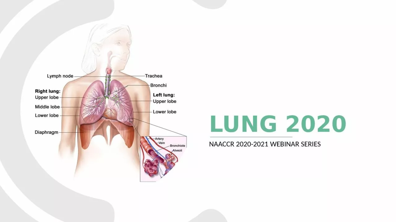
/
Lung 2020 NAACCR 2020-2021 Webinar Series Lung 2020 NAACCR 2020-2021 Webinar Series

Lung 2020 NAACCR 2020-2021 Webinar Series - PowerPoint Presentation

gelbero
gelbero . @gelbero
Follow
64 views
Uploaded On 2023-12-30

Lung 2020 NAACCR 2020-2021 Webinar Series - PPT Presentation

QampA Please submit all questions concerning the webinar content through the QampA panel If you have participants watching this webinar at your site please collect their names and emails ID: 1036001

grade tumor primary lung tumor grade lung primary code tumors eod invasive amp table predominant separate adenoca neoadjuvant unknown

Share:

Link:

Embed:

Download Presentation from below link

Download Presentation The PPT/PDF document "Lung 2020 NAACCR 2020-2021 Webinar Serie..." is the property of its rightful owner. Permission is granted to download and print the materials on this web site for personal, non-commercial use only, and to display it on your personal computer provided you do not modify the materials and that you retain all copyright notices contained in the materials. By downloading content from our website, you accept the terms of this agreement.


Presentation Transcript

1. Lung 2020NAACCR 2020-2021 Webinar Series

2. Q&APlease submit all questions concerning the webinar content through the Q&A panel. If you have participants watching this webinar at your site, please collect their names and emails. We will be distributing a Q&A document in about one week. This document will fully answer questions asked during the webinar and will contain any corrections that we may discover after the webinar. 2

3. 3Fabulous Prizes

4. Guest PresenterSDenise Harrison, CTRKelli Olsen, MS, CTR 4

5. Lung Cancer 2020Denise Harrison, BS, CTR

6. Case scenario56 y.o. woman w/persistent cough and chest painImaging:12/29/2015 CT Chest: Two "ground-glass" nodules (RUL and RLL) noted.04/19/2018 PET [OSF]: Suspicious for a low grade primary lung malignancy.09/12/2018 CT [Reporting Hosp]: 1.7cm sub-solid nodule in posterior RUL and 1.3cm ground-glass nodule in superior segment of RLL, both are "concerning for malignancy".Scopes: NoneSurgery: 10/22/2018 RUL wedge and RLL wedge w/two LNs.Path: Inv well diff Adenoca, acinar predominant, in RUL (2.2cm) and RLL (1.2cm); No visceral pleura invasion; LVI(-); surg margins(-); 0+/2 LNs.66

7. 7

8. MediastinumSuperior:Aortic arch, brachiocephalic veins, esophagus, phrenic nerves, superior vena cava, thoracic duct, (some) thymus, trachea, vagus nerves InferiorAnterior: thymus, sternopericardial ligamentsMiddle: heart, pericardiumPosterior: descending aorta, esophagus, sympathetic trunk, thoracic ducthttps://thegoofyanatomist.weebly.com

9. Solid Tumor Rules 2018Use Solid Tumor Rules by DATE OF DIAGNOSISDo NOT Use Rules for Metastases

10. Equivalent or Equal TermsAdenocarcinoma; carcinomaAnd; with (when describing multiple histologies within a single tumor)NSC-CA 8046 = all histo in Table 3 except NET 8041 and NET subtypes (and sarcomas)Simultaneous; existing at same time; concurrent; prior to FCOTSite; topographySquamous cell carcinoma; SCC; epidermoid carcinomaTumor; mass; lesion; neoplasm; noduleType; subtype; variant10

11. Terms NOT Equivalent/EqualBilateral ≠ single or multiple primariesBronchus ≠ mainstem bronchusComponent ≠ subtype/type/variantLung primaries ONLYMucinous ≠ colloid Mucin-producing/mucin-secreting 8481 ≠ mucinous carcinoma 8253 Multilocular ≠ multinodularPhenotype ≠ subtype/type/variant11

12. Table 1: Coding Primary Site Terminology Laterality Site Term and Code Bronchus intermedius Carina Hilus of lung Perihilar Bilateral Mainstem bronchus C340 Note: Bronchus intermedius is the portion of the Rt MSB between the upper lobar bronchus and the origin of the middle and lower lobar bronchi Lingula of lung Left Upper lobe C341 Apex Apex of lung Lung apex Pancoast tumor Superior lobar bronchus Upper lobe bronchi Bilateral Upper lobe C341 “Bilateral” means the structure occurs on both sides; do not use that terminology to code laterality!12

13. Table 1: Coding Primary Site Terminology Laterality Site Term and Code Base of lung Lower lobar bronchus Lower lobe Lower lobe bronchi Lower lobe segmental bronchi Bilateral Lower lobe C343 Overlapping lesion of lung Bilateral Overlapping lesion of lung C348 Note: One lesion/tumor which overlaps two or more lobes “Bilateral” means the structure occurs on both sides; do not use that terminology to code laterality!13

14. Table 1: Coding Primary Site Terminology Laterality Site Term and Code Bronchus NOS Bronchogenic Extending up to the hilum Extending down to the hilar region Lung NOS Pulmonary NOS Suprahilar NOS Bilateral Lung NOS C349 Note: Includes • Multiple tumors in different lobes of ipsilateral lung OR • Multiple tumors in ipsilateral lung; unknown if same lobe or different lobe OR • Tumor in bronchus, unknown if mainstem or lobar bronchus OR • Tumor present, unknown which lobe Lobar bronchi NOS Lobar bronchus NOS Bilateral Code the lobe in which the lobar bronchus tumor is present C34__ Note: When lobe of origin is not documented/unknown, code to lung NOS C349 “Bilateral” means the structure occurs on both sides; do not use that terminology to code laterality!14

15. Multiple Primary Rules (M1-M4)Unknown if Single or MultipleM1:Not possible to determine if there is a single tumor or multiple tumors = SPSingle TumorM2: Single tumor = SPMultiple Tumors (Do NOT code multiple primaries based on biomarkers!)M3: S/N-C* tumors with site codes different at 2nd CXxx or 3rd CxXx = MPM4: Subsequent tumor after clinically dz-free for > 3 after dx OR recurrence = MP If recurrence ≤ 3 years, keep reading the rules.*S/N-C = Separate, non-contiguous

16. Rule M5 – Multiple Tumors, cont.M5: ≥ 1 tumor small cell (8041) or variant AND another tumor non-small (8046) or variant = MPM6 – M8 refer us to Table 3M6: S/N-C tumors ≥ 2 different subtypes in column 3 of Table 3 = MPM7: Synchronous S/N-C tumors in the same lung in same row of Table 3 = SPM8: S/N-C tumors in different rows Table 3 = MPcombo code in Table 2 + code in Table 3 = MP*S/N-C = Separate, non-contiguous

17. Rules M6 - M8: Table 3M7: Same row = SP (any of the following in the same row)Same Histology:Col. 1 + Col.2; Col. 2 + Col. 2 -or-Col. 1 + 1 sub/var Col. 3 -or-Col. 2 + 1 sub/var Col. 3M6: Different subtypes = MPSame or Different NOSM8: Different Rows = MP(any column)12

18. Rules M9-M14: Multiple TumorsM9: Simultaneous multiple tumors = SP when: in both lungs OR in same lung OR single tumor in one lung; multi tumors in contralateral lungM10: In situ diagnosed after invasive AND tumors in same lung = SPM11: Single tumor in each lung = MPM12: Invasive tumor ≤ 60 days after in situ in same lung = SPM13: Invasive > 60 days after in situ in same lung = MPM14: None of the rules apply = SP

19. Histologic TypeGuidelines for ICD-O-3 Updates include:New histologiesChanges in behaviorNew preferred terminologySTR Editors recommend coding histo using: 2018 Solid Tumor RulesUpdated ICD-O histology codes and terms which can be found at: https://seer.cancer.gov/icd-o-3/ ICD-OAsk a SEER Registrar When preceding 3 bullets fail to ID a histology code

20. Important Notes for Coding Histology

21. Important Notes for Coding Histology

22. Documentation Priority to Identify Histology

23. Coding HistologyTerms A-C must describe a carcinoma or sarcoma  

24. Coding Histology

25. List of Ambiguous TermsApparentlyAppearsComparable withCompatible withConsistent withFavor(s)Malignant appearingMost likelyPresumedProbableSuspect(ed)Suspicious (for)Typical (of)Coding Histology

26. LungTissue/path from primary1FNA^2Tissue/path from mets3ScansCT > PET > MRI > CXR4 Physician Documentation5Summary: Coding HistologyCode histologyBefore neoadjuvant therapyUsing priority list & H rulesDo not change histo to stageMultiple HistologiesCode most specific histo or subtype/variant whether described as majority*, predominant*, minority*, or component*Code NOS w/ features or differentiation ONLY when there is a specific codeUse ambiguous terms ONLY when criteria metDo NOT code histology based on pattern architecture, focus/foci/focal*must describe a carcinoma or sarcoma^ from primary site, or pleural/pericardial fluid

27. Histology rulesSingleMultiple RuleH1H10 Code mucinous adenoca as follows (for lung only)8253/3 when behavior unk or invasive8257/3 when microinvasive or minimally invasive8253/2 when preinvasive or in situH2H11 Code non-mucinous adenoca as follows (for lung only)8256/3 when microinvasive or minimally invasive8250/2 when preinvasive or in situH3H12 Code specific histo when dx is NSCLC described by ANY ambiguous terminology when histo is:Clinically confirmed by MD (attending, pathologist, oncologist, pulmonologist, etc.) Patient is treated for the histology described by an ambiguous term Case accessioned based on single histo described by ambiguous terminology and no other histology information is available/documentedNote: Mucinous carcinoma mixed w/ another histo, code mucinous ONLY when mucinous is documented to be > 50% of the tumor.27

28. Adenocaspectrum lesionsCT appearance of peripheral lung adenocarcinomas encompasses a spectrum from ground glass nodules (GGN) to solid mass lesionsCT appearance reflects their heterogenous histologic subtypesSingle term, BAC, was not adequate to describe this spectrumLepidic growth manifests radiologically as GGOOn CT, parenchymal structures (airways and vessels) can be seen through the GGOOn pathology, lepidic features identified (w/ or w/o an invasive component)GGN (lepidic) can evolve to more solid (more likely invasive) Data from lung cancer screening literature showHigher rate of malignancy in incidental part-solid nodules compared to incidental solid nodules and the Majority of persistent GGNs represent adenocarcinoma spectrum lesionsRevised classification more clearly follows the multistep progression that many lung adenocarcinoma spectrum lesions are thought to take

29. The revised classification of lung adenocarcinoma(I) Preinvasive lesions     (i) Adenocarcinoma in situ (AIS) —mucinous 8253/2*, nonmucinous 8250/2*, or mixed (see H rules)     (ii) Atypical adenomatous hyperplasia (AAH)(II) Minimally invasive lesions     (i) Minimally invasive adenocarcinomas (MIA) —mucinous 8257/3*, nonmucinous 8256/3*, or mixed (see H rules)(III) Invasive adenocarcinoma     (i) Acinar predominant 8551/3*     (ii) Papillary predominant 8260/3     (iii) Micropapillary predominant 8265/3     (iv) Solid predominant with mucin production 8230/3     (v) Lepidic predominant adenocarcinoma (LPA) 8250/3*(IV) Variants of invasive adenocarcinoma     (i) Invasive mucinous adenocarcinoma 8253/3*     (ii) Colloid 8480/3, fetal 8333/3, and enteric 8144/3* New code

30. Table 3: Specific Histologies, NOS, and Subtype/Variants 

31. Histology rulesSingleMultiple RuleH4H13 Code histo when only one histo presentH5H14 Code invasive histo when in situ and invasiveH6H15 Code subtype/variant when NOS & single subtypeH7 Code histo that comprises greatest amount of tumor when 2 or more of the following histologies are present:Acinar adenoCA / AdenoCA, acinar predominant 8551 Lepidic adenoCA / AdenoCA, lepidic predominant 8250 Micropapillary adenoCA / AdenoCA, micropapillary predominant 8265 Papillary adenoCA / AdenoCA, papillary predominant 8260 Solid adenoCA / AdenoCA, solid predominant 8230 NOTE: If percentage unknown, continue through the rules31

32. Histology rulesSingleMultiple RuleH8H16 Code combo code (Table 2) when multi histologies AND Combination is listed in ORYou received a combo code from Ask A SEER RegistrarH9 Code adenoca with mixed subtypes 8255 forMultiple adenoca subtypes (includes adenoca + ≥ 2 subtypes) ORAny combo of histo NOT listed in Table 232

33. Adenocaspectrum lesionsCT appearance of peripheral lung adenocarcinomas encompasses a spectrum from ground glass nodules (GGN) to solid mass lesionsCT appearance reflects their heterogenous histologic subtypesSingle term, BAC, was not adequate to describe this spectrumLepidic growth manifests radiologically as GGOOn CT, parenchymal structures (airways and vessels) can be seen through the GGOOn pathology, lepidic features identified (w/ or w/o an invasive component)GGN (lepidic) can evolve to more solid (more likely invasive) Data from lung cancer screening literature showHigher rate of malignancy in incidental part-solid nodules compared to incidental solid nodules and the Majority of persistent GGNs represent adenocarcinoma spectrum lesionsRevised classification more clearly follows the multistep progression that many lung adenocarcinoma spectrum lesions are thought to take

34. The revised classification of lung adenocarcinoma(I) Preinvasive lesions     (i) Adenocarcinoma in situ (AIS) —mucinous 8253/2*, nonmucinous 8250/2*, or mixed (see H rules)     (ii) Atypical adenomatous hyperplasia (AAH)(II) Minimally invasive lesions     (i) Minimally invasive adenocarcinomas (MIA) —mucinous 8257/3*, nonmucinous 8256/3*, or mixed (see H rules)(III) Invasive adenocarcinoma     (i) Acinar predominant 8551/3*     (ii) Papillary predominant 8260/3     (iii) Micropapillary predominant 8265/3     (iv) Solid predominant with mucin production 8230/3     (v) Lepidic predominant adenocarcinoma (LPA) 8250/3*(IV) Variants of invasive adenocarcinoma     (i) Invasive mucinous adenocarcinoma 8253/3*     (ii) Colloid 8480/3, fetal 8333/3, and enteric 8144/3* New code

35. Table 3: Specific Histologies, NOS, and Subtype/Variants 

36. Table 2: Combo/Mixed Histo Codes

37. Exercise: # of Primaries, Primary site(s),  Histology(ies), behaviorCT Chest: 1.7cm sub-solid nodule in posterior RUL and 1.3cm ground glass nodule in superior segment of RLL, both concerning for malignancy. PET: Suspicious for a low grade primary lung malignancy.Wedge resection of RUL and RLL and LN dissection: Adenoca, acinar predominant, well diff, no invasion of visceral pleura; surg. margins negative;  TS 2.2cm and 1.2cm; 0+/2 LNs.# Primaries  _____  M7 (synchronous separate non-contiguous tumors in same row in same lung)Primary site  ____   There are 2 tumors, but we are basing the staging on the tumor in the RUL. If one the tumors were intrapulmonary mets, we would assign C34.9Histology  _____  H7 (single histology in all tumors) Adenocarcinoma, acinar predominantBehavior    _____ InvasiveC34.1855113

38. Staging lung CancersSummary Stage V2.0 (Effective with 2021 Diagnoses)Extent of Disease V2.0 (Effective with 2021 Diagnoses)AJCC 8th Edition (Effective with 2018 Diagnoses)

39. RemindersTimingAJCC staging classifications (clinical, pathological, and post-therapy) are based on distinct time-framesSS18 and EOD are clinicopathological staging/data collection systemsBased on the most extensive involvement, regardless of timing Use all available information from diagnosis through post-therapyConcordanceEOD is based on AJCC, so these systems are highly concordant (need TS Summary to derive correct EOD T at central registry)SS18, while generally concordant with AJCC and EOD, may classify T4 tumors as localized (code 1), or LNs that are regional in AJCC and EOD as distant

40. SS18 and EODNotes:Bronchopneumonia ≠ obstructive pneumonitisAtelectasis must be associated w/ an obstructing tumorInstructions to assist with coding VPI (PL1, PL2) and PPI (PL3) Separate tumor nodulesVC paralysis, SVC syndrome, compression of trachea or esophagusOccult carcinomaPleural/pericardial effusionsMinimally invasive and superficial spreading tumors (EOD only)

41. AJCC ClassificationscT and cNH&PImaging studies Staging procedures (scopes)Biopsies/CytologyExploratory thoracotomycMcM0 if no evidence of metscM1 if mets NOT microscopically proven during clinical timeframepM1 if mets microscopically proven during clinical timeframe414141pTcT + operative findings + pathological examination of primary OR (+) bxs confirming highest T and highest NpN ≥ 1 LN examined microscopically (also need pT)IASLC recommends at least 6 LN from 6 stationspMMicroscopic exam of distant LN, tissue, or fluid (found to be POSITIVE)cM0 and cM1 allowed in “pM” field when no positive microscopic findings of metsClinical ClassificationPathological Classification

42. AJCC ClassificationsycT and ycN AFTER completion of neoadjuvant therapy:H&PImaging studiesStaging procedures (scopes)Biopsies/CytologyExploratory thoracotomyNo ycMUse “cM” category assigned PRIOR to neoadjuvant therapycM0, cM1, and pM1 allowed424242AFTER completion of neoadjuvant therapy:ypTycT + operative findings + pathological examination of primary OR (+) bxs confirming highest T and highest NypN ≥ 1 LN examined microscopically (also need ypT)IASLC recommends at least 6 LN from 6 stationsNo ypM Use “cM” category assigned PRIOR to neoadjuvant therapycM0, cM1, and pM1 allowedyc Classificationyp Classification

43. Clinical Judgment of Non-Malignant Cause of EffusionPleural effusionCHFInfections (pneumonia, tuberculosis)Pulmonary embolismKidney failureAutoimmune (lupus, rheumatoid arthritis)Other diseasesPericardial effusionSame as pleural PLUSRT with heart in fieldHistory of chemoAdria, CytoxanHypothyroidismTrauma/puncture near heartCertain drugs (hydralazine, isoniazid, phenytoin)Special note in M1a about pleural effusion malignant vs non-malignant

44. AJCC T CategoryMultiple descriptions of in situLepidic pattern measurement importantCould be in situ or minimally invasiveSize matters!Cut points at ≤ 1 cm, 2 cm, 3 cm, 4 cm, 5 cm, 7 cmT2 – T4 based on size or extensionT2 has subcategories based on TST3 – T4 include additional descriptions for ipsilateral separate tumor nodules

45. Measuring TS: Lepidic Area Doesn’t CountJournal of Thoracic Oncology, Vol 8, Issue 1, Jan 2013

46.

47. SS18, EOD PRIMARY TUMOR, & EOD TEODSS2018Description0000 In situ, intraepithelial, noninvasive0000 Adenocarcinoma in situ (AIS): adenocarcinoma with pure lepidic pattern, ≤ 3 cm in greatest dimension0000 Squamous cell carcinoma in situ (SCIS)3001 Localized only (localized, NOS)3001 Confined to lung, NOS1001 Minimally invasive adenocarcinoma; Adenocarcinoma tumor W/ predominantly lepidic pattern (AIS) measuring ≤ 3 cm in greatest dimension W/ invasive component measuring ≤ 5 mm in greatest dimension2001 Superficial tumor, WITH invasive component limited to bronchial wall WITH or WITHOUT proximal extension to main stem bronchus (these types of tumors are uncommon)4001 Adjacent ipsilateral lobe4001 Confined to hilus4001 Main stem bronchus, NOS (without involvement of the carina) Including extension from other part of lung6001 Confined to carina, NOS

48. SS18, EOD PRIMARY TUMOR, & EOD TEODSS2018 Description4002 Atelectasis/obstructive pneumonitis that extends to hilar region, involving part or all of lung4502 Pleura, NOS4502 Pulmonary ligament4502 Visceral pleura invasion (PL1, PL2, or NOS) (SS18 includes PL3)5002 Brachial plexus (inferior branches or NOS)5002 Chest wall (thoracic wall) (separate lesion-see EOD Mets)5002 Diaphragm (separate lesion-see code 7) (separate lesion-see EOD Mets)5002 Pancoast tumor (superior sulcus syndrome), NOS5002 Parietal pericardium5002 Parietal pleura5002 Pericardium, NOS5002 Phrenic nerve5002 Separate tumor nodule(s) in same lobe as primary

49. SS18, EOD PRIMARY TUMOR, & EOD TEODSS2018Description6502 Code 600 (confined to carina) + (any of codes 100 through 500)6502 Blood vessel(s) (major)6502 Aorta6502 Azygos vein6502 Pulmonary artery or vein6502Superior vena cava (SVC syndrome)6502 Carina from lung (with involvement of any other parts of lung)6502 Cervical sympathetic (Horner's syndrome)6502 Compression of esophagus or trachea not specified as direct extension6502 Esophagus6502 Mediastinum, extrapulmonary or NOS6502 Nerve(s)6502 Recurrent laryngeal (vocal cord paralysis)6502 Separate tumor nodule(s) in a different ipsilateral lobe6502 Trachea6502 Vagus

50. SS18, EOD PRIMARY TUMOR, & EOD TEODSS2018Description6757 Adjacent rib6757 Rib6757 Skeletal muscle6757 Sternum7007 Heart7007 Inferior vena cava7007 Neural foramina7007 Vertebra(e) (vertebral body)7007 Visceral pericardium7007 Separate tumor nodule(s) in a different ipsilateral lobe7007 Further contiguous extension7007 Nerve(s)7007 Recurrent laryngeal (vocal cord paralysis)7007 Separate tumor nodule(s) in a different ipsilateral lobe

51. SS18, EOD PRIMARY TUMOR, & EOD TEODSS2018 Description8009 No evidence of primary tumor9809 Tumor proven by presence of malignant cells in sputum or bronchial washings but not visualized by imaging or bronchoscopy9999 Unknown; extension not stated9999 Primary tumor cannot be assessed9999 Not documented in patient record

52. Exercise: EOD Primary Tumor, AJCC T, and SS18CT Chest: 1.7cm sub-solid nodule in posterior RUL and 1.3cm ground glass nodule in superior segment of RLL, both concerning for malignancy. PET: Suspicious for a low grade primary lung malignancy.Wedge resection of RUL and RLL and LN dissection: Adenoca, acinar predominant, well diff, no invasion of visceral pleura; surg. margins negative;  TS 2.2cm and 1.2cm; 0+/2 LNs.EOD Primary Tumor _____ Any size tumor, confined to lungAJCC Clinical T _____ Largest TS = 1.7cm; no evidence of extension outside the lungAJCC Pathological T _____ Largest TS = 2.2cm, no VPI or extension outside the lungSS18 _____ Localized only1cT1bpT1c300

53. AJCC Regional LNsExtend from supraclavicular area to diaphragmLocation determines N descriptionThe farther away from the hilum the LN are, the higher the N descriptionContralateral raises N1 (hilar/interpulmonary) or N2 (mediastinal) to N3Any laterality supraclavicular/scalene LNs are N3Do NOT use Table 36.2 to code LN (Exploratory Subcategories)If surgeon uses IASLC LN Station descriptions or zones:1 = supraclavicular, 2 – 9 = mediastinal, 10 – 14 = hilarSee Fig. 36.1 and Table 36.1 in AJCC

54. Regional Lymph Node Zones and Stations Zone StationSupraclavicular1 Low cervical, SC, sternal notchSuperior mediastinal nodesUpper2 Upper paratracheal3a Prevascular3p Retrotracheal4 Lower paratrachealAortopulmonary (AP)5 Subaortic6 Para-aorticInferior mediastinal nodesSubcarinal7 SubcarinalLower8 Paraesophageal9 Pulmonary ligament54 Zone StationHilar/interlobar10 Hilar11 InterlobarPeripheral12 Lobar13 Segmental14 SubsegmentalSee the AJCC Cancer Staging Manual, Eighth Edition for Fig. 36.1 IASLC lymph node map and Table 36.1 Anatomic definitions for each lymph node station and station grouping by nodal zones in the map proposed by the IASLC.

55. Tumor Laterality and N Category by LN Station Tumor in Right Lung Tumor in Left Lung 10R – 14R 10L – 14L 2R, 4R, 7, 9R 2L, 4L, 5, 6, 7, 9L 3A (Rt of midline of trachea) 3A (Lt of midline of trachea) 3P --- 8 (Rt of midline of esophagus) 8 Lt of midline of esophagus) 1R, 1L, 2L, 4L, 5, 6, 9L, 10L – 14L 1L, 1R, 2R, 4R, 9R, 10R – 14R 3A (Lt of midline of trachea) --- 3A (Rt of midline of trachea) 3P 8 (Lt of midline of esophagus) 8 (Rt of midline of esophagus)

56.

57.

58. SS18, EOD REGIONAL NODES, & EOD NEODSS2018EOD NEODSS2018EOD NIPSI3003N14003N2BI/C7007N37007N3 Bronchial Carinal (tracheobronchial) (tracheal bifurcation) Hilar (bronchopulmonary) (proximal lobar) (pulmonary root) Mediastinal Anterior SuperiorAortic (above diaphragm), NOSParatracheal (Lt, Rt, upper, low, NOS) IntrapulmonaryPeri/para-aortic, NOSPrevascular Interlobar Ascending aorta (phrenic)Retrotracheal LobarSubaortic (A-P window) Periesophageal Segmental Inferior Pericardial SubsegmentalParaesophageal Peritracheal, NOS Peri/parabronchialPulmonary ligamentAzygos (lower peritracheal)Subcarinal Precarinal Posterior (tracheoesophageal) Pretracheal, NOSIPSI/CONTRAEOD - 600SS18 - 7EOD N - N3Low cervical; Proximal root; Scalene (inferior deep cervical); Sternal notch; Supraclavicular (transverse cervical)

59. Exercise: EOD Regional Nodes and , AJCC NcN0pN000

60. SS18, EOD Mets, & EOD MEODSS2018 Description007 No distant metastasis; Unknown if distant metastasis107 Pericardial effusion or pleural effusion (malignant) (ipsilateral, contralateral, bilateral, NOS) Pleural tumor foci or nodules on ipsilateral lung (separate from direct extension) or contralateral lung Pericardial nodules Contralateral lung/main stem bronchus Contralateral main stem bronchus Separate tumor nodule(s) in contralateral lung207 Single distant lymph node involvedCervicalDistant lymph node, NOS307 Single extrathoracic metastasis in a single organ

61. SS18, EOD Mets, & EOD MEODSS2018 Description507 Multiple extrathoracic metastases in a single organ or in multiple organs Abdominal organs Skin of chest Separate lesion in chest wall or diaphragm Multiple distant lymph node(s)CervicalDistant lymph node(s), NOS Carcinomatosis Distant metastasis WITH or WITHOUT distant LN(s)707 Distant metastasis, NOS999 Death Certificate Only

62. Exercise: EOD Mets and AJCC MCT Chest: 2.2cm spiculated nodule LUL; hilar lymphadenopathy. PET: FDG uptake in LUL lung mass & hilar LNs consistent w/ malignancy. MRI Brain: (-)LUL lobectomy & mediastinal LN dissection: Adenoca, mod diff, extending to visceral pleura (elastic stain performed); surg. margins negative; TS 2.5cm; 2+/5 peribronchial LN; all mediastinal LNs (-)EOD Mets _____ AJCC Clinical M _____ AJCC Pathological M _____ cM0cM000For all 3 fields: PE and CT negative for mets

63. IASLC Stage GroupingsN0N1N2N3M1aM1bM1cT1aIA1 (incl T1mi)IIBIIIAIIIB IVAIVAIVBT1bIA2IIBIIIAIIIB IVAIVAIVBT1cIA3IIBIIIAIIIB IVAIVAIVBT2aIBIIBIIIAIIIB IVAIVAIVBT2bIIAIIBIIIAIIIB IVAIVAIVBT3IIBIIIAIIIBIIICIVAIVAIVBT4IIIAIIIAIIIB IIICIVAIVAIVBIASLC 2015

64. Exercise: AJCC Stage groups and SS18AJCC Clinical Prognostic Stage Group _____ cT1b c0 cM0AJCC Pathological Prognostic Stage Group _____ pT1c pN0 cM0SS18 _____ Local Involvement Only1IA3IA2

65. Grade FieldsFor 2021, we have four grade fields!Grade ClinicalGrade PathologicalGrade Post Therapy Clin (yc)Grade Post Therapy Path (yp)65

66. Grade Timeframes ⏱ - 2021Grade ClinicalInfo during “clinical” time frameUsually bx or FNABefore any treatmentGrade Post-Therapy Clin (yc)Info after neoadjuvant or primary systemic/RTBx or FNAGrade PathologicalInfo from a primary tumor that has been resectedIncludes clinical infoGrade Post-Therapy Path (yp)Info from resected tumor POST neoadjuvantIncludes yc info66Resection must meet AJCC surgical criteria for cancer site to assign grade pathological and grade post therapyException for pM1

67. Grade Clinical Guidelines - 2021Cannot be BLANKHistological exam is done (FNA, biopsy, needle core biopsy, etc.)Assign highest grade from primary tumor during clinical time frameMultiple tumors w/ different grades abstracted as a SP, code the highest gradeCode 9 when:Grade from primary site not documentedClinical staging N/A (incidental finding)Grade checked N/A on CAP ProtocolIf only 1 grade available, and unknown grade time frame, assign it to grade clinical, 9 to grade pathological, and blank for grade post therapy clin and path

68. Grade Pathological Guidelines - 2021Cannot be BLANKSurgical resection performedWhen site has preferred grading system, butGrade clin uses preferred system and grade path does notUse generic grade category, if available for that siteCode 9 when no generic grade categories availableAssign highest grade from PRIMARY tumorMultiple tumors w/ different grades abstracted as a SP, code the highest grade68Guidelines are listed in priority order. Use the first one that applies.

69. Grade Pathological Guidelines - 2021Use Grade Clinical when:Surgical resection performed andClinical grade is higherAnd behavior for Clin and path dx are the sameClin is invasive and path is in situNo grade documented on surgical resectionNo residual cancerNo surgical resection of primary tumor, but (+) microscopic confirmation of distant mets during clin timeframe

70. Grade Pathological Guidelines - 2021Code 9 when:Grade from primary site not documented (and no grade clinical)No resection of primary tumor; clinical case only (except when (+) distant mets found during clin timeframe)Neoadjuvant therapy administeredGrade checked N/A on CAP Protocol and no other info availableClinical case onlyOnly 1 grade available & unknown if c, p, yc, or yp

71. Grade Post-Therapy Clin (yc) Guidelines - 2021 Leave BLANK when:No neoadjuvant therapyClinical or pathological case onlyOnly 1 grade available & unknown if c, p, yc, or ypAssign highest grade from microscopically sampled primary tumor following neoadjuvant or primary systemic/RT Multiple tumors w/ different grades abstracted as a SP, code the highest gradeCode 9 when microscopic exam done post neoadjuvant tx and:Grade from primary tumor not documentedNo residual tumorGrade checked N/A on CAP Protocol and no other info available

72. Grade Post-Therapy Path (yp) Guidelines - 2021Leave BLANK when:No neoadjuvant therapyClinical or pathological case onlyOnly 1 grade available & unknown if c, p, yc, or ypAssign highest grade from primary tumor that is resected AFTER neoadjuvant therapy completedMultiple tumors w/ different grades abstracted as a SP, code the highest gradeCode 9 when surgical resection done post neoadjuvant tx and:Grade from primary tumor not documented (and no yc grade?)No residual cancerGrade checked N/A on CAP Protocol and no other grade information is available72

73. Grade TableCodeDescription1G1: well differentiated2G2: moderately differentiated3G3: poorly differentiated4G4: undifferentiated, anaplastic9Grade cannot be assessed (GX), unkBlankPost-therapy grade fields ONLY

74. Exercise: Grade FieldsClinical information: No biopsyPathological information: Well differentiatedGrade Clinical ____Grade Pathological ____Grade Post-therapy (yc) ____Grade Post-therapy (yp) ____91BlankBlank

75. SSDI: Separate Tumor NodulesMD statement can be used when no other informationNote 3: Ipsilateral intrapulmonary mets; same histologic type via imaging or pathology (i.e. abstracted as a single primary)Note 4: Do NOT include second primary tumors, multifocal AIS or MIA, or diffuse pneumonic adenocarcinoma (assign code 0)If no mention of separate tumor nodules on resection or relevant imaging or , code 0Code 9 when no resection or relevant imaging of the tumor0 Single tumor; no separate tumor nodules of same histo; intrapulmonary mets not ID’d/present; multiple foci AIS or MIACodes 1-4 Separate tumor nodules of same histologic type in:1 Same lobe2 Different lobe3 Same and different lobe4 Unknown if same or different lobe7 Mult. nodules present, not classifiable per notes 3 & 48 N/A9 Not documented in med record; primary is in situ; separate nodules not assessed/unknown if assessed

76. SSDI: Visceral and Parietal Pleural InvasionV1.72021 Description (Based on surgical resection: do not use imaging)00 No evidence of visceral pleural invasion identified Tumor does not completely traverse the elastic layer of the pleura Stated as PL0; (Table notes instruct to code in situ Tumors here)1, 24 Invasion of visceral pleura present, NOS Stated as PL1 or PL235 Tumor invades into or through the parietal pleura OR chest wall Stated as PL366 Tumor extends to pleura, NOS; not stated if visceral or parietal88 Not applicable: Information not collected for this case99 Not documented in medical record; No surgical resection of primary site is performed; Visceral Pleural Invasion not assessed or unknown if assessed or cannot be determined; (Table notes instruct us to code FNA only here and when no mention of VPI/PPI is made on the surgical resection path)MD statement can be used when no other informationCode 0 for in situSurgical resection required to determine pleural involvementDo NOT use imagingCode 9 whenFNA only is performed Surgical resection performed and no mention of pleural invasion

77. SSDI: ALK Rearrangement – 2021CodeDescription0 Normal; ALK negative; Negative for rearrangement, no rearrangement identified, no mutations (somatic) identified, not present, not detected1 Abnormal Rearrangement identified/detected: EML4-ALK, KIF5B-ALK, TFG-ALK, and/or KLC1-ALK2 Rearrangement identified/detected: Other ALK Rearrangement not listed in code 14 Rearrangement, NOS7 Test ordered, results not in chart8 Not applicable: Information not collected for this case9 Not documented in medical record; ALK Rearrangement not assessed or unknown if assessedMD statement of ALK rearrangement for NSCCA can be used when no other informationCan be coded for all histologies and stages; primarily performed for NSCCAALK protein expression predicts ALK rearrangement gene which makes the tumor more likely to respond to targeted inhibitor treatmentMost common ALK rearrangements are: EML4-ALK, KIF5B-ALK, TFG-ALK, KLC1-ALKCode prior to neoadjuvant therapy; can base on post-neoadjuvant when no pre-neoadjuvant resultsCode 9 when insufficient tissue to perform test; results are equivocal; no micro confirmation of tumor; test not done, or unknown if done

78. SSDI: EGFR Mutational Analysis – 2021CodeDescription0 Normal; EGFR negative, EGFR wild type; Negative for mutations, no alterations, no mutations (somatic) identified, not present, not detected1 Abnormal (mutated)/detected in exon(s) 18, 19, 20, and/or 212 Abnormal (mutated)/detected but not in exon(s) 18, 19, 20, and/or 214 Abnormal (mutated)/detected, NOS, exon(s) not specified7 Test ordered, results not in chart8 Not applicable: Information not collected for this case9 Not documented in medical record; EGFR not assessed or unknown if assessedMD statement of EGFR can be used when no other informationCan be coded for all histologies and stages; primarily performed for NSCCAMost common EGFR mutations are: Exon 18 Gly719, Exon 19 deletion, Exon 20 insertion, Exon 20 Thr790Met, Exon 21 Leu858ArgCode prior to neoadjuvant therapy; can base on post-neoadjuvant when no pre-neoadjuvant resultsCode 9 when insufficient tissue to perform test; no micro confirmation of tumor; test not done, or unknown if done

79. Exercise: SSDI FieldsCT Chest: 1.7cm sub-solid nodule in posterior RUL; 1.3cm ground-glass nodule in superior segment of RLL.PET: Suspicious for low grade primary malignancy. Path: Adenoca, Acinar predominant, well diff, no VPI or LVI. Separate Tumor Nodules ____ (single tumor identified)*V/P Pleural Invasion ____ (no visceral pleura invasion)ALK Rearrangement ____ (no information)*EGFR Mutation ____ (no EGFR mutation)**Single primary per STR.  Coding based on predominant tumor.0090

80. Lung Cancer Treatment

81. Treatment over time1970s – Surgery +/- Radiation1980s – Chemotherapy1990s – Combination chemotherapy2000s – Targeted therapy +/- chemotherapyPresent – Next generation targeted therapy; immunotherapy

82. 82

83. targeted TherapiesMonoclonal antibodiesUsed when receptors are overexpressed on cancer cell surfacesAttach to cell surface receptors to prevent them from interacting with signaling molecules like growth factor receptorsDeliver radioactive molecules or toxins to the cell interior by attaching to cellular receptorsActivate the body’s natural immune responseSmall molecule inhibitorsTarget processes within the cellMust have sufficiently low molecular weight to enter the cell and interfere with proteins inside and outside the cellTarget proteins that code for or inhibit growth

84. Naming targeted TherapiesMonoclonal Antibodies-mab (ending letters)Source of antibodiesBullseye (target)Manufacturer’s choiceRi tu xi mabSmall molecule inhibitors-ib (ending letters)N/ABullseye (target)Manufacturer’s choiceIma tin ibSTEMSUBSTEMTARGETPREFIX

85. Common substems and bullseyesSubstems (–mabs ONLY)Identify the source on which the antibodies were generated or cloned-o- Nearly 100% mouse source -xi- Chimeric human mouse -zu- Humanized mouse -u- Fully human (mumab) – ipilimumabBullseyes (targets)-ci-: Cyclin-dependent kinase inhibitionDrugs that affect the circulatory or cardiovascular system-tin-: Tyrosine kinase inhibition -tu- or -tum-: drugs used to treat cancer-l(i)-: drugs that impact the immune system (immunomodulators)-zo-: Proteasome inhibition (break down proteins)

86. examplesMonoclonal Antibodies-mab: monoclonal antibody -xi: chimeric mouse source-tu: tumorRi: Manufacturer’s choiceRi tu xi mabSmall molecule inhibitors-ib: small molecule inhibitorN/A-tin: Tyrosine kinase inhibitionIma: Manufacturer’s choiceIma tin ibSTEMSUBSTEMTARGETPREFIX

87. Targeted Therapies - Chemotherapy Trademark Generic Description Category Tarceva erlotinib tyrosine kinase inhibitor for EGFR (+) tumors Chemo Gilotrif afitinib tyrosine kinase inhibitor for EGFR (+) tumors Chemo Iressa gefitinib tyrosine kinase inhibitor for EGFR (+) tumors Chemo Tagrisso osimeritinib tyrosine kinase inhibitor for EGFR (+) tumors Chemo Xalkori crizotinib tyrosine kinase inhibitor for ALK (+) tumors Chemo Zykadia ceritinib tyrosine kinase inhibitor for ALK (+) tumors Chemo Alecensa alectinib tyrosine kinase inhibitor for ALK (+) tumors Chemo Xalkori crizotinib tyrosine kinase inhibitor for Ros1 (+) tumors Chemo RXDX-101 entrectinib tyrosine kinase inhibitor for Ros1 (+) tumors Chemo

88. Targeted Therapies - immunotherapy TrademarkGeneric Description Category Avastin bevacizumab humanized angiogenesis inhibitor Immuno Cyramza ramucirumab human angiogenesis inhibitor Immuno Portrazza necitumumab human antitumor antibody for EGFR (+) tumors Immuno Opdivo nivolumab human immunomodulator (blocks PD-1) Immuno Keturda pembrolizumab humanized immunomodulator (blocks PD-1) Immuno Tecentriq atezolizumab humanized immunomodulator (blocks PD-1) Immuno Imfinzi Durvalumab human immunomodulator (blocks PD-1) Immuno Yervoy Ipilimumab human immunomodulator (blocks CTLA-4 Immuno

89. QUESTIONSDeniseCHarrisonLLC@gmail.com

90. 90Fabulous Prizes

91. 12/3/20 Thyroid 2020Melissa Riddle, CTRJim Hofferkamp, CTR1/7/21 Treatment 2021Wilson Apollo, CTRJennifer Ruhl, Chair SSDI WG, Public Health Analyst NIH/NCI SEERComing UP!91

92. PhraseLinkhttps://survey.alchemer.com/s3/5727427/Lung-2020 CE’s

93. jhofferkamp@naaccr.orghttps://www.naaccr.org/Thank you