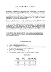
06072015 ISCHEMIC HEART DISEASE INTRODUCTION PATHOPHYSIOLOGY IMAGING IN ISCHAEMIC HEART DISEASE IHD COMPLICATION OF IHD ISCHEMIC HEART DISEASE INTRODUCTION Ischaemic heart disease IHD the leading cause of ID: 908832
Download Presentation The PPT/PDF document "ISCHEMIC HEART DISEASE DR. SACHIN RATHI" is the property of its rightful owner. Permission is granted to download and print the materials on this web site for personal, non-commercial use only, and to display it on your personal computer provided you do not modify the materials and that you retain all copyright notices contained in the materials. By downloading content from our website, you accept the terms of this agreement.
Slide1
ISCHEMIC HEART DISEASE
DR. SACHIN RATHI
06/07/2015
Slide2ISCHEMIC HEART DISEASE
INTRODUCTION
PATHOPHYSIOLOGY
IMAGING IN ISCHAEMIC
HEART
DISEASE (IHD)
COMPLICATION OF IHD
Slide3ISCHEMIC HEART DISEASE
INTRODUCTION
Ischaemic heart disease (IHD), the leading cause of
morbidity and mortality.
Survivors of a first myocardial infarction
are at
increased risk of death from IHD at later
ages.
Various contributing factors increasing risk
Need rational
, evidence-based use of
diagnostic and
therapeutic
means
Cardiac
computed tomography
(CCT) and cardiovascular
magnetic resonance
(CMR
) important in IHD
I
maging techniques to
study the highly complex
and heterogeneous
disease
entity of
IHD.
Slide4ISCHEMIC HEART DISEASE
PATHOPHYSIOLOGY
Coronary artery disease (CAD
)
usual cause of IHD
.
S
ignificantly impaired coronary blood flow leads to myocardial ischaemia .Chronic stable plaqueRuptured plaque (Acute Coronary Syndrome)An ischaemic cascade: metabolic disturbances regional dysfunction ECG changes anginal symptoms.20–30 min of sustained ischaemia leads to irreversible myocardial damage.Myocardial infarctions are usually classified by (a) location (b) size and (c) duration
Slide5Slide6Slide7ISCHEMIC HEART DISEASE
IMAGING
Conventional Film (X-ray)
Conventional Coronary Angiography
Echocardiography
Cardiac CT and Coronary CT angiography
Cardiac MRI and MR
Coronary Angiography Single-photon Emission Computed Tomography (SPECT)Positron Emission Tomography (PET)Nuclear Cardiac Imaging (perfusion studies)Stress Imaging
Slide8ISCHEMIC HEART DISEASE
Conventional Film (X-ray
)
Provide valuable information about
cardiac
size,
e
nlargement of a specific cardiac chamber pulmonary filling status,The chest X-ray help to excludepulmonary, pleural or aortic diseaseHelp to monitor therapy, demonstrate concomitant pulmonary disease , correct positioning of devices
Slide9ISCHEMIC HEART DISEASE
Conventional Coronary Angiography
S
elective
injection of contrast
medium into
the right and left coronary arteries and the
left ventricle.A percutaneous femoral arterial catheterization catheters are passed into each coronary artery and one for the left ventricle low osmolality contrast media with rapid filming at 25 frames/s Images are acquired in multiple orientations
Slide10ISCHEMIC HEART DISEASE
Coronary
angiography
provides information regarding
Severity
and length
of stenosis
, CA occlusions, Number of vessels affected, Stenosis configuration (smooth, ulcerated), Presence of thrombus, Collateral vessels, CA anatomy and variantsLimitations :Mild or nonstenotic CAD not visualised
No information about the plaque compositionD
egree of vascular remodelling.
Slide11ISCHEMIC HEART DISEASE
Slide12ISCHEMIC HEART DISEASE
Slide13Selective coronary arteriogram (right anterior oblique with cranial angulation) shows a significant left anterior descending coronary artery
stenosis
Slide14Slide15ISCHEMIC HEART DISEASE
Echocardiography
2D
echocardiography :
I
maging
of the
heart and great vessels through chest wall acoustic window.Doppler echocardiography : estimate blood flow and pressure gradientsContrast medium echocardiography : Margins of the ventricular cavity.Increase accuracy of assessing ventricular function Assess myocardial perfusionStress echocardiography :
Slide16ISCHEMIC HEART DISEASE
Echocardiography
Demonstrate effects
of IHD on
ventricular function.
Detect
structural
complicationsDemonstrate origins of the main coronary arteries and their anomalies
Slide17ISCHEMIC HEART DISEASE
Cardiac CT and Coronary CT
angiography
Electron beam CT (EBCT)
less
commonly
used and
uses an electronically steered beam of electrons allowing very rapid pulsing and rotation of the X-ray beamMultidetector CT (MDCT)Typically uses 16 rows of static detectors with fast tube rotations (with ECG triggering and control of the tube current modulation) Images obtained at end diastole (at the point of least movement artefact) Reliable imaging requires a regular, relatively slow heart rate (< 75 beats/min) may require
beta blockadeDual source cardiac CT : Contain 2
X-ray tubes and 2 sets of detectors, require a reduced rotation speed CTA
Dual head injection pumps allow contrast
injection to
be followed by saline injection (or a mixture of
contrast and
saline) using biphasic or
triphasic
injection
protocols (cf
. single head pumps) ▶ the typical volume of
contrast ranges
from 50 to 120ml with an injection rate of 4–7ml/s
Slide18ISCHEMIC HEART DISEASE
Cardiac CT and Coronary CT angiography
Post Processing :
Multiplanar
reconstruction (MPR
)
Maximum
intensity projection (MIP) Surface shaded display (SSD) Volume-rendered techniques
Slide19ISCHEMIC HEART DISEASE
Has two
major components:
U
nenhanced
coronary artery calcium scoring
Contrast enhanced
coronary artery angiography.Coronary Calcification : Agatston calcium scoreQuantifies high attenuation material within the coronary arteries. High calcium score predicts an increased risk of an adverse coronary eventDetect significant stenoses high sensitivity and specificity
Demonstrate normal coronary arteries and detect stenoses with a high positive and negative
predictive valueMDCT can detect significant non-cardiac diagnoses
Slide20Axial thin MIP reconstruction from a coronary CTA shows multiple areas of dense calcification in
the left
anterior descending (LAD) (curved arrow), ramus
intermedius
(arrowhead
), and
circumflex (arrow) coronary arteries.
Slide21Slide22ISCHEMIC HEART DISEASE
Cardiac MRI and MR Coronary
Angiography
Spin-echo
imaging (
‘Black blood’
imaging)
Gradient-echo imaging (White blood’ imaging)Phase shift velocity mapping (quantification of flow velocities)Myocardial perfusion imagingContrast-enhanced MRAMyocardial taggingCardiac gating
Slide23ISCHEMIC HEART DISEASE
CMR indications and applications
Myocardial
function at
rest
Myocardial
function during stress:
Myocardial perfusion and viability: Coronary arteries and bypass grafts: Valvular heart diseaseAcutemyocarditisCardiac infiltrationSarcoidosisAmyloidosis Myocardial iron overload
Slide24Axial MRI in mitral stenosis. (A) Axial
ECG-gated spin-echo image of patient
with mitral stenosis leading to right
heart failure
. Slow flow in the left atrium
has produced
high signal in that chamber
.
(B) Oblique coronal breath-hold cine-MRA image of the heart. There is dilatation
of the right atrium (RA) and right ventricle(RV). There is mild tricuspidregurgitation (jet of flow dephasing ¼ open
arrow).
(A)
(B)
Slide25Evaluation of cardiac perfusion
and viability
by CMR. (A)
Four-chamber long-axis
view from cine-FISP
MRA at
end systole shows persisting
apical myocardial thinning (arrows) while the basal myocardium thickens normally.
(B) Short-axis view obtained during a first pass of contrast (
gadopentetate) through the myocardium shows delayed enhancement of non-viable myocardium best shown at an inversion time of 182
ms
(arrows). * ¼ normal myocardium.*
(A)
(B)
Slide26ISCHEMIC HEART DISEASE
Single-photon Emission Computed Tomography (SPECT
)
N
uclear
medicine tomographic
imaging technique
using gamma rays.A gamma-emitting radioisotope (radionuclide) injected intravenously followed by multiple planar projections are acquired using gamma camera rotates and reconstructed into tomograms.Can combined with CT, which allow for attenuation correction and the provision of anatomical information
Slide27ISCHEMIC HEART DISEASE
POSITRON
EMISSION
TOMOGRAPHY (PET)
Positrons travel a very short distance in matter
followed by
an annihilation reaction with emission of two gamma photons in opposite directions are sensed simultaneously by opposing detectors.This inherent spatial information allows construction of a tomographic image without the need for collimators.Advantages over SPECT :better spatial resolution and higher sensitivitymost physiological molecules can be use as tracersmeasure tracer distribution in absolute terms as a function of time
Slide28ISCHEMIC HEART DISEASE
Nuclear Cardiac
Imaging
A
ssessment
of myocardial perfusion in CAD
patients and risk
stratification for major adverse cardiac events.Commonly used radionuclides are Thallium99m Technetium MIBI (SESTAMIBI)Myocardial perfusion imaging depends on differential flow delivering different amounts of activity to normal and ischaemic myocardium
Slide29ISCHEMIC HEART DISEASE
Thallium
:
M
yocardial
perfusion
agent.
Cyclotron produced and decays by electron capture.Higher myocardial accumulation than 99m technetiumPoor resolution , long t1/2 (3 days), high cost and limited availabilityIntracellular uptake and myocardial clearance proportional to the regional myocardial blood flow.Stress images (5-30 min post injection)Redistribution image (2-4 h post injection)
Slide30ISCHEMIC HEART DISEASE
99mTECHNETIUM MIBI (SESTAMIBI
)
High myocardial
accumulation (proportional to the
regional perfusion
) with a slow washout and a long
myocardial retention time. Therefore imaging requires separate injections for stress and rest studiesProtocols:1-day protocol: rest images stress images 4 h later2-day protocol: stress images (1st day) rest images (2nd day, if the original stress images are abnormal) A combined 99m technetium and thallium approach: an initial thallium injection
immediate 99mtechnetium injection (as its higher energy photons are unaffected by any residual thallium)Shorter
t1/2 (6 h) allows larger doses, improved resolution, low cost and easy availability
no redistribution
phase for viability assessment
Slide31ISCHEMIC HEART DISEASE
Stress
Imaging
A
natomical imaging (
conventional
coronary angiography
, CCT) provide limited information regarding the impact of a stenosis on the coronary flow.Stress testing help to assess the extent of myocardial ischaemia.Functional severity of a stenosis can be determined by the FFR During cardiac catheterisation.In stress test , reversible ischaemia is achieved by stressing the heart and evaluating whether during stress, symptoms of angina, ECG signs of myocardial ischaemia, ischaemia-induced myocardial wall motion abnormalities or myocardial perfusion disturbances occur.
Slide32ISCHEMIC HEART DISEASE
Stress Imaging
In stress imaging, perfusion of heart is evaluated in resting and under stressful condition.
To induce stress in cardiac muscle,
dobutamine
is infused.
Dobutamine
, β agonist which increases cardiac muscle contractility and heart rate leading to increase oxygen consumption. So blood supply to heart increases.
Slide33ISCHEMIC HEART DISEASE
Stress
Imaging
Normally
supplied myocardium shows a progressive increase in myocardial contractility.
Myocardium supplied by a flow-limiting coronary stenosis becomes ischaemic when the compensatory increase in coronary blood supply is insufficient to match the increased demand in oxygen.
This leads to various changes like wall motion abnormality, chest pain,
changes ECG, delayed perfusion on CMR, nuclear perfusion imaging.
Slide34Stress echocardiography. (A) End-diastolic apical
two-chamber view
at rest shows thinning of the apical myocardium (arrow
). (
B) End-systolic apical two-chamber view at rest shows
concentric contraction
of left ventricle, except for the cardiac apex. (C)
Enddiastolic apical two-chamber view during stress (dobutamine infusion) shows dilatation of the left ventricle when compared with (A). (D) End-systolic apical two-chamber view during stress (dobutamine infusion) shows dilatation of the left ventricle due to akinesia of the anterior wall to contract (arrows).
Slide35ISCHEMIC HEART DISEASE
Practical scheme for
dobutamine
stress CMR.
Per stress level, four short-axis and two long-axis cine studies
are obtained
in three consecutive breath-holds, making it possible to evaluate the left ventricle for new wall motion
abnormalities (WMAs). If termination criteria are not met at the highest dobutamine dose, atropine can be additionally administered.
Slide36MIBI SPECT shows reversible perfusion defect in lateral LV wall with an estimated ischaemia of 19% of LV myocardium
(*, A) (arrows, B). Stress perfusion CMR shows extensive stress-induced perfusion defect in lateral LV wall (arrows, C) and
subendocardial
perfusion in anterior LV wall and septum (arrowheads, C). At coronary angiography (D) severe two-vessel CAD is shown with distal
LCx
80% stenosis, 1st lateral 80% stenosis, and mid anterior LAD 70% stenosis and distal LAD 80% stenosis.
Slide37ISCHEMIC HEART DISEASE
IMAGING IN ISCHAEMIC HEART
DISEASE (IHD)
Myocardial Infarct Imaging
Most
patients usually have obstructive
coronary artery disease
Plaque rupture is important cause in <50% stenosisAssessment of the electrical cardiac activity using 12-lead electrocardiography and analysis of cardiac biomarkers are central diagnostic techniquesPlaque fissuring thrombosis develop on the exposed intima artery occlusion infarction or unstable angina
Slide38ISCHEMIC HEART DISEASE
Increase cardiac marker recent myocardial injury and EEG infarct related artery
However no one give inside in evolving process.
Cardiac USG and Cardiac catheterisation visualised acute MI indirectly by abnormal wall motion but no idea about necrotic myocardium
Gated SPECT show MI as fixed defect with loss of function. Small
subendothelial
infarct can be missed.
Slide39ISCHEMIC HEART DISEASE
Cardiac Magnetic Resonance Imaging :
Give idea about inside process. Better resolution so
subendocardial
infarct can be easily pick up
Increase water on ischemic myocardium prolongs T2 relaxation and is related duration of ischemia
Late gadolinium enhanced CMR : in this delayed imaging is done and
delay is of 10-25 min. High spatial and contrast resolution allows depiction of subtle myocardial damage.T2WI and LGE CMR images are used to determine area at risk and extent of infarction.Difference between these two images show salvageable myocardium
Slide40ISCHEMIC HEART DISEASE
Various parameter to be look on CMR :
Infarct size
Infarct
transmurality
Microvascular obstruction (CE CMR)
Postreperfusion
hemorrhage (T2WI)
Slide41ISCHEMIC HEART DISEASE
Conventional CXR
shows
moderate
cardiomegaly.
LV contrast
ventriculography
(RAO position, end-systolic time frame) shows extensive area of decreased contractility involving the anterior wall, apex and the apicoinferior LV wall (arrows, B). Cardiac ultrasound (longitudinal parasternal view) reveals similar information (arrows, C). LGE CMR shows extensive myocardial infarction involving the majority of the ventricular septum, apical two-thirds of the anterior wall, apex, and apical inferolateral wall (arrows, D). While the periphery of the infarct is strongly enhanced, centrally an extensive zone of microvascular obstruction remains on LGE CMR, reflecting severe microvascular damage. The functional consequences of the infarction can be well appreciated on cine CMR
Slide42Acute
laterobasal
myocardial infarction.
T2-weighted
imaging in short axis (A) and
horizontal long
axis (B). LGE CMR in short axis (C), and horizontal long axis (D). Sharply defined zone of myocardial oedema in
laterobasal (arrows, A, B). LGE CMR shows strong enhancement in anterior and lateral wall (arrows, C, D). The extent of enhancement coincides very well with the extent of myocardial oedema, which means that the majorpart of the jeopardised myocardium has been irreversibly damaged. In other words, myocardial salvage is very low.
Slide43A 47-year-old patient admitted with retrosternal
chest pain
and left hemiparesis. Slightly increased cardiac enzymes (troponin I: 0.5
μg
/L) and no evidence of obstructive CAD on coronary
angiography. LGE CMR in cardiac short axis (A), horizontal long axis (B) and vertical long axis (C
)
show subtle myocardial damage
Slide44Typical presentation of acute
transmural
anterior myocardial infarction
.
LGE
CMR in horizontal long axis (A), vertical long axis (B) and short axis (
C).
Transmural myocardial enhancement is shown in anterior myocardial
Slide45Acute
transmural
inferolateral
myocardial infarction with secondary pericardial inflammation.
T2-weighted
CMR (A
) and LGE CMR (B) in cardiac short axis. Presence of myocardial oedema (bright signal) in inferolateral LV wall (arrows, A), and presence of increased signal intensity of the pericardium mainly along the anterolateral part of the left ventricle (arrowheads, A). LGE CMR shows transmural enhancement of the inferolateral LV wall (arrows, B) and diffuse enhancement of a minimally thickened pericardium (arrowheads, B) reflecting infarct-related pericardial inflammation.
Slide46Slide47Short-axis LGE CMR (A) early post-infarction shows extensive
transmural
enhancement (arrows;
segments
with large zone of
microvascular
obstruction.
Cine CMR early (B–D) and 6 months (E–G) post-infarction. Note the important
wall thinning of the involved segments with aneurysm formation at 6-month follow-up (arrows, E–G).
Slide48ISCHEMIC HEART DISEASE
COMPLICATION OF
IHD
Cardiac Rupture
Left Ventricular Aneurysm
False or
pseudoaneurysm
Post-infarction VSDPost-infarction MRVentricular ThrombosisDressler’s syndrome
Slide49Thrombus formation post-infarction.
Contrast-enhanced inversion-recovery CMR early following contrast administration (A) showing myocardium is brightly hyperintense, and the blood pool is strongly enhanced. An
intracavitary
is visible as a hypointense structure (arrowhead, A). LGE CMR in horizontal long axis (B), vertical long axis (C) and apical short axis (D). On LGE CMR the irreversibly damaged myocardium in the LAD perfusion territory is strongly enhanced (arrows, B–D) and the adjacent mural thrombus is well visible
(arrowhead, B–D).
Slide50Chest radiograph (
A) showing presence
of severe cardiomegaly caused by lateral displacement of the left heart border. The abnormalities are caused by a moderate
to severe pericardial effusion as clearly visible on cardiac ultrasound (*, B).
CMR confirms the cardiac ultrasound findings
Slide51(A
)
Long-axis view from transthoracic echocardiography
shows
an 8mm
echo-free area (
arrow) in
the interventricular septum representing an infarct VSD.
(B) Equivalent view with colour Doppler shows variable colour (high-velocity flow, arrow) in the infarct VSD.*
(B)
(A)
Infarct VSD
Slide52False
aneurysm
(A)
Axial
s
pin-echo
MRI image
shows a large false aneurysm (FA) arising from the posterior wall of the left ventricle
(LV). Note the abrupt change in contour at the mouth of the FA (arrow).
(A)
(B) Axial cine-MRA
image
at
the same level as (A) shows
flow into the FA (
arrows)
(B)
Slide53THANK YOU