/
 Arrythmia Interpretation (cont’d)  Arrythmia Interpretation (cont’d)

Arrythmia Interpretation (cont’d) - PowerPoint Presentation

giovanna-bartolotta
giovanna-bartolotta . @giovanna-bartolotta
Follow
343 views
Uploaded On 2020-04-04

Arrythmia Interpretation (cont’d) - PPT Presentation

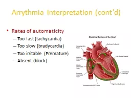
Rates of automaticity Too fast tachycardia Too slow bradycardia Too irritable Premature Absent block Interpreting Arrhythmias 1 Calculate the heart rate 2 Assess the rhythm 3 Identify the P waves ID: 775330

atrial heart normal block atrial heart normal block sinus complexes ventricular cardiac qrs rhythm fibrillation waves node premature rate

Share:

Link:

Embed:

Download Presentation from below link

Download Presentation The PPT/PDF document " Arrythmia Interpretation (cont’d)" is the property of its rightful owner. Permission is granted to download and print the materials on this web site for personal, non-commercial use only, and to display it on your personal computer provided you do not modify the materials and that you retain all copyright notices contained in the materials. By downloading content from our website, you accept the terms of this agreement.


Presentation Transcript

Slide1

Arrythmia Interpretation (cont’d)

Rates of automaticityToo fast (tachycardia)Too slow (bradycardia)Too irritable (Premature)Absent (block)

Slide2

Interpreting Arrhythmias

1. Calculate the heart rate

2. Assess the rhythm

3. Identify the P waves

4. Assess QRS shape and duration

5. Assess relationship between P waves and QRS complexes

6. Name the arrhythmia

Slide3

Normal Sinus Rhythm

Normal ECG tracing depicting a normal rhythm of electrical conductivity through the heart

Slide4

(Respiratory) Sinus Arrhythmia

All criteria of normal rhythm except heart and pulse rates increase with inspiration and decrease with expirationNormal finding in brachycephalic breeds and in chronic respiratory diseaseIncreased number of cardiac cycles during inspiration; decreased number during expirationOriginates in the SA node

Slide5

What is the normal HR for dogs and cats?

Dogs:

70 – 160 BPM

Cats:

150 – 210 BPM

Slide6

Sinus Bradycardia

Regular sinus rhythm but heart rate is below normalDogs under 45 lb: HR less than 70 bpmDogs >45 lb: HR < 60 BPMCats: 100 BPM or lessCS: weakness, hypotension, syncope

Slide7

Sinus Tachycardia

Regular sinus rhythm with increased ventricular rateDogs less than 45 lb; HR >180 BPMDogs more than 45 lb; HR >160 BPMCats: HR greater than 240 BPMCauses include: pain, fever, anemia, excitement, hyperthyroidism

Slide8

Atrial Premature Complexes

Premature atrial impulses originating from ectopic atrial site other than SA nodeSeen in dogs and cats with atrial enlargement, electrolyte disturbances, drug reactions, congenital heart disease, and neoplasia; a normal variation in older animalsPremature P waveQRS complexes are normal unless the P wave is so immature that it overlaps to varying degrees

Slide9

Atrial Premature contraction/complexes

Represent premature P wave/s

Slide10

Atrial Tachycardia

Rapid regular rhythm originating from an atrial site other than the sinus nodeMay be seen in dogs with severe heart disease and in cats with cardiomyopathy or hyperthyroidism

Slide11

Atrial Flutter

Appears as a regular, sawtooth formation between the QRS complexes.Occurs when the ventricular rate differs from the atrial rate.Atrial flutter is the precursor to atrial fibrillation.

Slide12

Fibrillation

is the rapid, irregular, and unsynchronized contraction of muscle fibers.

Slide13

Atrial Fibrillation

Caused by numerous disorganized atrial impulses frequently bombarding the AV node.Ventricular depolarization rate is irregular and rapid.No P waves are evident; replaced by numerous f waves.QRS complexes may be normal or wide and of varying amplitude

Slide14

Atrial Fibrillation

Slide15

Premature Ventricular Complexes (PVCs)

“Premature beats” - Cardiac impulses initiated within the ventricles instead of the sinus node

Ventricle discharges before the arrival of the next anticipated impulse from the SA node.

Can occur at any rate but pose a greater danger with tachycardia.

Associated with congenital defects, cardiomyopathy, GDV, drug reactions, cardiac neoplasia, anemia, acidosis, hyperthyroidism, hypokalemia

Slide16

PVCs (cont’d)

The P wave is often not seen on the ECG tracing.A wide, distorted QRS complex is also evidentThe beat preceding the PVC and the beat following are usually equal to the time of two normal beats.

Slide17

Ventricular Tachycardia

A series of four or more PVCs in a row . Potentially life threatening.

Slide18

Ventricular Fibrillation

The mechanical pumping of the heart is not evident on the ECG.The ECG has bizarre baseline with prominent undulations due to weak and uncoordinated ventricular contractions.Low to absent cardiac output.Associated with shock, trauma, electrolyte imbalances, drug reactions, electric shock, hypothermia, cardiac surgery.Rapidly fatal

Slide19

Ventricular Fibrillation

There are no recognizable P or QRS complexes.Irregular, chaotic, deformed reflections of varying width, amplitude, and shape.Unless controlled immediately, ventricular fibrillation will result in cardiac arrest.

Slide20

Sinus Arrest or Block

Normal sinus rhythm interrupted by an occasional prolonged failure of the SA node to initiate an impulse.Conduction disturbance in which normal sinus rhythm is interrupted by an occasional, prolonged failure of the impulse generated by the SA node to reach the atria.

Slide21

Heart Block

Electrical impulse is not transmitted through the heart.

Slide22

First Degree AV Block

Delay in conduction of an impulse through the atrioventricular junction and Bundle of His.The PR interval is longer than normal.This type of heart block is a result of a minor conduction defect.Seen in older patients secondary to degenerative changes in the conduction system.

Slide23

Second Degree AV Block

Some atrial pulses are not conducted through the AV node and therefore do not cause depolarization of the ventricles.There are two types:Type I (Mobitz type I or “Wenckebach” AV block): progressive lengthening of the PR interval on successive beats and then P waves occurring without QRS complexes.P waves occurring without QRS complexes are called “dropped beats”

Slide24

Second Degree AV Block (cont’d)

Type II: A constant PR interval that is usually of normal duration with random dropped beats..In the case of type 2 block, atrial contractions are not regularly followed by ventricular contraction

Slide25

Third degree AV block

The cardiac impulse is completely blocked in the region of the AV junction and/or all bundle branches. Also known as a complete heart block; the most severe heart block.No relationship between P waves and QRS complexes; atria and ventricles each beat independently.Atrial rate is normal.

Slide26

Heart Blocks

Slide27

Asystole (Flat line)

Cardiac Arrest: No cardiac electrical activity, no cardiac outputor blood flow. At this point the heart willnot respond to defibrillation. Causes: hypoxia, hypothermia, hypoglycemia, or an electrode has fallen off (hopefully)

Slide28

Asystole (Flat line)

Medications of choice: Epinephrine or Atropine

along with manual chest compressions.