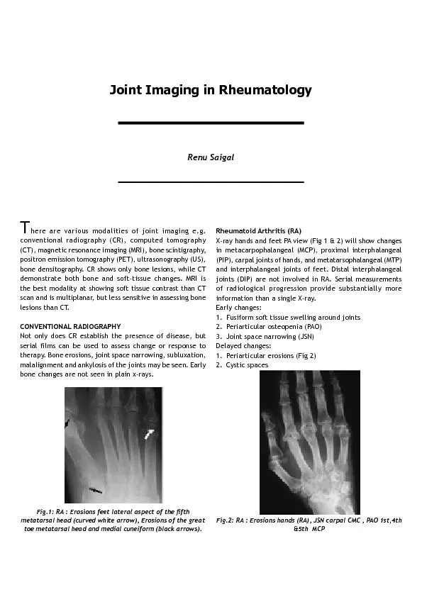
PDF-here are various modalities of joint imaging e.g. conventional radiogr
Author : giovanna-bartolotta | Published Date : 2016-12-06
Fig1 RA Erosions feet lateral aspect of the x00660069fth Rheumatoid Arthritis RAXray hands and feet PA view Fig 1 2 will show changes in metacarpophalangeal MCP
Presentation Embed Code
Download Presentation
Download Presentation The PPT/PDF document "here are various modalities of joint ima..." is the property of its rightful owner. Permission is granted to download and print the materials on this website for personal, non-commercial use only, and to display it on your personal computer provided you do not modify the materials and that you retain all copyright notices contained in the materials. By downloading content from our website, you accept the terms of this agreement.
here are various modalities of joint imaging e.g. conventional radiogr: Transcript
Fig1 RA Erosions feet lateral aspect of the x00660069fth Rheumatoid Arthritis RAXray hands and feet PA view Fig 1 2 will show changes in metacarpophalangeal MCP proximal interphalange. Traditionally clinical imaging exploits the contrast due to differences in spin density spinlattice relaxation T and spinspin relaxation T between normal tissues and pathology However other contrast mechanisms such as susceptibility magnetization Imaging modalities are often used based on the requests of referring physicians and the availability of equipment and experience of the radiologists. Most centers use ultrasound (US), computed tomogra of . Electromagnetic and Seismic . Wavefields. . International Symposium on Geophysical Imaging . with . Localized Waves. Sanya. , . Hainin. Island China. July 24-28, 2011. Gregory A. Newman. Earth Sciences Division. Chapter 1. Science of Modalities. energy. Transformed or transferred?. The capacity of a system for doing work and exists in various forms. Often transformed from one form to another. Often transferred from one location to another. Gregg C. Daversa. Director, Business Development. West . Physics. Objectives. Identify which JC-accredited facilities will be impacted by the 2014 standard changes, and the dates for full compliance. Dinesh Rao MD. Assistant Professor. Department of Radiology. Neuroradiology and Musculoskeletal Radiology. University of Florida College of Medicine - Jacksonville. Disclosures. I have no disclosures. EULAR recommendations for the use of imaging in the diagnosis and management of spondyloarthritis in clinical practice Target population/question 2 26/3/18 Target population: patients with suspected or diagnosed axial or peripheral Ilana Richman MD, Primary Care and Outcomes Research Fellow. Anne . Smeraglio. MD, Resident in Internal Medicine. Objectives. Differentiate patients with back pain due to a specific etiology versus those with nonspecific back pain. Computed tomography in oral and maxillofacial radiology. Computed tomography (CT) is today commonly used in imaging of the maxillofacial area. Conventional CT examinations are usually performed in medical X-ray departments. However, a relatively new technique named cone-beam computed tomography (CBCT) or digital volume tomography (DVT) has now also become available for dental purposes. The advantage with this technique is a lower radiation dose compared to conventional CT. Common examples when DVT is used are; for diagnosing the position of impacted canines and suspected root . Basic Radiograph . ROUTINE. ■ PA. ■ Lateral. ■ Scaphoid series – when there is tenderness in the anatomical snuff box.. PA ulnar deviation. note the disruption of arc I at the lunotriquetral joint.. injection or aspiration?These are procedures in which we use imaging 1. Pain relief: Your doctor may haverequested this procedure as a treatmentfor pain. Using image guidance, we injectmedicines into The theory of image formation in electron microscope s is very similar to image formation in light microscope s. However, the practice and design are completely different : The most crucial – An Introduction – . Dr . Rudra. . Pratap. . Pandey. Prof. VSR. Conventional radiography. Digital radiography (CR & DR). A. Equipment details. B. Safety issues – Radiation . Advantages . S. troke . T. reatment . R. esponse . Imaging Management Center Information. NIH . StrokeNet. Imaging Management Center Team . Achala Vagal, MD, MS. PI, NIH . StrokeNet. Imaging Management Center .
Download Document
Here is the link to download the presentation.
"here are various modalities of joint imaging e.g. conventional radiogr"The content belongs to its owner. You may download and print it for personal use, without modification, and keep all copyright notices. By downloading, you agree to these terms.
Related Documents