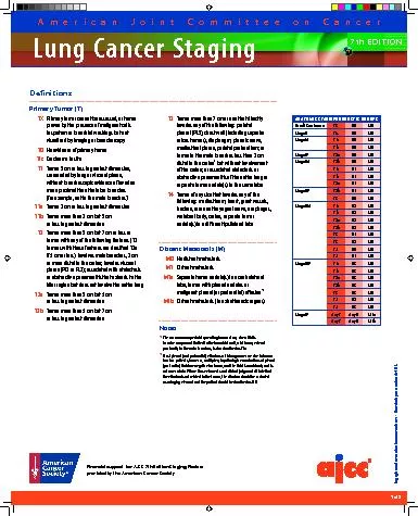
/
Primary Tumor (T)   TX Primary tumor cannot be assessed, or tumor prov Primary Tumor (T)   TX Primary tumor cannot be assessed, or tumor prov

Primary Tumor (T) TX Primary tumor cannot be assessed, or tumor prov - PDF document

giovanna-bartolotta
giovanna-bartolotta . @giovanna-bartolotta
Follow
476 views
Uploaded On 2015-10-31

Primary Tumor (T) TX Primary tumor cannot be assessed, or tumor prov - PPT Presentation

7th EDITION ANATOMIC STAGEPROGNOSTIC GROUPSOccult Carcinoma TX N0 Stage 0 Tis N0 NotesThe uncommon supercial spreading tumor of any size with its invasive component limited to the bronchial wall w ID: 178421

7th EDITION ANATOMIC STAGE/PROGNOSTIC GROUPSOccult Carcinoma

Share:

Link:

Embed:

Download Presentation from below link

Download Pdf The PPT/PDF document "Primary Tumor (T) TX Primary tumor can..." is the property of its rightful owner. Permission is granted to download and print the materials on this web site for personal, non-commercial use only, and to display it on your personal computer provided you do not modify the materials and that you retain all copyright notices contained in the materials. By downloading content from our website, you accept the terms of this agreement.


Presentation Transcript

7th EDITION Primary Tumor (T) TX Primary tumor cannot be assessed, or tumor proven by the presence of malignant cells in sputum or bronchial washings but not visualized by imaging or bronchoscopyT0 No evidence of primary tumorTis Carcinoma in situT1 Tumor 3 cm or less in greatest dimension, surrounded by lung or visceral pleura, without bronchoscopic evidence of invasion more proximal than the lobar bronchus (for example, not in the main bronchus)T1a Tumor 2 cm or less in greatest dimensionT1b Tumor more than 2 cm but 3 cm or less in greatest dimensionT2 Tumor more than 3 cm but 7 cm or less or tumor with any of the following features (T2 tumors with these features are classied T2a if 5 cm or less): involves main bronchus, 2 cm or more distal to the carina; invades visceral pleura (PL1 or PL2); associated with atelectasis or obstructive pneumonitis that extends to the hilar region but does not involve the entire lungT2a Tumor more than 3 cm but 5 cm or less in greatest dimensionT2b ANATOMIC STAGE/PROGNOSTIC GROUPSOccult Carcinoma TX N0 Stage 0 Tis N0 NotesThe uncommon supercial spreading tumor of any size with its invasive component limited to the bronchial wall, which may extend proximally to the main bronchus, is also classied as T1a.Most pleural (and pericardial) eusions with lung cancer are due to tumor. In a few patients, however, multiple cytopathologic examinations of pleural T3 Tumor more than 7 cm or one that directly invades any of the following: parietal pleural (PL3), chest wall (including superior sulcus tumors), diaphragm, phrenic nerve, mediastinal pleura, parietal pericardium; or tumor in the main bronchus less than 2 cm distal to the carina but without involvement of the carina; or associated atelectasis or obstructive pneumonitis of the entire lung or separate tumor nodule(s) in the same lobeT4 Tumor of any size that invades any of the following: mediastinum, heart, great vessels, trachea, recurrent laryngeal nerve, esophagus, vertebral body, carina, separate tumor nodule(s) in a dierent ipsilateral lobeDistant Metastasis (M)M0 No distant metastasisM1 Distant metastasisM1a Separate tumor nodule(s) in a contralateral lobe, tumor with pleural nodules or malignant pleural (or pericardial) eusionM1b American Joint Committee on CancerLung Cancer StagingCopyright 2009 American Joint Committee on Cancer Printed with permission from the AJCC. Financial support for AJCC 7th Edition Staging Posters provided by the American Cancer Society 7th EDITION American Joint Committee on CancerLung Cancer Staging Copyright 2009 American Joint Committee on Cancer Printed with permission from the AJCC. Regional Lymph Nodes (N)NX Regional lymph nodes cannot be assessedN0 No regional lymph node metastasesN1 Metastasis in ipsilateral peribronchial and/or ipsilateral hilar lymph nodes and intrapulmonary nodes, including involvement by direct extensionN2 Metastasis in ipsilateral mediastinal and/or subcarinal lymph node(s)N3 Metastasis in contralateral mediastinal, contralateral hilar, ipsilateral or contralateral scalene, or supraclavicular lymph node(s) Financial support for AJCC 7th Edition Staging Posters provided by the American Cancer Society ILLUSTRATIONThe IASLC lymph node map shown with the proposed amalgamation of lymph into zones.(© Memorial Sloan-Kettering Cancer Center, 2009.)