/
Recurrent  exertional  compartment syndrome – Recurrent  exertional  compartment syndrome –

Recurrent exertional compartment syndrome – - PowerPoint Presentation

giovanna-bartolotta
giovanna-bartolotta . @giovanna-bartolotta
Follow
391 views
Uploaded On 2018-03-22

Recurrent exertional compartment syndrome – - PPT Presentation

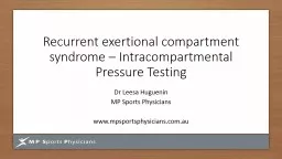
Intracompartmental Pressure Testing Dr Leesa Huguenin MP Sports Physicians wwwmpsportsphysicianscomau RECS 95 lower leg 45 anterior compartment Lateral deep posterior and superficial posterior ID: 661317

compartment pressure exercise min pressure compartment min exercise studies postexercise step resting patient posterior normal raised pressures anterior slow

Share:

Link:

Embed:

Download Presentation from below link

Download Presentation The PPT/PDF document "Recurrent exertional compartment syndr..." is the property of its rightful owner. Permission is granted to download and print the materials on this web site for personal, non-commercial use only, and to display it on your personal computer provided you do not modify the materials and that you retain all copyright notices contained in the materials. By downloading content from our website, you accept the terms of this agreement.


Presentation Transcript

Slide1

Recurrent

exertional compartment syndrome – Intracompartmental Pressure Testing

Dr Leesa Huguenin

MP Sports Physicians

www.mpsportsphysicians.com.auSlide2

RECS

95 % lower leg45% anterior compartmentLateral, deep posterior and superficial posterior

follow

85- 95% are bilateral

Also possible;Forearm (kayak, motorcross, sailboard)thigh, hands and rarely feetSlide3

Risk factors

Increased muscle swelling – genetic/ overuse/ biomechanical control

Increased

fascial

thickness/ toughness (?????)Reduced muscle capillary density? Creatine supplementation (transient)Slide4

Table 3 Diagnostic criteria of

intracompartmental pressure measurementAuthors Year Diagnostically relevant pressure values

French and Price" 1962

Postexercise

fall time <30 min in normal subjects,> 100 min in patientsReneman"7 1975 At six minutes postexercise pressure >15 cm H2O (11

mm Hg) above resting pressure

Puranen

" 1981 Mean pressure of 50 mm Hg during running. Resting

of no value, but did observe slow

postexercise

fall

McDermott3' 1982 Mean pressure of 85 mm Hg during running

Mubarak and Hargens5" 1982 Resting pressure >15 mm Hg. Exercise pressure >75

mm Hg. Pressure remains >30 mm Hg for >5 min after

exercise

Qvarfordt

et al" 1983 Pressures raised before, during, and after exercise.

Postexercise

decline 40 min. Ti 6

minWallensten

" 1983 No difference at rest, still raised 10 min

postexercise

(

anterior), returned to normal <10 min (deep posterior)

Detmer

et al" 1985 At rest, normal pressure < 15 mm Hg

Styf

and

Korner

" 1986 Muscle relaxation pressure

Styf

and Korner34 1986

Postexercise

pressure >35 mm Hg remained raised for >6

min (Also muscle relaxation pressure was raised during

exercise. >20 mins to return to normal)

Allen and Barnes" 1986 Exercise pressure >50 mm Hg anterior, >40 mm Hg deep

posterior No difference in resting pressures

Fronek

et al" 1987 Resting pressure .10 mm Hg and/or .25 mm Hg 5 min

after exercise

Rorabeck

et al'2 1988

Pre-exercise

pressure > 10 mm Hg and

postexercise

> 15 mm Hg for > 15 min

Styfs3 1988 Relaxation pressure >35 mm Hg, resting pressure >30

mm Hg,

postexercise

return to normal >6 min

Turnispeed

et al" 1989 > 20 mm Hg at rest (

postexercise

increase and slow

decline > 10 min)

Pedowitz

et

alr

1990

Pre-exercise

pressure .15 mm Hg, or

postexercise

pressure .30 mm Hg at

imin

or .20 mm Hg at 5 minSlide5

My guidelines

Resting pressures (post 1st exercise bout) 15-20mmHg +

1 min post exercise 30 - 35mmHg +

Slow recovery patterns, 5 mins > 20 mmHg

Fast recovery ?? Surgical successSlide6

COMPARTMENT PRESSURE STUDIES

STEP 1

PATIENT IS SENT OUT TO

EXERCISE TO ONSET OF SYMPTOMSSlide7

COMPARTMENT PRESSURE STUDIES

STEP 2

PATIENT RESTS ON BED –

ultrasound identification of popliteal

blood vessels

Injection of local anaesthetic down

to

periosteumSlide8

COMPARTMENT PRESSURE STUDIES

STEP 3

Insertion of split catheter

Plastic cannula inside needle remains in

Compartment as needle withdrawn,

Needle split and removedSlide9
Slide10

COMPARTMENT PRESSURE STUDIES

STEP 4

Cannula is tested for flow and

Secured in place to avoid

kinkingSlide11

COMPARTMENT PRESSURE STUDIES

STEP 5

PATIENT IS EXERCISED –

SINGLE LEG HEEL RAISES TO FAILURESlide12

COMPARTMENT PRESSURE STUDIES

STEP 6

PATIENT LIES SUPINE, ANKLE SUPPORTED AT

90 DEGREES

PRESSURES MEASURED AT 1, 3 AND 5 MINS

EXTRA MEASURES IF SLOW RECOVERYSlide13

COMPARTMENT PRESSURE STUDIES

STEP 7

ADDITIONAL COMPARTMENTS TESTED

ANTERIOR/ LATERAL/ SUPERFICIAL

POSTERIORSlide14

COMPARTMENT PRESSURE STUDIES

STEP 8

COMPRESSION DRESSINGS APPLIED

PATIENT INSTRUCTED RE

ACTIVITY OVER NEXT 48 HOURS

AND DISCHARGED HOME WITH

EMERGENCY CONTACT NUMBERSSlide15

COMPARTMENT PRESSURE STUDIES

FEET!

ESSENTIALLY SAME PROTOCOLSlide16

COMPARTMENT PRESSURE STUDIES

COMPRESSION DRESSINGS STILL IMPORTANTSlide17

COMPARTMENT PRESSURE STUDIES

IT IS USUAL TO BE SORE 48 HOURS

= DOMS

COMPLICATIONS ARE RARE

BRUISE/ LOCAL BLEEDING

ACUTE COMPARTMENT SYNDROME

=SEVERE WORSENING PAIN

=

EARLY SURGERYSlide18

Thanks for your attentionSlide19
Slide20