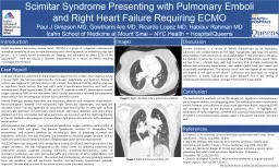
/
Scimitar Syndrome Presenting with Pulmonary Emboli Scimitar Syndrome Presenting with Pulmonary Emboli

Scimitar Syndrome Presenting with Pulmonary Emboli - PowerPoint Presentation

grewhypo
grewhypo . @grewhypo
Follow
342 views
Uploaded On 2020-06-23

Scimitar Syndrome Presenting with Pulmonary Emboli - PPT Presentation

and Right Heart Failure Requiring ECMO Paul J Simpson MD Gowthami Are MD Ricardo Lopez MD Habibur Rahman MD Icahn School of Medicine at Mount Sinai NYC Health HospitalQueens Partial ID: 785033

anomalous pulmonary scimitar ecmo pulmonary anomalous ecmo scimitar heart vein return papvr patient cardiovascular syndrome septal left normal hypertension

Share:

Link:

Embed:

Download Presentation from below link

Download The PPT/PDF document "Scimitar Syndrome Presenting with Pulmon..." is the property of its rightful owner. Permission is granted to download and print the materials on this web site for personal, non-commercial use only, and to display it on your personal computer provided you do not modify the materials and that you retain all copyright notices contained in the materials. By downloading content from our website, you accept the terms of this agreement.


Presentation Transcript

Slide1

Scimitar Syndrome Presenting with Pulmonary Emboli and Right Heart Failure Requiring ECMOPaul J Simpson MD, Gowthami Are MD, Ricardo Lopez MD, Habibur Rahman MDIcahn School of Medicine at Mount Sinai – NYC Health + Hospital/Queens

Partial anomalous pulmonary venous return (PAPVR) is a group of congenital cardiovascular anomalies comprising of one or more pulmonary veins draining directly or indirectly to the right atrium.  It is usually found incidentally on imaging, and estimated to affect 0.7% of the population[1].  Here we discuss a dramatic presentation of a variant of PAPVR requiring remarkable interventions.

A 24 year old woman presented to the emergency department for sudden onset dyspnea during a 6-hour flight. She had associated pleuritic chest pain, palpitations, and dizziness. Review of systems was negative for calf pain, fever or sick contacts. She denied smoking, alcohol, drug use, or significant family history. She was G2P1 with a spontaneous abortion, and took oral contraceptives. Blood pressure was 107/68, pulse 99, respiratory rate 30, temperature normal, oxygen saturation 94% on non-rebreather mask. Cardiac examination revealed left parasternal heave without JVD. Breathing was labored. Exam otherwise normal.Arterial blood gas showed hypoxemia and respiratory alkalosis with metabolic compensation. Electrocardiogram revealed sinus tachycardia, right ventricular hypertrophy, and right axis deviation. Chest x-ray was significant for prominent right heart border. CT chest with contrast showed right interlobar and pulmonary artery emboli, right heart dilatation, pulmonary artery dilatation to 4.7cm, and partial anomalous pulmonary venous return with her right lung draining to the IVC. Bedside echocardiogram showed dilated right atrium and ventricle, with paradoxical septal motion.Anticoagulation was initiated.  Her dyspnea increased and oxygen saturation deteriorated, at which time BiPAP was given. She became hypotensive, resistant to intravenous fluid resuscitation, and received alteplase for thrombolysis. Work of breathing increased and hypoxemia continued. She was intubated and paralyzed to decrease oxygen demand. Due to continued cardiovascular decompensation, alteplase again given without improvement.The ECMO team was contacted, who initiated veno-arterial (VA) ECMO, with improvement in her oxygen saturation. She was transported to the ECMO center for definitive treatment and underwent multiple surgeries including pulmonary thromboendarterectomy, repair of anomalous pulmonary vein with pericardial baffle and reduction of atrial septal defect, and ultimately ECMO VA decannulation. The patient had tracheostomy, and was gradually weaned from the ventilator. Her left ventricular function and pulmonary hypertension gradually improved. She was able to return to her home country some time after recovery.

Scimitar Syndrome is a variant of PAPVR characterized by an anomalous pulmonary vein predominantly on the right, hypogenetic right lung, and cardiac abnormalities, most commonly an atrial septal defect[2]. The anomalous vein gives the disorder its name due to its resemblance to the Middle-Eastern sword. There are two forms – infantile type, with high mortality from multiple cardiovascular defects and pulmonary hypertension, and pediatric/adult type, less severe and occasionally asymptomatic. The severity varies with the degree of the anomaly and degree of the right to left shunt[3]. Indications and techniques for surgical intervention in stable individuals are under debate[4], but in the setting of cardiovascular decompensation, emergent intervention is warranted.

This patient led a relatively normal life despite her significant cardiopulmonary and circulatory defects, including giving birth. The additional burden of pulmonary emboli, however, was sufficient to cause acute right heart failure, requiring ECMO and emergent repair of the anomalous pulmonary vein.  It highlights the importance of early recognition of PAPVR and successful intervention application of ECMO in a hemodynamically unstable patient. The patient ultimately recovered well.

1. Healey JE Jr. An anatomic survey of anomalous pulmonary veins: their clinical significance. J Thorac Surg 1952; 23:433.2. Najm HK, Williams WG, Coles JG, et al. Scimitar syndrome: twenty years' experience and results of repair. J Thorac Cardiovasc Surg 1996; 112:1161.3. Sears EH, Aliotta JM, Klinger JR. Partial anomalous pulmonary venous return presenting with adult-onset pulmonary hypertension. Pulmonary Circulation. 2012;2(2):250–255.4. Chan DY, Skillington P, Laufer E. Adult scimitar syndrome: a surgical approach. Heart Lung Circ. 2015 Jul;24(7):e115-7. The authors have no financial disclosures to report.

Image 1:

Right interlobar artery pulmonary embolus

.

Image 2: Right pulmonary scimitar vein.

Introduction

Case Report

Discussion

Conclusion

References

Images