PDF-(BOOS)-Clinical Hematology Atlas, 3rd Edition
Author : gwendaraatz | Published Date : 2022-06-24
Ideal for identifying cells at the microscope this atlas covers the basics of hematologic morphology including examination of the peripheral blood smear basic maturation
Presentation Embed Code
Download Presentation
Download Presentation The PPT/PDF document "(BOOS)-Clinical Hematology Atlas, 3rd Ed..." is the property of its rightful owner. Permission is granted to download and print the materials on this website for personal, non-commercial use only, and to display it on your personal computer provided you do not modify the materials and that you retain all copyright notices contained in the materials. By downloading content from our website, you accept the terms of this agreement.
(BOOS)-Clinical Hematology Atlas, 3rd Edition: Transcript
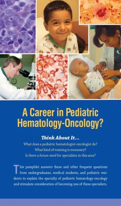
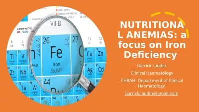
Ideal for identifying cells at the microscope this atlas covers the basics of hematologic morphology including examination of the peripheral blood smear basic maturation of the blood cell lines and discussions of a variety of clinical disorders Over 400 photographs schematic diagrams and electron micrographs illustrate hematology from normal cell maturation to the development of various pathologiesNumerous illustrations include excellent schematic diagrams photomicrographs and electron micrographsIntroductory chapters succinctly describe the peripheral blood smear its preparation and examination and hematopoiesis in generalCoverage of cellular maturation includes schematics that illustrate the maturation of each cell line individually and highlight the cell in questionDescriptions for each cell type include size reference intervals and nuclear and cytoplasmic characteristicsBody Fluids chapter covers the other fluids found in the body besides blood using images from cytocentrifuged specimensWhite blood cell differential table shows cells found in a normal white blood cell differentialOverview of hematopoiesis includes a schematic drawing along with a detailed presentation of each cell demonstrating the relationship between individual stages of hematopoiesis and the overall development schemeMorphologic abnormalities are presented in chapters on erythrocytes and leukocytes along with a schematic description of each cell to provide correlations to various disease statesCoverage of common cytochemical stains along with a summary chart for interpretation aids in classifying malignant and benign leukoproliferative disordersSpiral binding and a compact size make this book easy to use in a laboratory settingNEW Normal Newborn Peripheral Blood Morphology chapter covers the normal cells found in neonatal bloodMore examples of specific cells and disorders allow you to compare abnormal cells to each other and to normal cells differentiating those that are similarExpanded Evolve resources include case studies study questions links to related websites and content updates. computing in Geneva. 26. 8 CPU cores (login + batch). 180 TB for data. the analysis facility for Geneva grou. p. grid batch production for ATLAS. special features:. direct line to CERN at 10 . Gb/s. . Current Controversies in Hematology and Oncology he cost of cancer drugs is at an all-time high, with several lifesaving agents costing more than $100,000 a year. Much of the discussion related to hea - . June 2015. 1. ATLAS Software Infrastructure :. Requirements and Goals at Run 2 Period. Alex Undrus. . Alex Undrus – U.S. . ATLAS . S. &. C. . Planning . Meeting – . June 2015. 2. Outline. Nick Barlow. (University of Cambridge). on behalf of the ATLAS collaboration. Contents. Motivation . for searching for long-lived particles.. Very. quick look at a couple of . signal models. .. The ATLAS detector and the 2011 dataset.. IBL – . Insertable. B-Layer. Tobias Flick. University Wuppertal. 17.09.2009, VERTEX 2009 . Putten. , Netherlands. Preliminary. Overview. Current ATLAS pixel detector. What is the IBL and why do we need it?. Reference Sources. Conventions of Standard English . CCSS.ELA-Literacy.L.3.2g . Consult reference materials, including beginning dictionaries, as needed to check and correct spellings.. . Vocabulary Acquisition and Use . Reference Sources. Conventions of Standard English . CCSS.ELA-Literacy.L.3.2g . Consult reference materials, including beginning dictionaries, as needed to check and correct spellings.. . Vocabulary Acquisition and Use . Susan . Claster. MD. Division of Hematology Oncology. UCI Medical Center. Thank you. What is Benign Hematology?. Clotting/thrombophilia. Bleeding disorders. Hemoglobinopathies. Non malignant disorders of . Outline. Anemia. Thrombocytopenia. Neutropenia. Coagulation disorders. Lymphoma. Terese. Winslow, Lydia . Kibiuk. , http://. stemcells.nih.gov. /info/. scireport. /pages/chapter5.aspx. Terese. Winslow, Lydia . Barbara A. Konkle, M.D.. Medical Director, Hemostasis Reference Laboratory. Puget Sound Blood Center. Professor of Medicine/Hematology. University of Washington. Seattle, WA. Disclosures. Nothing to disclose. Pediatric Hematology/Oncology Prepared by The American Society of Pediatric Hematology/Oncology ASPHO 8735 W. Higgins Rd., Suite 300, Chicago, IL 60631 847.375.4716 aspho.org ABOUT ASPHO The America What does a pediatric hematologist-oncologist do? What kind of training is necessary? Is there a future need for specialists in this area? T his pamphlet answers these and other frequent questions ATLAS Surveillance Program. Pfizer ATLAS Program flyer (. front). . ATLAS Surveillance Program. Pfizer ATLAS Program exhibit . r. enders. . Garrick Laudin . Clinical Haematology. CHBAH: Department of Clinical Haematology . Garrick.laudin@gmail.com. . Case 1. Lab Parameter. 14 Feb 2021. Lab parameter. 14 Feb 2021. WCC x 10. 9. /L. N = . 3.90 - 12.60.
Download Rules Of Document
"(BOOS)-Clinical Hematology Atlas, 3rd Edition"The content belongs to its owner. You may download and print it for personal use, without modification, and keep all copyright notices. By downloading, you agree to these terms.
Related Documents