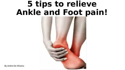
/
Chapter 14-15 The Foot, Ankle, and Lower Leg (FALL) Unit Chapter 14-15 The Foot, Ankle, and Lower Leg (FALL) Unit

Chapter 14-15 The Foot, Ankle, and Lower Leg (FALL) Unit - PowerPoint Presentation

hadley
hadley . @hadley
Follow
343 views
Uploaded On 2022-02-24

Chapter 14-15 The Foot, Ankle, and Lower Leg (FALL) Unit - PPT Presentation

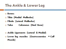
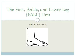
The foot has stresses that exceed the demands placed on any other area of the body The foot stabilizes and supports the rest of the body while standing walking running or jumping Whether the impact with the ground is on the heel the ball of the foot or the toe the foot responds by absorbin ID: 909787

ankle foot ligaments bones foot ankle bones ligaments anatomy joint lateral talus medial arch ligament side malleoli body tibia

Share:

Link:

Embed:

Download Presentation from below link

Download Presentation The PPT/PDF document "Chapter 14-15 The Foot, Ankle, and Lower..." is the property of its rightful owner. Permission is granted to download and print the materials on this web site for personal, non-commercial use only, and to display it on your personal computer provided you do not modify the materials and that you retain all copyright notices contained in the materials. By downloading content from our website, you accept the terms of this agreement.


Presentation Transcript

Slide1

Chapter 14-15

The Foot, Ankle, and Lower Leg (FALL) Unit

Slide2

The foot has stresses that exceed the demands placed on any other area of the body.

The foot stabilizes and supports the rest of the body while standing, walking, running, or jumping.

Whether the impact with the ground is on the heel, the ball of the foot, or the toe, the foot responds by absorbing several hundred pounds of force up to three times the body’s weight.

THE FOOT

Slide3

THE FOOT

The foot is the site of some of the most minor yet some of the most debilitating conditions suffered by athletes.

Examples of these conditions include:

- blisters - calluses - athlete’s foot - turf toe - ingrown toenails

Slide4

THE FOOT

Left untreated, these conditions can develop into serious foot problems, such as:

- heel bruises

- arch strains - ankle sprains - fractures

Slide5

Individually, the parts of the foot (bones, muscles, ligaments) are weak. As a whole, the foot is strong enough to withstand most of its demands.

ANATOMY OF THE FOOT

Slide6

ANATOMY OF THE FOOT: ARCHES

The key to the foot’s function is a set of 4 arches, which help in absorbing the impact of walking , running, and jumping.

The arches are called:

- metatarsal arch - transverse arch - medial (inner) longitudinal arch - lateral (outer) longitudinal arch

Slide7

Arches of the Foot

Slide8

ANATOMY OF THE FOOT: BONES

The feet contain about ¼ of the total number of bones in the body. (There are 206 total bones in an adult human body.)

Each foot has 26 bones (7 tarsal bones, 5 metatarsal bones, and 14 phalanges) and 38 joints.

Slide9

ANATOMY OF THE FOOT: BONES

The hind (back) foot is made up of

7 Tarsal bones

: - Talus - Calcaneus - Navicular - Cuboid - Medial, Intermediate, and Lateral CuneiformThe mid-foot region is made up of

5 Metatarsal bones

. (1

st

-5

th

from medial to lateral).

The forefoot (front) is where the toes are located. It is made of 14 bones known as

Phalanges

.

The

Hallux

(Big Toe) has 2 phalanges. The other toes have 3 each.

Slide10

Foot Anatomy-Let’s Color!

Slide11

FUN FACTS about the FALL

Whether the impact is on the heel, the ball of the foot, or the toes, the foot responds by absorbing several hundred pounds of force up to three times your body weight.

The foot may be in a abnormal position. Abnormal positions can create added stress on the FALL and lead to injury.

Supination = high archPronation=flat foot or fallen arch

Slide12

The talocrural joint (ankle joint) is the most commonly injured major joint in an athlete. The ankle is also called the Ankle mortise.

It is considered a modified Hinge joint.

The talocrural joint or ankle mortise is formed by two joints:Talocrural joint (tibia, fibula, talus)

Subtalar joint (talus and calcaneus)

ANATOMY OF THE ANKLE

Slide13

The Ankle Joint

Slide14

ANATOMY OF THE ANKLE: BONES

The tibia and the fibula are the two bones of the lower leg; while the talus and the calcaneus are the two largest bones of the foot.

The tibia is the main weight bearing bone of the lower leg. The main reason for this is because of its location on the talus. The tibia is mounted directly over top of the talus and it extends over its medial side.

The fibula is a non-weight bearing bone that extends down the lateral side of the talus forming the lateral

malleoli

.

Slide15

ANATOMY OF THE ANKLE: BONES

The malleoli are the two large, bony prominences (bumps) on either side of the ankle.

The medial malleoli is the distal end of the tibia.

The lateral malleoli is the distal end of the fibula.

Slide16

ANATOMY OF THE ANKLE: LIGAMENTS

After the bony structure, the first line of defense against ankle sprains is the joint’s strong

ligamentous

support.Just as with the bony structures, ligaments help the ankle joint be more stable. Remember ligaments hold bones together.Most of the ligaments involved in supporting the ankle are attached to the rough edges of the malleoli.The ligaments at the ankle are named for the bones they connect or bring together.

Slide17

ANATOMY OF THE ANKLE: LIGAMENTS

The ligaments most commonly injured on the lateral side of the ankle are:

- Anterior

talofibular (ATF) ligament - Calcaneofibular (CF) ligament - Posterior talofibular

(PTF) ligament

-Anterior tibiofibular ligament

-Posterior tibiofibular ligament

The ligaments most commonly injured on the medial side of the ankle are:

- Deltoid ligament (anterior

tibiotalar

, posterior

tibiotalar

,

tibiocalcaneal

, and

tibionavicular

ligaments)

Slide18

Ligaments of the Ankle