/
Erythropoiesis and general aspects of Erythropoiesis and general aspects of

Erythropoiesis and general aspects of - PowerPoint Presentation

hadly
hadly . @hadly
Follow
66 views
Uploaded On 2023-05-27

Erythropoiesis and general aspects of - PPT Presentation

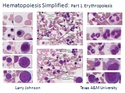
anaemia AssistProf DrMaysem M Alwash Erythropoiesis is the generation of red blood cells All the circulating blood cells derive from pluripotential stem cells in the marrow ID: 999627

cell haemoglobin anaemia marrow haemoglobin cell marrow anaemia red cells blood erythropoiesis erythropoietin bone normal erythroid ratio peripheral curve

Share:

Link:

Embed:

Download Presentation from below link

Download Presentation The PPT/PDF document "Erythropoiesis and general aspects of" is the property of its rightful owner. Permission is granted to download and print the materials on this web site for personal, non-commercial use only, and to display it on your personal computer provided you do not modify the materials and that you retain all copyright notices contained in the materials. By downloading content from our website, you accept the terms of this agreement.


Presentation Transcript

1. Erythropoiesis andgeneral aspects ofanaemiaAssist.Prof. :Dr.Maysem M. Alwash

2. Erythropoiesis is the generation of red blood cells ..-All the circulating blood cells derive from pluripotential stem cells in the marrow.

3. This process is governed by complex transcriptional and epigenetic programmes, in response to extracellular signalling

4. -We each make approximately 1012 new erythrocytes(red cells) each day by the complex and finely regulated process of erythropoiesis .Erythropoiesis passes from the stem cell through the progenitor cells, colony‐forming unit granulocyte, erythroid, monocyte and megakaryocyte (CFUGEMM), burst‐forming unit erythroid (BFUE) and erythroid CFU (CFUE) , to the first recognizable erythrocyte precursor in the bone marrow, the pronormoblast.

5. Diagrammatic representation of the bone marrow pluripotent stem cell and the cell lines that arise from it.

6. This process occurs in an erythroid niche in whichabout 30 erythroid cells at various stages of development surround a central macrophage.

7. An erythroblastic island with its central macrophage surrounded by erythroid progenitors at various stages of differentiation.

8. sequences Erythrocyte maturation

9. Pronormoblast (Rubriblast)SIZE: 12 to 20 μmNUCLEUS: Round to slightly ovalNucleoli: 1 to 2Chromatin: FineCYTOPLASM: Dark blue; may have prominent GolgiN:C RATIO: 8:1REFERENCE INTERVAL:Bone Marrow: 1%Peripheral Blood: 0%.

10. Pronormoblast (rubriblast

11. Basophilic Normoblast (Prorubricyte)SIZE: 10 to 15 μmNUCLEUS: Round to slightly ovalNucleoli: 0 to 1Chromatin: Slightly condensedCYTOPLASM: Dark blueN:C RATIO: 6:1REFERENCE INTERVAL:Bone Marrow: 1% to 4%Peripheral Blood: 0%.

12. Basophilic normoblast

13. Polychromatic ErythroblastRubricyteSIZE: 10 to 12 μmNUCLEUS: RoundNucleoli: NoneChromatin: Quite condensedCYTOPLASM: Gray-blue as a result of hemoglobinizationN:C RATIO: 4:1REFERENCE INTERVAL:Bone Marrow: 10% to 20%Peripheral Blood: 0%

14. Polychromatic normoblast.

15. Orthochromic normoblast.MetarubricyteSIZE: 8 to 10 μmNUCLEUS: RoundNucleoli: 0Chromatin: Fully condensedCYTOPLASM: More pink or salmon than blueN:C RATIO: 0.5:1REFERENCE INTERVAL:Bone Marrow: 5% to 10%Peripheral Blood: 0%

16. Orthochromic normoblast

17. Polychromatic erythrocyteSIZE: 8 to 8.5 μmNUCLEUS: AbsentNucleoli: NAChromatin: NACYTOPLASM: Color is slightly more blue/purple than the mature erythrocyteN:C RATIO: NAREFERENCE INTERVAL:Bone Marrow: 1%Peripheral Blood: 0.5% to 2.0%

18.

19. When stained with supravital stain (e.g., new methylene blue), polychromatic erythrocytes appear as reticulocytes (contain precipitated ribosomal (materialReticulocytes

20. Reticulocyte which still contains some ribosomalRNA and is still able to synthesize haemoglobin .This cell is slightly larger than a mature red cell, and circulates in the peripheral blood for 1–2 days before maturing, when RNA is completely lost. A completely pink‐staining mature erythrocyte results which is a non‐nucleated biconcave disc.

21. ERYTHROCYTESIZE: 7 to 8 μmNUCLEUS: AbsentNucleoli: NAChromatin: NACYTOPLASM: Salmon with central pallor of about one-third of the diameter of the cellN:C RATIO: NAREFERENCE INTERVAL:Bone Marrow: Peripheral Blood: Predominant cell type

22.

23.

24. -One pronormoblast usually gives rise to 16 mature red cells -Nucleated red cells (normoblasts) are not presentin normal human peripheral blood .They appear inthe blood if erythropoiesis is occurring outside the marrow(extramedullary erythropoiesis) and also with some marrowdiseases

25. ErythropoietinErythropoiesis is regulated by the hormone erythropoietin.Erythropoietin is a heavily glycosylated polypeptide. Normally, 90% of the hormone is produced in the peritubular interstitial cells of the kidney and 10% in the liver and elsewhere. -There are no preformed stores and the stimulus to erythropoietin production is the oxygen (O2) tension in the tissues of the kidney .

26. Hypoxia induces synthesis of hypoxia inducible factors (HIF‐1α and β), which stimulate erythropoietinproduction and also new vessel formation and transferrin receptor synthesis, and reduces hepcidin synthesis, increasing iron absorption. Von Hippel‐Lindau (VHL) protein breaks down HIFs and PHD2 hydroxylates HIF‐1α allowing VHL binding .Abnormalities in these proteins may cause Polycythaemia .

27. Erythropoietin production therefore increases in : 1-anaemia.2 - when haemoglobin for some metabolic or structurareason is unable to give up O2 normally . 3-atmospheric O2 is low 4-defective cardiac or pulmonary function .5- damage to the renal circulation affects O2 delivery to the kidney.

28. Erythropoietin stimulates erythropoiesis by increasing the number of progenitor cells committed to erythropoiesis. The transcription factor GATA‐2 is involved in initiating erythroid differentiation from pluripotential stem cells. Subsequently the transcription factors GATA‐1 and FOG‐1 are activated by erythropoietin receptor stimulation and are important in enhancing expression of erythroid‐specific genes (e.g. globin, haem biosynthetic and red cell membrane proteins) and also enhancing expression of anti‐apoptotic genes and of the transferrin receptor (CD71)

29. Late BFUE and CFUE, which have erythropoietin receptors, are stimulated to proliferate, differentiate and produce haemoglobin. The proportion of erythroid cells in the marrow increases and, in the chronic state, there is anatomical expansion of erythropoiesis into fatty marrow and sometimes into extramedullary sites. In infants, the marrow cavity may expand into cortical bone resulting in bone deformities with frontal bossing and protrusion of the maxilla.

30.

31. Conversely, increased O2 supply to the tissues (because of an increased red cell mass or because haemoglobin is able to release its O2 more readily than .normal) reduces the erythropoietin drive

32. The marrow requires many other precursors for effective erythropoiesis.These include metals such as iron and cobalt, vitamins(especially vitamin B12, folate, vitamin C, vitamin E, vitamin B6, thiamine and riboflavin) and hormones such as androgens and thyroxine.Deficiency in any of these may be associated with anaemia.

33. Plasma erythropoietin levels can be valuable in clinical diagnosis. They are high in anaemia unless this is due torenal failure and if a tumour secreting erythropoietin is present, but low in severe renal disease or polycythaemia vera.

34. Indications for erythropoietin therapyAnaemia of chronic renal diseaseMyelodysplastic syndromeAnaemia associated with malignancy and chemotherapyAnaemia of chronic diseases, e.g. rheumatoid arthritisAnaemia of prematurity Perioperative uses It is given subcutaneously either three times weekly or once every 1–2 weeks or every 4 weeks, depending on the indication and on the preparation used (erythropoietin alpha or beta, darbepoetin alpha (a heavily glycosylated longer‐acting form), or Micera the longestactingpreparation)

35. Side‐effects include a rise in blood pressure,thrombosis and local injection site reactions. It has been associated with progression of some tumours which express Epo receptors.

36. Haemoglobin Haemoglobin synthesis :Each molecule of normal adult haemoglobin A (Hb A) (the dominant haemoglobin in blood after the age of 3–6 months) consists of four polypeptide chains, α2β2, each with its own haem group.

37. Normal haemoglobins in adult blood.

38. Haem synthesis occurs largely in the mitochondria by a series of biochemical reactions commencing with the condensation of glycine and succinyl coenzyme A under the action of the key rate‐limiting enzyme δ‐aminolaevulinic acid (ALA) synthase .Pyridoxal phosphate (vitamin B6) is a coenzyme for this reaction. Ultimately,protoporphyrin combines with iron in the ferrous (Fe2+) state to form haem .A tetramer of four globin chains each with its own haem groupin a ‘pocket’ is then formed to make up a haemoglobin molecule

39.

40.

41. Haemoglobin functionAs the haemoglobin molecule loads and unloads O2 the individual globin chains move on each other .. The α1β1 and α2β2 contacts stabilize the molecule.When O2 is unloaded the β chains are pulled apart, permittingentry of the metabolite 2,3‐diphosphoglycerate (2,3‐DPG) resulting in a lower affinity of the molecule for O2. This movement is responsible for the sigmoid form of the haemoglobin O2 dissociation curve .The P50 (i.e. the partial pressure of O2 at which haemoglobin is half saturated with O2) of normal blood is 26.6 mmHg. With increased affinity for O2, the curve shifts to the left (i.e. the P50 falls) while with decreased affinity for O2, the curveshifts to the right (i.e. the P50 rises).

42. The normal position of the curve depends on the concentrationof 2,3‐DPG, H+ ions and CO2 in the red cell and on the structure of the haemoglobin molecule. High concentrations of 2,3‐DPG, H+ or CO2, and the presence of sickle haemoglobin (Hb S), shift the curve to the right (oxygen is given up more easily) whereas fetal haemoglobin (Hb F) – which is unable to bind 2,3‐DPG – and certain rare abnormal haemoglobins associated with polycythaemia shift the curve to the left because they give up O2 less readily than normal.

43.

44.

45. Red cell metabolismThe red cell has two biochemical pathways for metabolizing glucose, the Embden–Meyerhof, which generates ATP needed for maintenance of red cell shape and flexibility and NADH which prevents oxidation of haemoglobin, and the hexose monophosphate pathway, which generates NADPH, important for maintaining glutathione which keeps cell proteins in themembrane and haemoglobin in the reduced state.

46. Red cell membraneThe red cell membrane comprises a lipid bilayer, integralmembrane proteins and a membrane skeleton .Approximately 50% of the membrane is protein, 20% phospholipids,20% cholesterol molecules and up to 10% is carbohydrate.Carbohydrates occur only on the external surface while proteins are either peripheral or integral, penetrating the lipid bilayer.

47. The structure of the red cell membrane. Some of the penetrating and integral proteins carry carbohydrate antigens; other antigens are attached directly to the lipid layer

48. AnaemiaThis is defined as a reduction in the haemoglobin concentration of the blood below normal for age and sex.The WHO defines anaemia in adults as a haemoglobin less than 130 g/L in males and less than 120 g/L in females.

49. -Typical values would be less than 135 g/L in adult males and less than 115 g/L in adult females .-From the age of 2 years to puberty, less than 110 g/L indicates anaemia. -As newborn infants have a high haemoglobin level, 140 g/L is taken as the lower limit at birth.

50. Anaemia was most frequent in South Asia, and Central, Westand East Sub‐Saharan Africa. The main causes are iron deficiency (hookworm, schistosomiasis), sickle cell diseases, thalassaemia,malaria and the anaemia of chronic disorders

51. Clinical features of anaemiaThe major adaptations to anaemia are in the cardiovascularsystem and in the haemoglobin O2 dissociation curve..The presence or absence of clinical features can be consideredunder four major headings:1. Speed of onset Rapidly progressive anaemia causes moresymptoms than anaemia of slow onset.2. Severity Mild anaemia often produces no symptoms orsigns but these are usually present when the haemoglobinis less than 90 g/L. Even severe anaemia (haemoglobin concentration as low as 60 g/L) may produce remarkably few symptoms, when there is very gradual onset in a young subject who is otherwise healthy

52. 3. Age The elderly tolerate anaemia less well than the youngbecause normal cardiovascular compensation is impaired.4. Haemoglobin O2dissociation curve Anaemia, in general, isassociated with a rise in 2,3‐DPG in the red cells and a shiftin the O2 dissociation curve to the right so that oxygen is givenup more readily to tissues. This adaptation is particularlymarked in some anaemias that either raise 2,3‐DPG directly(e.g. pyruvate kinase deficiency )or that are associatedwith a low‐affinity haemoglobin (e.g. Hb S

53. Symptomsshortness of breath, particularly on exertion, weakness, lethargy, palpitation and headaches. In older subjects, symptoms of cardiac failure, angina pectoris or intermittent claudication or confusion may be present. Visual disturbances in very severe anaemia, particularly of rapid onset.

54. Signs General signs: -pallor of mucous membranes or nail beds, which occursif the haemoglobin level is less than 90 g/L .Conversely,skin colour is not a reliable sign. -A hyperdynamic circulation with tachycardia, a bounding pulse, cardiomegaly and a systolic flow murmur especially at the apex. -Particularly in the elderly, features of congestive heart failure .

55. Specific signs : -koilonychia (spoon nails) with iron deficiency,-jaundice with haemolytic or megaloblastic anaemias - leg ulcers with sickle cell and other haemolytic anaemias.- bone deformities with thalassaemia major

56. The association of features of anaemia with excess infectionsor spontaneous bruising suggest that neutropenia orthrombocytopenia may be present, possibly as a result of bonemarrow failure

57. Classification and laboratory findings in anaemiaRed cell indicesThe most useful classification is that based on red cell indices

58.

59. Other laboratory findings-Leucocyte and platelet counts :-Subnormal levels of red cells, neutrophils and platelets which suggests a more general marrow defect or destruction of cells (e.g. hypersplenism). -In anaemias caused by haemolysis or haemorrhage, the neutrophil and platelet counts are often raised- In infections and leukaemias, the leucocyte count is also often raised and there may be abnormal leucocytes or neutrophil precursors present.

60. Reticulocyte countThe normal percentage is 0.5–2.5%, and the absolutecount 50–150 × 109/L .-This should rise inanaemia because of erythropoietin increase, and be higher the more severe the anaemia. This is particularly so when there has been time for erythroid hyperplasia to develop in the marrow as in chronic haemolysis.-.

61. After an acute major haemorrhage there is an erythropoietin response in 6 hours the reticulocyte count rises within 2–3 days, reaches a maximum in 6–10 days and remains raised until the haemoglobin returns to the normal level -If the reticulocyte count is not raised in an anaemic patient this suggests impaired marrow function or lack of erythropoietin stimulus .

62.

63. Blood filmIt is essential in all cases of anaemia.Abnormal red cell morphology or red cell inclusions may suggest a particular diagnosis. During the blood film examination, white cell abnormalities are sought, platelet number and morphology are assessed and the presence or absence of abnormal cells (e.g. normoblasts, granulocyte precursors or blast cells) is noted

64. Bone marrow examinationThis is needed when the cause of anaemia or other abnormalityof the blood cells cannot be diagnosed from the blood count,film and other blood tests alone. It may be performed by aspiration or trephine biopsy.Special tests, e.g. immunology, cytogenetics, can be performed on the cells obtained

65. Indications for bone marrow aspiration and trephine biopsy.

66. Ineffective erythropoiesisErythropoiesis is not entirely efficient because approximately10–15% of developing erythroblasts die within the marrowwithout producing mature cells. This is termed ineffective erythropoiesis and it is substantially increased in a number of chronic anaemia.

67. when ineffective erythropoiesis is marked,the followings are seen: -Raised serum unconjugated bilirubin (derived from breaking down haemoglobin) -Raised lactate dehydrogenase (LDH, derived from breaking down cells) .-Reticulocyte count is low in relation to the degree of anaemiaand to the proportion of erythroblasts in the marrow.

68. Summary of the regulation of erythropoiesis with mass the key points for assessment boxed in blue. ME denotes assessment of the myeloid/erythroid ratio in the bone marrow.Hct, haematocrit.

69. Assessment of erythropoiesisTotal erythropoiesis and the amount of erythropoiesis that is effective in producing circulating red cells can be assessed by: examining the bone marrow, haemoglobin level and reticulocyte count.

70. -Total erythropoiesis is assessed from the marrow cellularityand the myeloid : erythroid ratio(normal M:E ratio is 2:1 to 4:1). This ratio falls and may be reversed when total erythropoiesis is selectively increased.Effective erythropoiesis is assessed by the reticulocyte count. This is raised in proportion to the degree of anaemia when erythropoiesis is effective, but is low when there is ineffective erythropoiesis or an abnormality preventing normal marrow response

71. The relative proportions of marrow erythroblastic activity, circulating red cell mass and red cell lifespan in normal subjectsand in three types of anaemia

72. THANK YOU