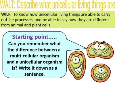
/
Atypical Bacteria Bacterial Taxonomy:  How are these  unicellular organisms classified? Atypical Bacteria Bacterial Taxonomy:  How are these  unicellular organisms classified?

Atypical Bacteria Bacterial Taxonomy: How are these unicellular organisms classified? - PowerPoint Presentation

hanah
hanah . @hanah
Follow
342 views
Uploaded On 2022-06-15

Atypical Bacteria Bacterial Taxonomy: How are these unicellular organisms classified? - PPT Presentation

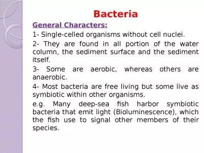
complex system of classification based on shape amp size oxygen pH and temperature requirements laboratory characteristics biochemical analyses serology tests nucleic acid and protein analysis techniques ID: 919006

bacteria acid cell fast acid bacteria fast cell chlamydia stain host cells fungus pneumonia spp nocardiosis mycobacterium tuberculosis wax

Share:

Link:

Embed:

Download Presentation from below link

Download Presentation The PPT/PDF document "Atypical Bacteria Bacterial Taxonomy: H..." is the property of its rightful owner. Permission is granted to download and print the materials on this web site for personal, non-commercial use only, and to display it on your personal computer provided you do not modify the materials and that you retain all copyright notices contained in the materials. By downloading content from our website, you accept the terms of this agreement.


Presentation Transcript

Slide1

Atypical Bacteria

Slide2

Bacterial Taxonomy: How are these unicellular organisms classified?

complex system of classification

based on shape & size; oxygen, pH, and temperature requirements; laboratory characteristics, biochemical analyses, serology tests, nucleic acid and protein analysis techniques ..…

Classification is now based on similarities in nucleotide sequences in rRNA

The

Bergey

s Manual

is considered the official listing of all recognized bacteria

see appendix A in your text for the details (don

t memorize!)

Slide3

Reminder

The majority of Bacteria and

Archaea

have not been cultured

Scientists estimate that only 1% of these microbes have been discovered!

And…of more than 2600 species of bacterial names, fewer than ____ are human pathogens.

Slide4

Some atypical bacteria

not

typical

will discuss

1. fungus-like bacteria

2. acid fast bacteria3. Mycoplasmas4. Chlamydias5. Rickettsias

Slide5

Fungus-like (filamentous) bacteria

myc

in word refers to fungus or something like a fungus (filamentous)

Streptomyces spp

.: most important source of antibiotics

More than half of our _____________

are produced by species of

Streptomyces

.

Antibiotics are substances

produced by microbes

that in small amountsinhibits anothermicrobe.

Slide6

Slide7

Actinomyces

branching filaments and spore-case (sporangium)

Actinomyces spp

.: cause actinomycosis (lumpy jaw)

large abscess in jawsulfur granules in pusFigure 11.22

Slide8

Actinomycosis

Fungal-like bacterial disease

Infection follows trauma to body tissues

lumpy jaw

Slide9

Actinomycosis

Slide10

Actinomyces

sulfur

granules

in pus

Slide11

2.

____________bacteria

gram positive bacilli with wax coat on cell wall

wax increases survival in environment

Nocardia

spp. (weakly acid fast; require special acid-fast stain)nocardiosis: infection of lungs, skin or other tissue

Mycobacterium

spp.

M. tuberculosis

(tuberculosis)

M.

leprae (leprosy)many other species

Slide12

Mycobacterium tuberculosis

fig. 24.8

Slide13

nocardia

Procaryotic,

acid-fast,

but grows much

like a fungus

Slide14

Nocardia

:

nocardiosis

,

mycetoma

Follows penetration

from the soil via

a wound

Slide15

Nocardiosis growing back

through ribs from lungs to

skin

When inhaled, the disease

strikes the lungs, where

multiple abscesses form.

Symtoms can be similar to

tuberculosis

Reports of death from

nocardiosis have been

linked to AIDS

Slide16

Acid-fast stain: pp. 70-71, fig. 3.13

Primary stain:

_

______________

Mordant: heat or detergent (cold method we will use in lab) accompanying primary stain, to force stain through wax coat on cells

Decolorization: acid alcohol

3%

HCl

in 95% ethanol

acid-fast bacteria hold primary stain because wax resists acid alcohol

non acid-fast bacteria decolorize

Counterstain: ________________red = acid-fast; blue = nonacid-fast

Slide17

Mycobacterium leprae

Red = acid-fast

Slide18

Mycobacterium ulcerans:

Buruli ulcer

Reservoir = swamps & slow-flowing rivers

Increasing in world

Global threat to public health (WHO)

Page 594 in your text

Slide19

3.

____________

smallest known cells (“submicroscopic”)

never have cell walls

don’t rupture because of small size and tough cell membrane

myco

= filamentous; plasma = fluid

pleomorphic

= variable shape

will grow on culture media

“fried egg” colonies

Genera: Mycoplasma and

Ureaplasma

Slide20

Mycoplasmas cause:

pneumonia in humans and animals

walking pneumonia

15-20% of cases human pneumonia

Can be severe

mastitis in cowsurethritis in humans stunting of plant growth

Slide21

Mycoplasma pneumoniae

attached to surface of host cells

Slide22

4.

Chlamydias

Submicroscopic

Have cell walls: oval shape, gram negative

Obligate intracellular parasites

= will grow only inside of living host cells

energy parasites

Genus

Chlamydia

trachoma

genital

chlamydia

http://www.cdc.gov/std/Chlamydia/STDFact-Chlamydia.htm

Pneumonia

Chlamydophila

psittaci

In pathogen group 4

figure 11.24

Slide23

2 forms of chlamydia cells

_______________:

0.3 µm

rigid cell wall

can survive outside of host cell

Infectious_______________:0.5 to 1.0µmfragile cell wall

Can’t survive outside of host cell

not infectious

adapted for growth

Slide24

chlamydia reproduction

red = elementary body; green = reticulate body

entering reproducing escaping

See figure 11.24

Slide25

5.

Rickettsias

tiny oval to rod-shaped bacteria; just visible with light microscope

See figure 11.1 on page 304

obligate intracellular parasites

But now widely separated from the chlamydia (see table 13.1 on page 368)

most have arthropod vectors

Genera

Rickettsia

and

Ehrlichia

Cause:

Rocky Mountain spotted fever (ticks are vectors)

Typhus fevers (various vectors)

Will study in pathogen group 9!

Slide26

Rickettsia in host cell

Figure 11.1

Slide27

End of Unit 1 – Exam 1 Covers everything up to this point!