/
Sterilization of biological tissues using low energy electron irradiation Sterilization of biological tissues using low energy electron irradiation

Sterilization of biological tissues using low energy electron irradiation - PowerPoint Presentation

hazel
hazel . @hazel
Follow
0 views
Uploaded On 2024-02-16

Sterilization of biological tissues using low energy electron irradiation - PPT Presentation

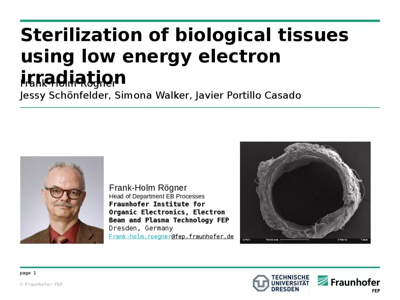
FrankHolm Rögner Jessy Schönfelder Simona Walker Javier Portillo Casado FrankHolm Rögner Head of Department EB Processes Fraunhofer Institute for Organic Electronics Electron Beam and Plasma Technology FEP ID: 1046349

vascular leei sterilization µm leei vascular µm sterilization electron penetration depth irradiation 200 energy 500 tissue 100 untr assessment

Share:

Link:

Embed:

Download Presentation from below link

Download Presentation The PPT/PDF document "Sterilization of biological tissues usin..." is the property of its rightful owner. Permission is granted to download and print the materials on this web site for personal, non-commercial use only, and to display it on your personal computer provided you do not modify the materials and that you retain all copyright notices contained in the materials. By downloading content from our website, you accept the terms of this agreement.


Presentation Transcript

1. Sterilization of biological tissues using low energy electron irradiationFrank-Holm RögnerJessy Schönfelder, Simona Walker, Javier Portillo CasadoFrank-Holm RögnerHead of Department EB ProcessesFraunhofer Institute for Organic Electronics, Electron Beam and Plasma Technology FEPDresden, GermanyFrank-holm.roegner@fep.fraunhofer.de

2. Sterilization procedures for biological tissuesProcedureLimitations for biological tissuesAutoclavingDenaturation of proteins / loss of tissue architectureChemical SterilizationImpaired biocompatibilityIonizing Irradiation Gamma irradiationImpairment of mechanical properties high-energy electron irradiationHigh shielding necessary

3. Low Energy Electron Irradiation - LEEIOn-site sterilization possibleIrradiation inside the final packaging Short irradiation timeIrradiation under atmospheric pressureNo heat development inside the sample (< 40 °C)Sterilization of medical products according to DIN EN ISO 11137https://www.sterihealth.de/

4. Part I: Sterilization of Vascular GraftsIntroductionReplacement of dysfunctional vessels or infected graftsLarge diameter vessels are often replaced by polymeric vascular graftsSmall diameter vessels (< 8 mm) are preferentially replaced by autologous veinsAllografts and xenografts are infrequently used: Challenge - SterilityVarious vascular prosthesis(Source: Rotes Kreuz Krankenhaus Gefäßzentrum Bremen; http://www.gefaesszentrum-bremen.de/lexikon/gefaessprothese)

5. IntroductionWall structure of blood vessels Source: S.A.I. Ghesquiere (2006) The Role of Phospholipases in Atherosclerosis, UPM Universitaire Pers Maastricht, ISBN 9789052785301Challenge:Adjusting the penetration depth so that cells in the media and intima remain viableessentiell for vascular function

6. Experimental approachGoal: Sterilisation and maintanance of the tissue functionSterilisation by electron irradiation→ Variation of the dose → Variation of the penetration depthRat aortaModel for vascular graftsVessel functiona) Wall stressb) ACh-induced Relaxationc) SNP-induced RelaxationSterilitya) Turbidity testb) ImpedanceDosimetry

7. Experimental approachLow-energy electron irradiation:Sample 1: penetration depth of max. 18 µm only irradiation of the Adventitia (outer layer)Sample 2: penetration depth ca. 110 µmirradiation of the complete vessel wall2 doses: - ca. 21 kGy - ca. 42 kGy - untreated control (FEP) - physiological control (4 °C)Treatment from both sidessubsequently:Assessment of the sterility and vessel functionCooling unit (4 °C)Electron gunPackaged non-sterile sampleLEEISterile sampleSchematic diagram of the LEEIAortaPE-RingpackagingSchematic diagram of the packaged aorta

8. Sterilization concept of aortic vascular graftsParameter optimization, so thatHomogenous irradiationSurface dose 21 kGyPenetration depth < 18 µm 0 - 18 µm 19 - 36 µm 37 – 54 µm 5 kGy25 kGy50 kGyAortaDosimeterS.A.I. Ghesquiere (2006), UPM Universitaire Pers Maastricht, ISBN 97890527853013 layers dosimeter foilCalibration

9. Sterility assessment - MethodsTurbidity testing and impedance measurements (Bac Trac 4100, SY-LAB Geräte):Metabolic activity and growth of bacteria  CO2lowering of the impedanceSterile (left) and non-sterile (right) samplesMeasured curves of sterile and non-sterile samples

10. Sterility assessment - ResultsSterility assessment: growth delay of non-sterile samplesChallenge:Untr. LEEI - 21 LEEI - 42

11. Sterility assessment - ResultsSterility assessment: Increase of sterile samples after LEEI Up to 50% of the samples sterileSterilization only on the surface of the sample possibleSterile Impedance measurementsSterile – turbidity testingNon-sterile – Impedance measurementsUntr. LEEI - 21 LEEI - 42

12. Vascular function – Experimental approachMyograph 610 M (DMT) at 37 °CFixation of the aorta on the grip which is coupled to a force measurement unit Measurement of the vessel function by stimulation of the cells with the following reagents:contraction: high potassium-ion concentration (K+) Noradrenaline (NA)Relaxation: Acetylcholine (ACh) Natriumnitroprussid (SNP)Myograph

13. Vascular function - ResultsVascular function: Wall stress as a measure for vascular contraction penetration depth 18 µm penetration depth110 µm→ no changes→ decrease with increasing doses***** P < 0,05 compared to untreated4°C Untr. LEEI - 21 LEEI - 424°C Untr. LEEI - 21 LEEI - 4218 µm110 µm

14. Vascular function - ResultsVessel function: Relaxation penetration depth 18 µm penetration depth 110 µm→ SNP-induced: no changes→ ACh-induced: decrease after 42 kGy→ SNP-induced: no changes→ ACh-induced: decrease after 42 kGy**4°C Untr. LEEI - 21 LEEI - 424°C Untr. LEEI - 21 LEEI - 4218 µm110 µm

15. Vascular functionality after LEEI Rat-Aorta Penetration depth18 µm110 µmDose21 kGy42 kGy21 kGy42 kGySterility++O+Wall stressOO--Endothelial independent relaxationO-O(-)Endothelial dependent relaxationO(-)(-)-+ positive impactO no impact- negative impact(-) not significantSterilization of rat aortae by LEEI possible. However, the penetration depth needs to be adjusted to keep the endothelial layer unaffected

16. Part II: Sterilization of Pericardial TissuePericardium-based tissue transplantsClinical ApplicationsCardiac surgery (Heart valve prosthesis, Cardiac reconstruction, closure of pericardial sac)Vascular reconstructionAbdominal wall and diaphragm defectsOrbital repairDural repairTendon and ligament augmentation Phelan et al., Pericardial Disease, 2015: http://www.clevelandclinicmeded.com/medicalpubs/diseasemanagement/cardiology/pericardial-disease/Herzzentrum Dresden

17. Heart Valve ReplacementsBiological heart valvesBioprosthetic heart valvesPatients > 65 yearsMechanical heart valvesPatients < 65 years HomograftsPatients 35 – 65 years~250 000 heart valve replacements annually worldwide

18. Crosslinking of collagen-rich tissuesAccelerated biodegradation or insufficient mechanical strengthGold standard: pre-treatment with glutaraldehydeMajor limitationsCytotoxicityCalcium depositionAlternative treatmentsCrosslinkers - carbodiimides, genipinRadical scavengers - glucose, ascorbate, riboflavinLow energy electron irradiation

19. Tissue preparationSamples: untr. Untreated control GA Glutaraldehyde-crosslinking (0,2 % 30 min)LEEI 10 Low-energy electron irradiation with a dose of 10 kGy ● ● ●LEEI 500 Low-energy electron irradiation with a dose of 500 kGy Native pericardium removal of Pl. pericardia removal of cells dissected pericardiumHandling of the samples during irradiation

20. Sterility assessment - Resultsuntreated samples non-sterileReproducible sterilization above 25 kGyn = 5Non-sterilesterileUntr.

21. Change of Appearance- ResultsPericardium becomes stiffer with increasing dose (indication of cross-linking)Color change: more yellowish and more transparent with increasing doseUntreatedEB 100 kGyEB 200 kGyEB 250 kGyEB 500 kGyGA

22. Morphological analysis - ResultsUntr.GALEEI 200 kGyLEEI 500 kGy→ physiologic structure→ unorganized structure→ loss of structure→ oriented structure2000x

23. Experimental approach – in vitro biodegradabilityOne hour incubation in collagenase solution:  0.6 U/ml collagenase in TRIS-Buffer, 37 °C, pH 7.4Analysis of the wet weight before and after incubationCalculation of the mass relative to the initial mass before digestion

24. In vitro biodegradability - ResultsEB 100 und EB 200 are rapidly digested, EB 500 is stableGA very stable, even after 5 h digestionAn extra treatment should be addedUntr. GA LEEI LEEI LEEI LEEI 25 100 200 500Untr. GA LEEI LEEI LEEI LEEI 25 100 200 500

25. Experimental Approach – Cytocompatibility Testwell plateGlass ringpericardiumFibroblast cell seeding20 / 40 hours cell incubationMetabolic activitycell numbercell morphology

26. Cytocompatibility testing - resultsUntr.GALEEI 500 kGy→ almost confluent cell layer→ separated cells→ almost confluent cell layerLEEI 200 kGy→ almost confluent cell layer100x

27. GA: no proliferationuntr + EB: proliferationCytocompatibility - ResultsUntr. GA LEEI LEEI LEEI 100 200 500

28. Determination of the secreted pro-inflammatory Interleukins IL-6 und IL-8 in the cell culture supernatantUntr. > GAUntr. < EBImmunogenicity- Results LPS Glas untr. GA LEEI LEEI LEEI 100 200 50020 hours

29. DecellularizationModification and sterilization by low energy electron irradiationUVA treatment, pericardium soaked in riboflavinNovel SULEI procedure for sterilization and crosslinking of transplantable tissuesIP Reference number: 10 2016 121 982.7Example: pericardial tissue

30. Bioburden determination sterility assessmentWalker et al., in preparationBioburden: 5.1*105 ± 4.6*105 CFU/cm2 (n=20)TreatmentsterileNon-sterilenGlutaraldehyde28 %72 %29Riboflavin / UV0 %100 %30SULEI100 %0 %30

31. Sterilization depthn = 3, n.a. – not applicableWalker et al., in preparationn = 4> 106 CFU / cm2 b. pumilus spores240 µm / 312 µm HDPE foil stackpackagingThickness [µm]Thickness HDPE stack [µm]Inocculum [CFU/cm2]CFU/cm2 after LEEILog-Reduction Turbidity2402.2*106 < 1n.a.-3122.2*106 5.7*105± 8.1*1050.5+SULEI

32. Biomechanical propertiesn = 8Walker et al., in preparationGlutaraldehydeDecellularizedSULEI

33. CytocompatibilityDecellularizedSULEI 48 h 96 h 100 µmWalker et al., in preparationGlutaraldehyde

34. Morphology50 µm20 µmDecellularizedSULEIGlutaraldehyde

35. Conclusions and outlookLow-energy electron beam tissue treatment  targeted modification:Crosslinking predominates until 100 kGy:Pericardium: increase bending stiffness, shrinkage and elasticityAorta: reduction of wall tension by crosslinking in the vessel wallDegradation predominates from 200 kGy:Pericardium: tears at low tension in tensile testSensitive control of the penetration depth

36. Conclusions and outlookFeasibility of sterilization of biological tissues using LEEI was shown using two modelsRat aorta as a model for vascular graftsPorcine pericardium as a model for pericardium-based tissue transplantsMajor functional parameters of the tissue could be maintainedValidation of the process in progress

37. AcknowledgementsFraunhofer FEP Prof. Dr. Volker KirchhoffDr. Christiane WetzelDr. Jessy SchönfelderSimona WalkerJavier Protillo CasadoDr. Gaby GotzmannLysann KennerMarleen DietzeAndré PorembaJuliane SchötzInstitute of Physiology, TU Dresden Prof. Dr. Andreas Deußen Pharmaceutical Technology, Universität LeipzigProf. Dr. Michaela Schulz-SiegmundDr. Michael Hacker Institute of Anatomy, TU Dresden Prof. Dr. Richard Funk Department of Cardiac Surgery, TU DresdenProf. Dr. Sems Malte TugtekinDr. Katrin PlötzeAline JakobDr. Claudia Dittfeld