/
the brain parenchyma and this requires prompt adminBrain abscesses f the brain parenchyma and this requires prompt adminBrain abscesses f

the brain parenchyma and this requires prompt adminBrain abscesses f - PDF document

heavin
heavin . @heavin
Follow
342 views
Uploaded On 2022-10-13

the brain parenchyma and this requires prompt adminBrain abscesses f - PPT Presentation

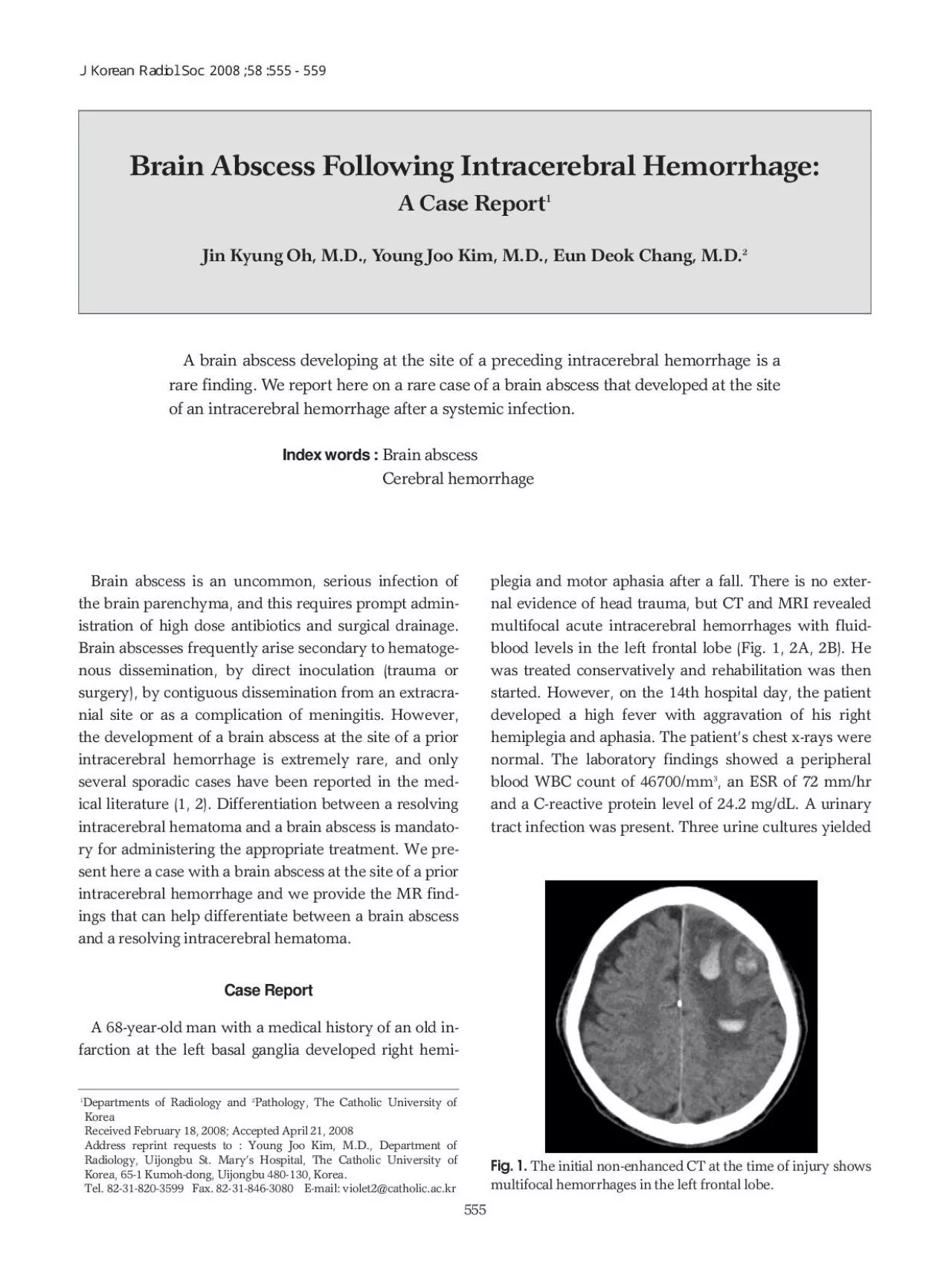
Brain Abscess Following Intracerebral Hemorrhage A Case ReportJin Kyung Oh MD Young Joo Kim MD Eun Deok Chang MD Tel 82318203599 Fax 82318463080 Email violet2catholicackr ID: 959803

abscess brain hemorrhage intracerebral brain abscess intracerebral hemorrhage ring signal hematoma case fig high dwi weighted edema hematomas hemorrhages

Share:

Link:

Embed:

Download Presentation from below link

Download Pdf The PPT/PDF document "the brain parenchyma and this requires p..." is the property of its rightful owner. Permission is granted to download and print the materials on this web site for personal, non-commercial use only, and to display it on your personal computer provided you do not modify the materials and that you retain all copyright notices contained in the materials. By downloading content from our website, you accept the terms of this agreement.


Presentation Transcript

the brain parenchyma, and this requires prompt admin-Brain abscesses frequently arise secondary to hematoge-surgery), by contiguous dissemination from an extracra-several sporadic cases have been reported in the med-intracerebral hematoma and a brain abscess is mandato-ry for administering the appropriate treatment. We pre-intracerebral hemorrhage and we provide the MR find-A 68-year-old man with a medical history of an old in-farction at the left basal ganglia developed right hemi-plegia and motor aphasia after a fall. There is no exter-䨠䭯牥慮⁒慤楯氠卯挠㈰〸㬀㔸㨀㔀㔵ⴀ㔵㤀 Brain Abscess Following Intracerebral Hemorrhage: A Case ReportJin Kyung Oh, M.D., Young Joo Kim, M.D., Eun Deok Chang, M.D. Tel. 82-31-820-3599 Fax. 82-31-846-3080 E-mail: violet2@catholic.ac.kr A brain abscess developing at the site of a preceding intracerebral hemorrhage is arare finding. We report here on a rare case of a brain abscess that developed at the site Fig. 1. The initial non-enhanced CT at the time of injury showsmultifocal hemorrhages in the left frontal lobe. Fig. 1. The initial non-enhanced CT at the time of injury showsmultifocal hemorrhages in the left frontal lobe. Brain Abscess Following Intracer

ebral Hemorrhage: A Case ReportJin Kyung Oh, M.D., Young Joo Kim, M.D., Eun Deok Chang, M.D. Departments of Radiology and 2Pathology, The Catholic University ofKorea sites of the preceding intracerebral hemorrhages, in ad-bands of heterogeneous signal intensity with an inverse-㨠⁂牡楮⁁扳捥獳⁆潬汯睩湧⁉湴牡捥牥扲慬⁈敭潲牨慧攀 A D Fig. 2. A, B. ) show the layered, acute stage hematomas with minimal per-The MR images taken 3 weeks after the hemorrhage. The axial T2-weighted im-) demonstrates uniform peripheral wall enhancements, which accurately cor-) reveals layered heterogeneous signal intensities with inversely heteroge-marked hypointense rim at the periphery. Ring enhancement with a complete hy-indicate that brain abscess developed at the site of the preceding hemorrhage. G periphery on the DWI and T2-weighted images accu-material was aspirated and three well capsulated ab-macrophages, a mononuclear infiltrate, revasculariza-a brain abscess (Fig. 3). The cultures of the surgical spec-was treated with broad-spectrum antimicrobial cover-brain tissue susceptible to infection by blood-borne bac-teria with subsequent abscess formation (1). In the pre-which indicated systemic infection and b

acteremia, oc-curred 0-90 days after the onset of the intracereberal he-was considered to be the source of hematogenous seed-specific finding for a brain abscess and it must be distin-guished from a necrotic neoplasm and other cystic le-sions. Intracerebral hematomas usually resolve sponta-the hemorrhage evolves, different characteristic appear-blood-brain barrier in the vascularized capsule that sur-rounds the hematoma (3) and this mimics the appear-ance of an abscess. Because a brain abscess is an emer-high dose antibiotics and surgical drainage, it is manda-hematoma. However, this differentiation can be diffi-More recently, DWI has demonstrated significant po-high signal intensity in the central cavity and a corre-restricted diffusion in brain abscesses are the microscop-pus thatÕs caused by a high protein level and the differ-molecules are bound to amino acid groups on the sur-other brain diseases like hemorrhagic primary or sec-high signal intensities on the DWI with an inversely het-demonstrated that the central heterogeneous signal in-䨠䭯牥慮⁒慤楯氠卯挠㈰〸㬀㔸㨀㔀㔵ⴀ㔵㤀 Photomicrograph of the histologic specimen reveals fi-with hemosiderin pigments in the abscess capsule, and amor- pr

operties have been ascribed to collagen and the para-magnetic free radicals that are released from the phago-as subacute and chronic hematomas, metastases, granu-hemosiderin-laden macrophages begin to take up resi-subacute stage of an intracerebral hematoma, the pres-have been the primary cause of a complete T2 hy-manifested as a ring-enhancing lesion with complete hy-a rare condition with ring enhancement and a T2 hy-pointense border (8). The histology for our case con-firmed the presence of macrophages, a mononuclear in-leading to edema formation in the extracellular compo-nent (9). This edema usually subsides and the mass ef-fect gradually diminishes in cases with a resolving hem-orrhage. It is known that pronounced or persistent ede-ma is one of the signs of hemorrhagic intracranial neo-much of the associated mass effect. In spite of encapsu-barrier persisted, and this was responsible for the belat-hematoma cavity should be considered in the differen-the T2-weighted images and DWI and the unusual ex-diagnose a brain abscess rather than a resolving suba-1. Chen ST, Tang LM, Ro LS. Brain abscess as a complication of2. Siatouni A, Mpouras T, Boviatsis EJ, Gatzonis S, Stefanatou M,3. Zimmerman RD, Leeds NE, Naidich

TP. Ring blush associated4. Hartmann M, Jansen O, Heiland S, Sommer C, Munkel K, Sartor K.Restricted diffusion within ring enhancement is not pathognomon-5. Mishra AM, Gupta RK, Jaggi RS, Reddy JS, Jha DK, Husain N, etresonance spectroscopy in the differential diagnosis of ring-en-6. Kang BK, Na DG, Ryoo JW, Byun HS, Roh HG, Pyeun YS.Diffusion-weighted MR imaging of intracerebral hemorrhage.8. Schwartz KM, Erickson BJ, Lucchinetti C. Pattern of T2 hy-9. Wagner KR, Xi G, Hua Y, Kleinholz M, de Courten-Myers GM,rapid edema development in perihematomal white matter. 10. Wallenfang T, Bohl J, Kretzschmar K. Evolution of brain abscess in㨠⁂牡楮⁁扳捥獳⁆潬汯睩湧⁉湴牡捥牥扲慬⁈敭潲牨慧攀 䨠䭯牥慮⁒慤楯氠卯挠㈰〸㬀㔸㨀㔀㔵ⴀ㔵㤀듫쟑뾵믳샇쟐좸쇶㈰〸㬀㔸㨀㔀㔵ⴀ㔵㤀 돺뷇쇺뎻쟷솾냺떿망뗈돺돳뻧㨠쇵럊몸냭낡엧뢯듫쟐놳뾵믳샇쟐냺Ⱐ돺뷇쇺뎻쟷솾샌샖뻺듸삧쒡뾡돺돳뻧샌몴맟뗇듂냦뿬듂쟶샧뇮쇶ㄳ뾹낡몸냭뗈룅뿬뗥릮쇵럊샌듙⸠샺샚뗩삺돺뷇쇺뎻쟷솾샌샖뻺듸삧쒡뾡샼뷅원쟷쇵삸럎샎쟑돺돳뻧샌몴맟뗇뻺듸좯샚ㆿ릸ꘀ냦쟨쟏뾴뇢뾡뾵믳볒냟냺쟔늲몸냭쟏듂맙샌듙â¸