/
May 2019 Review May 2021 May 2019 Review May 2021

May 2019 Review May 2021 - PDF document

isabella
isabella . @isabella
Follow
342 views
Uploaded On 2022-08-26

May 2019 Review May 2021 - PPT Presentation

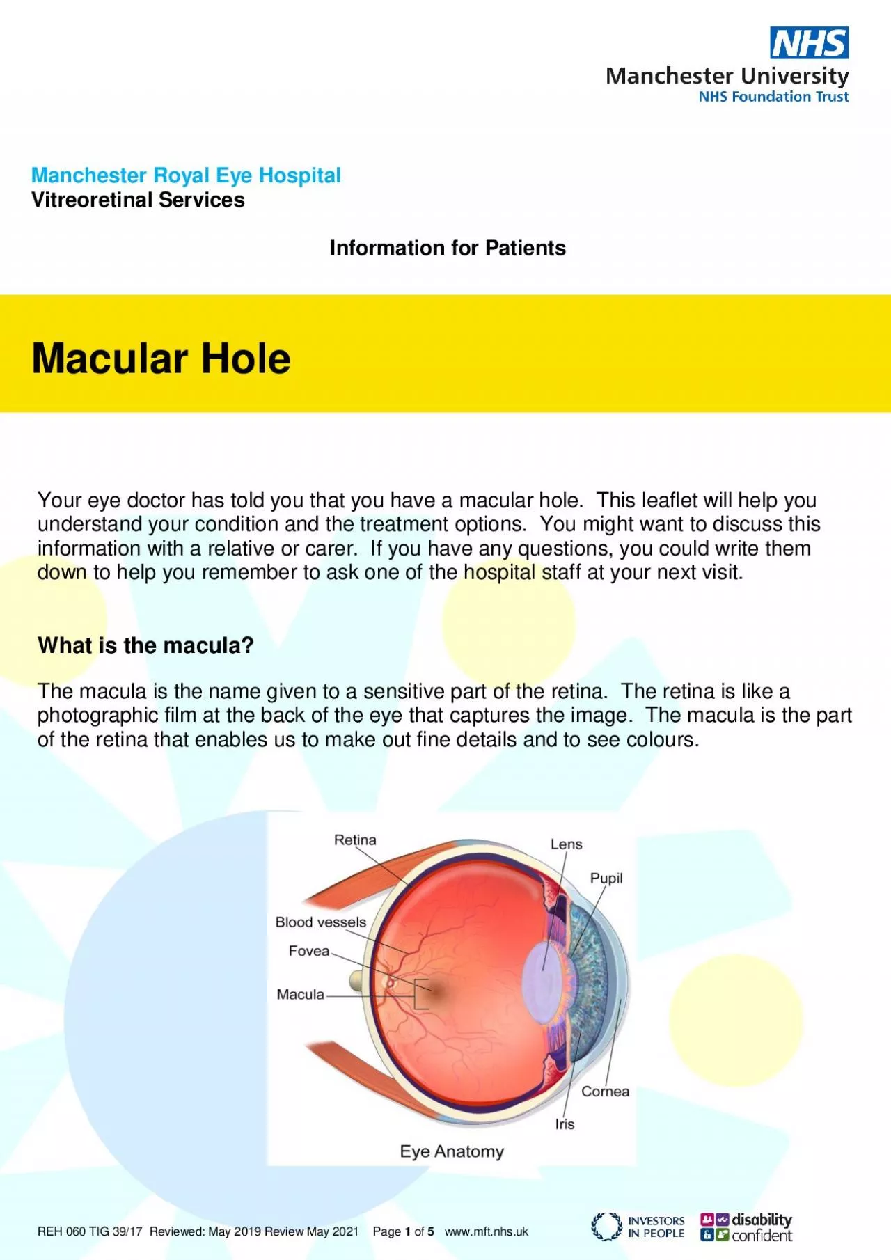
REH 060 TIG 3917 R eview ed Page 1 of 5 wwwmftnhsuk Manchester Royal Eye Hospital Vitreoretinal Services Information for Patients Macular Hole Your eye doctor has told you that you have a macu ID: 942230

surgery eye macula operation eye surgery operation macula hole gas macular bubble vision retina vitreous doctor occur important staff

Share:

Link:

Embed:

Download Presentation from below link

Download Pdf The PPT/PDF document "May 2019 Review May 2021" is the property of its rightful owner. Permission is granted to download and print the materials on this web site for personal, non-commercial use only, and to display it on your personal computer provided you do not modify the materials and that you retain all copyright notices contained in the materials. By downloading content from our website, you accept the terms of this agreement.


Presentation Transcript

REH 060 TIG 39/17 R eview ed : May 2019 Review May 2021 Page 1 of 5 www.mft.nhs.uk Manchester Royal Eye Hospital Vitreoretinal Services Information for Patients Macular Hole Your eye doctor has told you that you have a macular hole. This leaflet will help you understand your condition and the treatment options. You might want to discuss this information with a relative or carer. If you have any questions, you could write them down to help you remember to ask one of the hospital staff at your next visit. What is the macula ? The macula is the name given to a sensitive part of the retina. The retina is like a photographic film at the back of the eye that captures the image. The macula is the part of the retina that enables us to make out fine details and to see colours. REH 060 TIG 39/17 R eview ed : May 2019 Review May 2021 Page 2 of 5 www.mft.nhs.uk What is a macular hole ? A macular hole is as it sounds ‘a hole in the macula.’ This is caused by a separation or loss of cells in the macula. You will have a scan taken of the back of your eye called Optical Coherence Tomography (OCT). This provides very detailed pictures of t he retina and macula that helps your doctor to diagnose and manage your condition. The OCT uses light waves to generate a cross - sectional image of your eye and make a map of the retina at the back of your eye to show up any damaged areas. Normal macula on OCT scan: Macular hole on OCT scan : Why does it happen? A macular hole is caused by an ageing process in the eye and is most commonly found in patients aged between 60 – 75 years. It is more common in females.

The eye contains a clear jelly like substance called ‘vitreous humour’, which is attached, in parts to the retina. Because of ageing, the vitreous humour shrinks and this can pull on thin areas of the retina in the macula area and cause a hole to appear. REH 060 TIG 39/17 R eview ed : May 2019 Review May 2021 Page 3 of 5 www.mft.nhs.uk What is the treatment? An ope ration called vitrectomy is required to remove the vitreous humour and to peel off the inner limiting membrane (a thin layer on the macula) in order to reduce the pulling force on the macula. A bubble of gas is injected into this space by the surgeon. In addition, if you have an early cataract (changes in the lens of your eye) the doctor will often remove this as part of the same procedure. How is the surgery performed? You could have your operation under a general anaesthetic (while you are asleep) or a local anaesthetic (while you are awake). Your eye doctor will discuss this with you. Your surgeon makes three very small incisions through the sclera (the white part of your eye) and using very fine instruments removes the vitreous jelly from inside. A special dye is then used to stain the scar tissue and the membrane is then peeled off and removed. Does the vitreous gel get replaced naturally? No, the vitreous does not naturally replace itself and a gas bubble is usually used at the end of surger y. The gas bubble is absorbed naturally within 1 - 6 weeks. The eye produces its own clear fluid known as aqueous humour that gradually fills the vitreous cavity as the gas is absorbed. It is important that you do not fly whilst you have gas in your eye, as the reduced atmospheric pressure in the aircraft may cause the bubble to

expand, leading to a dangerous rise in your eye pressure. It is also important to alert other medical staff if a general anaesthetic is required in the future, so that the anaesth etic teams know which gas to avoid. You will be given an ID wrist bracelet which will indicate the gas used and the approximate date. Please keep this safe. What are the risks of surgery? Complications are not very common and in most cases they can be treated. Very rarely complications can lead to reduced vision or loss of vision. Please remember that the overall benefits of surgery far outweigh the risks, but your surgeon will discuss this with you further. The important potential risks are: REH 060 TIG 39/17 R eview ed : May 2019 Review May 2021 Page 4 of 5 www.mft.nhs.uk  A cataract will develop in virtually everybody who undergoes macular hole surgery that has not already had a cataract operation. It is not possible to put a time on when it might occur, however, if this happens you will require another operation to remove t he cataract. Sometimes cataract surgery is performed at the same time as the vitrectomy operation.  Infection and bleeding in the eye can occur. The risk of these occurring is very low but they are serious if they occur and can result in a permanent, seve re visual loss.  Inflammation in the eye, which is treated with eye drops.  A retinal detachment could occur if tears develop on the retina. If this happens the doctor will laser the tears and put a bubble of gas into the eye.  Raised pressure can occur in y our eye. It is usually a temporary problem occurring in the first few days after surgery. In most cases this can be successfully treated with eye drops or tablets

. A very small proportion of patients might need a surgical procedure to treat high pressur e following surgery. Low pressure can also occur in the eye after surgery. Depending on the cause of the low pressure, some patients might require surgery to correct the problem.  Failure of surgery/need for further surgery. In some cases, more than one surgery is required. Very rarely after eye surgery, inflammation can develop in the other eye which can damage your vision. This is called sympathetic ophthalmia. The chance of this happening is greater if you have multiple operations on the same eye, b ut it is still very rare. What happens after the operation? Following your operation, you will most likely be required to adopt a ‘face down’ position, which we call posturing. This position helps the bubble float towards the macular hole and flatten it. Posturing is an extremely important part of your treatment. You will need to posture for between 1 - 14 days, or until your surgeon advises you to stop. Further information on posturing is available and the staff will support you in achieving the best posturing position. Do I need to stay in the hospital? Surgery can be done as a day case so it is not necessary to stay in the hospital, however, you should not drive yourself home after the operation and you might wish to be accompanied by a friend or family member. A member of nursing staff will discuss this with you at your pre - assessment appointment. REH 060 TIG 39/17 R eview ed : May 2019 Review May 2021 Page 5 of 5 www.mft.nhs.uk What to expect after your surgery The most important factor determining your rate of recovery, and the final outcome, is the condition of your eye before

surgery. Another important factor is your ability to follow instructions/recommendations following your surgery.  Your eye will be mildly gritty, sensitive, red and swollen.  You will be prescribed a combination of eye drops to use when you go home. These will help to prevent infection, reduce inflammation and rest your eye following surgery.  You will be reviewed the day after your surgery where the doctor or nurse specialist will examine you and discuss your progress. Further appointments wil l normally be required. What will my vision be like after the operation? Your vision will be very blurred for a few weeks following the operation whilst the gas/air bubble is in your eye. The gas/air bubble will gradually get absorbed, which will appea r as a wobbly black ring in your line of vision. The bubble will move as you move and gradually get smaller or break into smaller bubbles, and eventually disappear. You may or may not notice a significant improvement in your vision after the operation. The main aim of the operation is to prevent your vision from getting worse. Most people will notice an improvement, but it may take several months before this happens. What else do I need to know? There is a small risk of you developing a macular hol e in your other eye. If you notice similar symptoms in your vision in your un - operated eye you should contact your Optician or GP. Please remember that each patient is different and the information contained in this leaflet is only a general guide. For more information and support or if you have any questions relating to the information in this leaflet please contact staff on (0161) 276 5543 Monday - Friday, 8.00am – 5.00pm and ask for a member of nursing staf