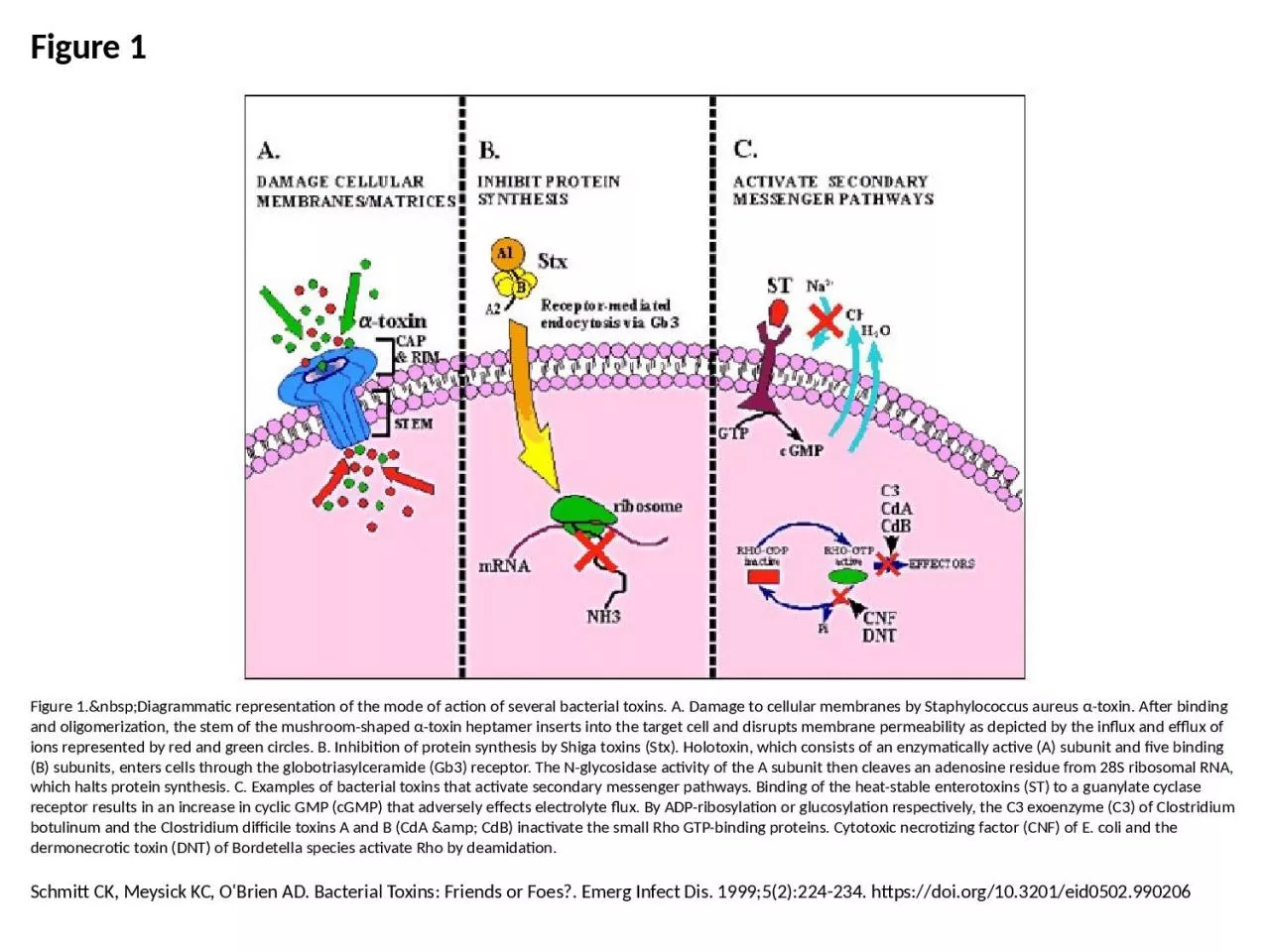
/
Figure 1 Figure 1. Diagrammatic representation of the mode of action of several Figure 1 Figure 1. Diagrammatic representation of the mode of action of several

Figure 1 Figure 1. Diagrammatic representation of the mode of action of several - PowerPoint Presentation

isla
isla . @isla
Follow
68 views
Uploaded On 2023-08-30

Figure 1 Figure 1. Diagrammatic representation of the mode of action of several - PPT Presentation

Schmitt CK Meysick KC OBrien AD Bacterial Toxins Friends or Foes Emerg Infect Dis 199952224234 httpsdoiorg103201eid0502990206 ID: 1014828

binding toxins amp bacterial toxins binding bacterial amp toxin subunit protein activate synthesis receptor rho clostridium

Share:

Link:

Embed:

Download Presentation from below link

Download Presentation The PPT/PDF document "Figure 1 Figure 1. Diagrammatic..." is the property of its rightful owner. Permission is granted to download and print the materials on this web site for personal, non-commercial use only, and to display it on your personal computer provided you do not modify the materials and that you retain all copyright notices contained in the materials. By downloading content from our website, you accept the terms of this agreement.


Presentation Transcript

1. Figure 1Figure 1. Diagrammatic representation of the mode of action of several bacterial toxins. A. Damage to cellular membranes by Staphylococcus aureus α-toxin. After binding and oligomerization, the stem of the mushroom-shaped α-toxin heptamer inserts into the target cell and disrupts membrane permeability as depicted by the influx and efflux of ions represented by red and green circles. B. Inhibition of protein synthesis by Shiga toxins (Stx). Holotoxin, which consists of an enzymatically active (A) subunit and five binding (B) subunits, enters cells through the globotriasylceramide (Gb3) receptor. The N-glycosidase activity of the A subunit then cleaves an adenosine residue from 28S ribosomal RNA, which halts protein synthesis. C. Examples of bacterial toxins that activate secondary messenger pathways. Binding of the heat-stable enterotoxins (ST) to a guanylate cyclase receptor results in an increase in cyclic GMP (cGMP) that adversely effects electrolyte flux. By ADP-ribosylation or glucosylation respectively, the C3 exoenzyme (C3) of Clostridium botulinum and the Clostridium difficile toxins A and B (CdA & CdB) inactivate the small Rho GTP-binding proteins. Cytotoxic necrotizing factor (CNF) of E. coli and the dermonecrotic toxin (DNT) of Bordetella species activate Rho by deamidation.Schmitt CK, Meysick KC, O'Brien AD. Bacterial Toxins: Friends or Foes?. Emerg Infect Dis. 1999;5(2):224-234. https://doi.org/10.3201/eid0502.990206