PPT-Surgical anatomy of the esophagus
Author : jaena | Published Date : 2023-07-09
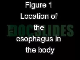
Asses Prof Dr Rafid majeed Esophagus The esophagus is the connecting tube between the pharynx and stomach that functions to transport food fluids and saliva
Presentation Embed Code
Download Presentation
Download Presentation The PPT/PDF document "Surgical anatomy of the esophagus" is the property of its rightful owner. Permission is granted to download and print the materials on this website for personal, non-commercial use only, and to display it on your personal computer provided you do not modify the materials and that you retain all copyright notices contained in the materials. By downloading content from our website, you accept the terms of this agreement.
Surgical anatomy of the esophagus: Transcript
Asses Prof Dr Rafid majeed Esophagus The esophagus is the connecting tube between the pharynx and stomach that functions to transport food fluids and saliva The 3 portion of the esophagus . John L. Dalrymple, MD. Division Director, Gynecologic Oncology. Department of Obstetrics and Gynecology. UT Southwestern Austin Programs. I have nothing to disclose.. Objectives. Describe basic abdominal and pelvic anatomy . or ambulatory surgical centers for calendar year 2016. The Congress should also require ambulatory surgical Report to the Congress: Medicare Payment Policy Ambulatory surgical center servicesChapte – . II. For undergraduate. Staff Members of Cardio-thoracic Surgery Departments. Egypt. HIATUS HERNIA. Definition. :. Protrusion of any part of the stomach through the esophageal hiatus into the thorax. . Alan Chu. March 13, 2013. Anatomy. 18 – 26cm from UES to LES. Esophageal wall layers. Mucosa, . submucosa. , . muscularis. . propia. , adventitia. Proximal 33% skeletal muscle, middle 35-40% mixed, distal 50-60% smooth muscle. La gamme de thé MORPHEE vise toute générations recherchant le sommeil paisible tant désiré et non procuré par tout types de médicaments. Essentiellement composé de feuille de morphine, ce thé vous assurera d’un rétablissement digne d’un voyage sur . 2017 - 18 1651 ). Posted: April 2017. 2017 - 2018 PROGRAM REQUIREMENTS D egree : Associate of Applied Science Major: Surgical Technology About This Major . . . The Associates of Applied Science Su Barretts Esophagus Introduction Barretts esophagus is a condition in which columnar cells replace the usual squamous cell in the of theesophagus The condition is recognized as a complication o Corrosive stricture. &. Perforation of Esophagus . Dr . Saurabh. . Pathak. Professor. Dept. of Surgery. The primitive foregut forms during the fourth week of gestation by a longitudinal folding and incorporation of the dorsal part of the yolk sac into the embryo.. Esophageal Cancer: Introduction The incidence of esophageal cancer is on the rise with over 12,000 Americans developing this disease each year(Figure 2). Variations in the incidence of esoph MANAGING FAILED ANTI-REFLUX THERAPYpH monitoring while the patient is taking med-ications,may help identify which patients aremore likely to respond to surgery.Patients with Airway Manifestations of G DEFINITION. Is a radiologic examination of the Upper GI tract. It consists of a series of X-ray images of the esophagus, stomach and duodenum by . usinng. C.M. Anatomy:. When the stomach is empty. The internal lining is thrown into numerous longitudinal folds called . Congenital. Infectious. Traumatic. Inflammatory. Perforation. Diverticula. Narrowing. Motility disorders. Neoplasms. Miscellaneous. Tracheoesophageal fistula. Esophageal Atresia. Stricture. Dysphagia lusoria. Rafid. . Majeed. . Naeem. . Indication. 1. foreign . bodies (potatoes, turnips…. ect. ) in horses and cattle lodged in the cervical part of the esophagus…….in dogs and cats bones are lodged in the cervical and thoracic parts of the... Books. Part A:. Pastest. MRCS Part A: Essential Revision Notes Book 1 & 2. Part B:. DrExam. Part B MRCS OSCE Revision Guide Books 1 & 2. Kanani. , Surgical Critical Care . Vivas. Lowe, Surgical Pathology Revision.
Download Document
Here is the link to download the presentation.
"Surgical anatomy of the esophagus"The content belongs to its owner. You may download and print it for personal use, without modification, and keep all copyright notices. By downloading, you agree to these terms.
Related Documents