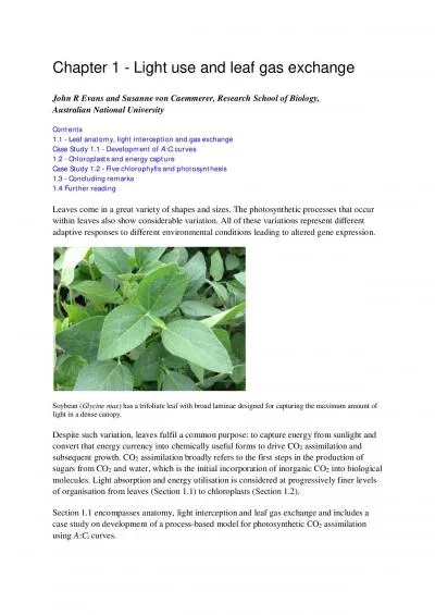
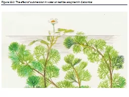
/
Figure 35.0  The effect of submersion in water on leaf deve Figure 35.0  The effect of submersion in water on leaf deve

Figure 35.0 The effect of submersion in water on leaf deve - PowerPoint Presentation

jane-oiler
jane-oiler . @jane-oiler
Follow
391 views
Uploaded On 2016-09-07

Figure 35.0 The effect of submersion in water on leaf deve - PPT Presentation

Cabomba Figure 350x The effect of wind on plant form in fir trees Figure 352 Morphology of a flowering plant an overview Figure 351 A comparison of monocots and dicots Figure 353 Radish root hairs ID: 462060

plant figure layer plants figure plant plants layer growth water leaf transport overview soil left secondary cells stem phloem

Share:

Link:

Embed:

Download Presentation from below link

Download Presentation The PPT/PDF document "Figure 35.0 The effect of submersion in..." is the property of its rightful owner. Permission is granted to download and print the materials on this web site for personal, non-commercial use only, and to display it on your personal computer provided you do not modify the materials and that you retain all copyright notices contained in the materials. By downloading content from our website, you accept the terms of this agreement.


Presentation Transcript

Slide1

Figure 35.0 The effect of submersion in water on leaf development in CabombaSlide2

Figure 35.0x The effect of wind on plant form in fir treesSlide3

Figure 35.2 Morphology of a flowering plant: an overviewSlide4

Figure 35.1 A comparison of monocots and dicotsSlide5

Figure 35.3 Radish root hairsSlide6

Figure 35.4 Modified shoots: Stolons, strawberry (top left); rhizomes, iris (top right); tubers, potato

(bottom left); bulb, onion (bottom right)Slide7

Figure 35.5 Simple versus compound leavesSlide8

Figure 35.6 Modified leaves: Tendrils, pea plant (top left); spines, cacti (top right); succulent (bottom left); brightly-colored leaves, poinsettia (bottom right)Slide9

Figure 35.6x Lithops, a stone-mimicking plant from South African desertsSlide10

Figure 35.7 The three tissue systemsSlide11

Figure 35.8 Water-conducting cells of xylemSlide12

Figure 35.9 Food-conducting cells of the phloemSlide13

Figure 35.10 Review of general plant cell structureSlide14

Figure 35.11 The three major categories of plant cellsSlide15

Figure 35.12 Locations of major meristems: an overview of plant growthSlide16

Figure 35.13 Morphology of a winter twigSlide17

Figure 36.18 Tapping phloem sap with the help of an aphidSlide18

Figure 35.14 Primary growth of a rootSlide19

Figure 35.15 Organization of primary tissues in young rootsSlide20

Figure 35.16 The formation of lateral rootsSlide21

Figure 35.17 The terminal bud and primary growth of a shootSlide22

Figure 35.18 Organization of primary tissues in young stemsSlide23

Figure 35.19 Leaf anatomySlide24

Figure 35.20 Production of secondary xylem and phloem by the vascular cambiumSlide25

Figure 35.21 Secondary growth of a stem (Layer 1)Slide26

Figure 35.21 Secondary growth of a stem (Layer 2)Slide27

Figure 35.21 Secondary growth of a stem (Layer 3)Slide28

Figure 35.22 Anatomy of a three-year-old stemSlide29

Figure 35.22x Secondary growth of a stemSlide30

Figure 35.23 Anatomy of a tree trunkSlide31

Figure 35.24 A summary of primary and secondary growth in a woody stemSlide32

Figure 36.0 Eucalyptus treesSlide33

Figure 36.0x TreesSlide34

Figure 36.1 An overview of transport in whole plants (Layer 1)Slide35

Figure 36.1 An overview of transport in whole plants (Layer 2)Slide36

Figure 36.1 An overview of transport in whole plants (Layer 3)Slide37

Figure 36.1 An overview of transport in whole plants (Layer 4)Slide38

Figure 36.2 A chemiosmotic model of solute transport in plant cellsSlide39

Figure 36.3 Water potential and water movement: a mechanical modelSlide40

Figure 36.4 Water relations of plant cellsSlide41

Figure 36.5 A watered tomato plant regains its turgorSlide42

Figure 36.6 Compartments of plant cells and tissues and routes for lateral transportSlide43

Figure 36.7 Lateral transport of minerals and water in rootsSlide44

Figure 36.8 Mycorrhizae, symbiotic associations of fungi and rootsSlide45

Figure 36.9 GuttationSlide46

Figure 36.12x Stomata on the underside of a leafSlide47

Figure 35.19 Leaf anatomySlide48

Figure 36.10 The generation of transpirational pull in a leafSlide49

Figure 36.11 Ascent of water in a treeSlide50

Figure 36.12 An open (left) and closed (right) stoma of a spider plant (Chlorophytum colosum) leafSlide51

Figure 36.13a The mechanism of stomatal opening and closingSlide52

Figure 36.13b The mechanism of stomatal opening and closingSlide53

Figure 36.13b The mechanism of stomatal opening and closingSlide54

Figure 36.14 A patch-clamp study of guard cell membranesSlide55

Figure 36.15 Structural adaptations of a xerophyte leafSlide56

Figure 36.15x Structural adaptations of a xerophyte leafSlide57

Figure 36.16 Loading of sucrose into phloemSlide58

Figure 36.17 Pressure flow in a sieve tubeSlide59

Figure 36.18 Tapping phloem sap with the help of an aphidSlide60

Figure 35.25 The proportion of Arabidopsis genes in different functional categoriesSlide61

Figure 37.0 HyacinthSlide62

Figure 37.1 The uptake of nutrients by a plant: an overviewSlide63

Figure 37.2 Using hydroponic culture to identify essential nutrientsSlide64

Table 37.1 Essential Nutrients in PlantsSlide65

Figure 37.3 Magnesium deficiency in a tomato plantSlide66

Figure 37.4 Hydroponic farmingSlide67

Figure 37.5 Soil horizonsSlide68

Figure 37.6 The availability of soil water and mineralsSlide69

Figure 37.7 Poor soil conservation has contributed to ecological disasters such as the Dust BowlSlide70

Figure 37.8 Contour tillageSlide71

Figure 37.9 The role of soil bacteria in the nitrogen nutrition of plants (Layer 1)Slide72

Figure 37.9 The role of soil bacteria in the nitrogen nutrition of plants (Layer 2)Slide73

Figure 37.9 The role of soil bacteria in the nitrogen nutrition of plants (Layer 3)Slide74

Figure 37.10 Root nodules on legumesSlide75

Figure 37.10x NodulesSlide76

Figure 37.11 Development of a soybean root noduleSlide77

Figure 37.12 Crop rotation and “green manure”Slide78

Figure 37.13 Molecular biology of root nodule formationSlide79

Figure 37.14 MycorrhizaeSlide80

Figure 37.15a Parasitic plants: Cross section of dodderSlide81

Figure 37.15b Parasitic plants: Indian pipeSlide82

Figure 37.16 Carnivorous plants: Venus fly trap (left), pitcher plant (right)Slide83

Figure 37.16x Sundew with fruit flySlide84

Figure 35.25x Arabidopsis thalianaSlide85

Figure 35.26 The plane and symmetry of cell division influence development of formSlide86

Figure 35.27 The preprophase band and the plane of cell divisionSlide87

Figure 35.28 The orientation of plant cell expansionSlide88

Figure 35.29 A hypothetical mechanism for how microtubules orient cellulose microfibrilsSlide89

Figure 35.30 The fass mutant of

Arabidopsis confirms the importance of cortical microtubules to plant growthSlide90

Figure 35.31 Establishment of axial polaritySlide91

Figure 35.32 Too much “volume” from a homeotic geneSlide92

Figure 35.33 Example of cellular differentiationSlide93

Figure 35.34 Phase change in the shoot system of EucalyptusSlide94

Figure 35.35 Organ identity genes and pattern formation in flower developmentSlide95

Figure 35.36 The ABC hypothesis for the functioning of organ identity genes in flower development