/
NATURE BIOTECHNOLOGY VOL  FEBRUARY  httpbiotech NATURE BIOTECHNOLOGY VOL  FEBRUARY  httpbiotech

NATURE BIOTECHNOLOGY VOL FEBRUARY httpbiotech - PDF document

jane-oiler
jane-oiler . @jane-oiler
Follow
459 views
Uploaded On 2015-03-08

NATURE BIOTECHNOLOGY VOL FEBRUARY httpbiotech - PPT Presentation

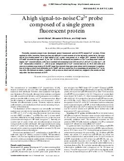
naturecom 137 The measurement of intracellular Ca 2 concentration Ca 2 became practical and common after chemically synthesized Ca 2 indicators were developed However these indicators have signifi cant limitations For example although these indicator ID: 42839

naturecom 137 The measurement

Share:

Link:

Embed:

Download Presentation from below link

Download Pdf The PPT/PDF document "NATURE BIOTECHNOLOGY VOL FEBRUARY http..." is the property of its rightful owner. Permission is granted to download and print the materials on this web site for personal, non-commercial use only, and to display it on your personal computer provided you do not modify the materials and that you retain all copyright notices contained in the materials. By downloading content from our website, you accept the terms of this agreement.


Presentation Transcript

cence increase in response to ATP was similar for G3 and G22(Table 1).G3 and G22 showed a marked fluorescence decrease at the begin-ning of excitation (Fig. 2B). Subsequently, both Caprobes recov-ered fluorescence after several tens of seconds in the dark, indicatingthat the fluorescence decrease results from photoisomerization, butnot from bleaching. This photoisomerization, however, did notoccur for a number of other cpEGFPs that we tested (data notshown). Comparison of amino acid sequences of these othercpEGFPs with those of G3 and G22 indicated that residue 148 mightbe responsible for the photoisomerization. To test this hypothesis,the connecting amino acid sequence between M13 andcpEGFP149Ð144 of G3, which contained residue 148, was changedto other amino acids (Table 1). Analysis of these mutants confirmedthe hypothesis that when residue 148 has a hydroxyl side chain (G3,G17, G22, G47, and G77), the probes exhibit photoisomerization.Because crystallographic analysis has shown that the side chain ofresidue 148 projects toward the interior of the GFP -barrel andinteracts with the fluorophore, hydrogen bonding between thehydroxyl side chains of residue 148 and Tyr66, which is a componentof the fluorophore, may be responsible for the photoisomerization.The photoisomerization we observed might relate to the on/offblinking of GFPs reported previouslyFrom our results and the earlier crystallographic study, we canpredict that other residues (Gln94, Arg96, and Glu222) may alsointeract with the fluorophore and modify GFP fluorescence intensity.Examination of residue 148 also gave us some information aboutthe responsiveness to Ca(Table 1). Whereas acidic side chains(aspartic acid in G52, glutamic acid in G18 and G79), and hydroxylside chains (serine in G3 and G17, threonine in G22, tyrosine inG47) were acceptable for function, basic side chains (lysine in G62,arginine in G19 and G75) abolished responsiveness to Ca. We alsofound that residue 147 is important. For example, G3 response toATP was about two times stronger than that of G17, although G3and G17 differ only at position 147 (threonine in G3, glycine inG17). Among the Caprobes so far tested, G18 produced the bestATP responses (Table 1).For further tuning of G18, we examined the effect of deletingglycine in the connecting sequence between cpEGFP and CaM (G72and G85). G72 and G85, which were identical except for the N-terminal sequences, showed Caresponsiveness similar to that ofG18 (Table 1), but G72 and G85 were much brighter than G18 (datanot shown). Because brighter probes are preferable to identifyexpressing cells, we chose G85 (hereafter named G-CaMP) for fur-ther characterization.Responses of G-CaMP to agonists are shown in Figure 2C. Incomparison with G3 (Fig. 2B), G-CaMP exhibited no photoisomer-ization and responded to agonists about three times better than G3.The responses of G-CaMP were also faster than those of G3, becauseinitial spikes recorded from G-CaMP were sharper than those of G3.Intracellular pH (pHi) was also measured (data not shown), becauseG-CaMP is sensitive to pH (see below). During the application ofATP or carbachol, there was no change in pHi (pHi 7.3), indicatingthat fluorescent changes responded not to pHi but to [Ca. In thepresence of 2 mM extracellular Ca, the addition of ionomycinincreased both [Ca(as seen by an increase in fluorescence) andpHi (from 7.3 to 8.0). The addition of ionomycin plus ethylenedi-amine tetraacetic acid (EDTA) in the absence of extracellular Caresulted in a marked decrease in both [Caand pHi (whichdropped from 8.0 to 7.0). After correction for the effects of pH, therewas a 4.3-fold difference between maximum and minimum fluores-cence. We estimated resting [Cato be between at 50 and 100 nM,and peak [Caduring ATP application at ~250 nM, in good agree-ment with previous studiesIn vitrocharacterization of the G-CaMP. Figure 3A shows fluo-rescence excitation and emission spectra of G-CaMP purified frombacteria. Excitation and emission maxima were 489 nm and 509 nm,respectively, similar to those of EGFP. Addition of Caincreasedfluorescence up to 4.5-fold. It is known that GFP forms homo-dimers at high protein concentrations. Gel filtration experiments athigh (54.3 M) and low (5.4 M) protein concentrations revealedno shift in apparent molecular weight as a function of protein con-centration, suggesting that G-CaMP exists as a monomer.Figure 3B shows the Catitration of G-CaMP, which was fittedwith a monophasic curve yielding an apparent for Caof 235 nMand a Hill coefficient of 3.3 (at 0.3 M protein concentration). Theapparent 30 times lower than that of camgaroo1 M; ref. 7). To determine whether the CaÐCaMÐM13 interaction NATURE BIOTECHNOLOGY VOL 19 FEBRUARY 2001http://biotech.nature.com Figure 1. Schematic representation of GFP-based Caprobes. (A) Allprobes consist of the M13 fragment from myosin light chainkinase (M13), a circularly permutated EGFP (cpEGFP) andcalmodulin (CaM) located N to C terminally. The start- and end-residue numbers are shown in parentheses. Linkers are indicatedabove the structures in one-letter amino acid code. (B) Schematicprobes. Figure 2. Expression of GFP-based Caprobes in HEK-293 cells. (A) HEK-293 cell expressing cytosolic G3. (B, C) Fluorescent changes inresponse to drugs for cells expressing G3 (B) and G-CaMP (C). Applications of 100 M ATP, 100 are indicated by shaded bars. ABC NATURE BIOTECHNOLOGY VOL 19 FEBRUARY 2001http://biotech.nature.comis intramolecular or intermolecular, Catitration was done at bothlow (0.3 M) and high (30 M) protein concentrations. Figure 3Brevealed that there was no concentration-dependent difference in theapparent of G-CaMP. In addition, gel filtration analysis revealedno shift in apparent molecular weight at 1 mM and zero Caconcen-trations. These results suggest that the CaÐCaMÐM13 interaction isintramolecular and that G-CaMP functions as a monomer.Figure 3C illustrates the pH sensitivity of G-CaMP. ApparentpKa values with and without Cawere 7.1 and 8.1, respectively.Figure 3D shows the Ca-binding kinetics of G-CaMP. The dissoci-ation time constant (200 ms) did not depend on [CaM are shown in Fig. 3D inset right), whereas the associationtime constant is faster at increasing [Ca]. Thus, these data indicatethat association kinetics of G-CaMP are fast enough at high [Ca&#x 10 ;&#xms f;&#xor [;쨀] 500 nM) to make the probe suitable for mon-itoring [Cain excitable cells.Application of the Caprobe to myotubes. We next tested G-CaMP in myotubes, in which [Carises quickly upon depolar-ization. Figure 4A illustrates that electrical stimulation elicited alocal depolarization-induced increase in [Cawithin a restrictedregion near the stimulating electrode (image 2), followed by a Cawave that propagated to an adjacent region (image 3). Carbacholand caffeine, which are known to increase [Cain myotubes, alsoinduced substantial increases in fluorescence (Fig. 4B). pHi did not Figure 3. characterization of G-CaMP. (A) Fluorescence excitation and emission spectra inthe presence of 1 mM Caor 5 mM ethyleneglycol tetracetic acid (EGTA). (B) CapH 7.5. Protein concentrations were 30 ). Apparent and Hill coefficient forM protein concentration were 234 nM and 3.4 or 235 nM and 3.3, respectively. (C) pHtitration of normalized fluorescence obtained in the presence of 100 ) or 5 mM EGTA (The data points in (B) and (C) represent the averages ( s.d.) of three independent experiments. (D)Stopped-flow fluorometry at pH 7.5. Association (inset left) and dissociation (inset right) kinetics ofbinding recorded in 1 . Fluorescence (F) is shown in arbitrary units. Data were fittedaccording to monoexponential curves. Table 1. Identity and fluorescent responses for GFP-based CaprobesProbeN-terminal Sequence cpEGFPSequence ATPnumbersequenceconnecting connecting response M13 with cpEGFP with ( cpEGFPCaMG3MGTTS149Ð144GTR0.728+++G17MGTGS149Ð144GTR0.311+++G18MGTLE149Ð144GTR1.678ÐG19MGTPR149Ð144GTR012ÐG41MGTTI149Ð144GTR018ÐG44MGTTP149Ð144GTR021ÐG46MGTTA149Ð144GTR012ÐG47MGTTY149Ð144GTR0.719++G49MGTTQ149Ð144GTR018ÐG50MGTTN149Ð144GTR018ÐG52MGTTD149Ð144GTR0.320ÐG54MGTTC149Ð144GTR0.321ÐG55MGTTW149Ð144GTR020ÐG56MGTTG149Ð144GTR020ÐG58MGTTV149Ð144GTR023ÐG61MGTTF149Ð144GTR0.522ÐG62MGTTK149Ð144GTR028ÐG72MGTLE149Ð144TR1.667ÐG75MVDTR149Ð144GTR029ÐG76MVDTM149Ð144GTR0.623ÐG77MVDTT149Ð144GTR0.110+++G79MVDTE149Ð144GTR0.526ÐG80MVDTH149Ð144GTR0.323+G81MVDTL149Ð144GTR0.68ÐG85(G-CaMP)MVDLE149Ð144TR1.516Ð G22MGTTS150Ð144GTR0.626+++Amino acids are indicated using one-letter code. Residues 147 and 148 of cpEGFP149Ð144 and residue 148 (threonine) of cpEGFP150Ð144 are included in thelinker sequences connecting M13 with cpEGFP.Number of cells tested.ABC change during the experiments (pHi 7.3; data not shown). Thedepolarization-induced increase in [Cawas further analyzed byconfocal line-scan imaging (Fig. 4C). The fluorescence increaseshowed an initial rapid phase and a subsequent slow phase of [Caincrease, followed by recovery to the basal Calevel. The rapidphase lasted 60 ms and [Careached its maximum within 200ms. The time course of the fluorescence increase measured with G-CaMP is similar to that previously found in myotubes with Fluo 3(ref. 24).In the work described here, we developed a high signal-to-noiseprobe based on a single GFP protein (G-CaMP). The improvedsignal over background ratio due to the increased affinity is a majoradvantage of G-CaMP. Although G-CaMP exhibits pH sensitivityand the requirement of folding at temperatures below 37¡C, Caresponses are sufficiently large and fast that G-CaMP should proveto be a powerful tool in studies of Cadynamics in excitable cells.On the other hand, accurate measurements of micromolar [Camight best be achieved using camgaroo1 because of its moderateaffinity and broad dynamic range. By introducing the cDNAwith specific promoters into animals and plants, expression of theprobe could be controlled spatially and temporally.Experimental protocolPlasmid construction. The original circular permutation of EGFP(cpEGFP145Ð144) was made using pEGFP-N1 (Clontech, Palo Alto, CA) byPCR. Amplification of cpEGFP149Ð144 and cpEGFP150Ð144 fromcpEGFP145Ð144 was by PCR, both of which had a SpeI site (encoding aminoacid residues Thr-Ser) at the 5end and GGG (encoding glycine) with a site (Thr-Arg) at the 3end. The complementary DNA (cDNA) encoding thechicken smooth muscle M13 (SSRRKWNKTGHAVRAIGRLSS; refs 5, 17),which was preceded by a I site, Kozak sequence (CGCCACC), startcodon (ATG), and a KpnI site (Gly-Thr), was connected to cpEGFP149Ð144SpeI site. The 3end of cpEGFP149Ð144 was connected to the PCRfragment encoding amino acid residues 2Ð148 of rat calmodulin (pRCaM;ref. 18) at the I site. The calmodulin sequence was followed by a stopcodon and a NotI site. The entire coding sequence was inserted into the NotI sites of pEGFP-N1 to yield pN1-G3. We used PCR to make pN1-G17, 18, 19 or 21, and replaced the connecting sequence between M13 andcpEGFP149Ð144 of pN1-G3 (the SpeI site) by BamI (Leu-II (Pro-Arg), or I (Arg-Ser) site, respectively (G21 did not pro-duce fluorescence.) pN1-G22 includes cpEGFP150Ð144 instead ofcpEGFP149Ð144. In pN1-G72, the GGG (glycine) sequence preceding theI site of pN1-G18 was deleted. For pN1-G75, 76, 77, 79, 80, 81, and 85,KpnI sites before and after the start codon of pN1-G72weresubstituted by BglI (Val-Asp) sites, respectively. Site-directed muta-tions were also done by PCR.Bacterial expression and in vitrofluorescence measurements. NotI cDNA fragment of pN1-G85 was inserted into pET23d(+) orpET32a (Novagen, Madison, WI), resulting in pET23d-G85 or pET32a-G85,respectively. pET32a-G85 produced a construct that possessed a polyhisti-dine tag at the N terminus, whereas pET23d-G85 resulted in a nontaggedconstruct. There was no difference in behavior with and without the tag.Escherichia coliBL21(DE3)pLysS transformed with the plasmids were cul-tured at 28¡C for 4 h after protein induction with 1 mM isopropyl-thiogalactoside (IPTG). Cells were lysed by freezing (for 30 min at Ð20¡C)and thawing (for 30 min at room temperature) three times and suspendedwith a solution of 25 mM Tris-HCl (pH 8), 1 mM -mercaptoethanol, andprotease inhibitors. After centrifugation the supernatant was dialyzed againstKM buffer containing (in mM) 100 KCl and 20 MOPS (pH 7.5). A NiNTAresin (Qiagen, Hilden, Germany) was used for purification of the tagged pro-tein. Gel filtration was performed using Sephadex G-100.Excitation spectra (detected at 520 nm) and emission spectra (excited at470 nm) were taken with the fluorescent spectrophotometer F4500 (Hitachi,Tokyo, Japan). For Catitration experiments, free Caconcentrations with20 mM BAPTA were calculated using MaxChelator program. The fluores-cence excited at 480 nm was monitored at 510 nm. Data were fit according tothe Hill equation. For pH titration experiments at 1.1 M protein concen-tration, MOPS (for pH 6 to 8) in KM buffer was replaced with either citrate(for pH 5 to 6) or glycine (for pH 8 to10). Stopped-flow fluorometry RSP-601 (Unisoku, Osaka, Japan) was done at 26¡C. Fluorescence at pH 7.5 wasexcited at 488 nm and emission was detected at 510 nm. Concentrationsquoted are those before 1:1 mixing. Mixing dead time was 1 ms. Data wereaveraged at least three times. For measurements of association kinetics, thepurified protein (30 M) in 2 mM BAPTAwas mixed with various amountof Ca. Identical results were obtained using 5 mM BAPTA (data notshown). For measurements of the dissociation kinetics, the purified proteinM) in 1 mM BAPTA with 1, 10, or 50 M free Cawas mixed with 17mM BAPTA. NATURE BIOTECHNOLOGY VOL 19 FEBRUARY 2001http://biotech.nature.com Figure 4. Expression of G-CaMP in myotubes. (A and B) Lower panelsshow the time course of fluorescent signal from a region indicated bya black oval in each image 1. Images were taken at the indicatedstimulation (st) induced an increase in [Caelectrode was placed in the right side of the middle portion of the(CCH)- and 10 mM caffeine-induced increases in [Camonitoredwith G-CaMP. The relatively slow onset of the responses was due toesponses was due to2+]iincrease. The lower panel shows the time course of thechange in fluorescence of the above image. Extracellular electricalstimulation (st) was delivered at the indicated time. ABC NATURE BIOTECHNOLOGY VOL 19 FEBRUARY 2001http://biotech.nature.comFluorescence measurements in HEK cells. HEK-293 cells were culturedand transfected with the plasmids as described. Cells were incubated at 28¡Cfor two to four days before testing. Fluorescence excited at 488 nm was detect-ed at 525 25 nm with a confocal laser scanning microscope (interval time 2 s). A solution containing (in mM) 135 NaCl, 5.4 KCl, 2 CaCl10 glucose, 5 HEPES (pH 7.4) or HEPES-buffered salineexperiments.We estimated the physiological pHi to be 7.3, with pHi 8.0 and pHi 7.0corresponding to the maximum fluorescence () and minimum fluores-cence (), respectively, as described elsewhere. For estimation of [Cafluorescent signals were corrected for pHi using the pH titration curves inFigure 3C. pCa was subsequently calculated using the following equation:ing equation:(Fmax,corrmin,corr, where is the Hill coef-ficient obtained from Figure 3B, max,corrmin,corrare the maximum andminimum fluorescence values corresponding to pH 7.3, respectively, experimental fluorescence, and is the apparent dissociation constantobtained from Figure 3B.Fluorescence measurements in myotubes. Preparation of mousemyotubes (BALB/c) was as described. After introduction of cDNA intocells, they were cultured at 28¡C for an additional two to four days beforetesting. Extracellular electrical stimulation was done as describedFluorescent images were recorded with the confocal microscope. For line-scan images, a scan speed of 4.5 ms/line was used and the raw data were sub-sequently filtered using a box filter.We thank Masayuki Mori for the rat calmodulin cDNA and Michiyo Murataand Mitsutoshi Ono for technical assistance. We also thank ToshihikoNagamura and Tatsuo Nakagawa of Unisoku Co., Ltd. for technical assistance.The work was supported by grants from the Ministry of Education, Science,Sports and Culture, by "the Research for the Future Program" of the JSPS, andby the JSPS Research Fellowships for Young Scientists. 1.Inouye, S. Cloning and sequence analysis of cDNA for the luminescentprotein aequorin. 2.Prasher, D.C., Eckenrode, V.K., Ward, W.W., Prendergast, F.G. & Cormier, M.J.Primary structure of the Aequorea victoriagreen-fluorescent protein. 3.Miyawaki, A. Fluorescent indicators for Ca based on green fluorescentproteins and calmodulin. Nature 4.Miyawaki, A., Griesbeck, O., Heim, R. & Tsien, R.Y. Dynamic and quantitativemeasurements using improved cameleons. 5.Romoser, V.A., Hinkle, P.M. & Persechini, A. Detection in living cells of Cadependent changes in the fluorescence emission of an indicator composed oftwo green fluorescent protein variants linked by a calmodulin-binding sequence.A new class of fluorescent indicators. 6.Persechini, A., Lynch, J.A. & Romoser, V.A. Novel fluorescent indicator proteinsfor monitoring free intracellular Ca7.Baird, G.S., Zacharias, D.A. & Tsien, R.Y. Circular permutation and receptorinsertion within green fluorescent proteins. 8.Baubet, V. Chimeric green fluorescent proteinÐaequorin as bioluminescentreporters at the single-cell level. 9.Allen, G.J. Cameleon calcium indicator reports cytoplasmic calciumguard cells. 10.Emmanouilidou, E. concentration changes at the secretoryvesicle surface with a recombinant targeted cameleon. Curr. Biol.11.Fan, G.Y. Video-rate scanning two-photon excitation fluorescencemicroscopy and ratio imaging with cameleons. 12.Jaconi, M. Inositol 1,4,5-trisphosphate directs Cachondria and the endoplasmic/sarcoplasmic reticulum: a role in regulating car-13.Foyouzi-Youssefi, R. Bcl-2 decreases the free Caconcentration within theendoplasmic reticulum. 14.Yu, R. & Hinkle, P.M. Rapid turnover of calcium in the endoplasmic reticulum dur-ing signaling: studies with cameleon calcium indicators. 15.Kerr, R. Optical imaging of calcium transients in neurons and pharyngeal16.Allen, G.J. Alteration of stimulus-specific guard cell calcium oscillations andArabidopsisdet317.Rhoads, A.R. & Friedberg, F. Sequence motifs for calmodulin recognition. FASEB18.Mori, M. Novel interaction of the voltage-dependent sodium channel(VDSC) with calmodulin: does VDSC acquire calmodulin-mediated CaBiochemistry19.Bischof, G., Serwold, T.F. & Machen, T.E. Does nitric oxide regulate capacitative20.Yang, F., Moss, L.G. & Phillips, G.N. Jr. The molecular structure of green fluores-cent protein. 21.Dickson, R.M., Cubitt, A.B., Tsien, R.Y. & Moerner, W.E. On/off blinking andswitching behavior of single molecules of green fluorescent protein. Nature22.Okada, T. sient receptor potential protein homologue TRP7. 23.Cormack, B.P., Valdivia, R.H. & Falkow, S. FACS-optimized mutants of the greenfluorescent protein (GFP). 24.Nakai, J. Functional nonequality of the cardiac and skeletal ryanodinereceptors. 25.Bers, D.M., Patton, C.W. & Nuccitelli, R. A practical guide to the preparation ofbuffers. 26.James-Kracke, M.R. Quick and accurate method to convert BCECF fluores-cence to pHi: calibration in three different types of cell preparations. 27.Tanabe, T., Beam, K.G., Powell, J.A. & Numa, S. Restoration of excitationÐcon-traction coupling and slow calcium current in dysgenic muscle by dihydropyri-dine receptor complementary DNA. Nature NATURE BIOTECHNOLOGY VOL 19 FEBRUARY 2001http://biotech.nature.comThe measurement of intracellular Caconcentration, [Cabecame practical and common after chemically synthesized Caindicators were developed. However, these indicators have signifi-cant limitations. For example, although these indicators are loadedinto cells as acetoxymethyl esters, such loading does not enable tar-geting to specific cells. A means of Cameasurement that allowsgenetic targeting to specific cells is the use of aequorin ©2001 Nature Publishing Group http://biotech.nature.com ©2001 Nature Publishing Group http://biotech.nature.com