/
NOTES: Skeletal System NOTES: Skeletal System

NOTES: Skeletal System - PowerPoint Presentation

jane-oiler
jane-oiler . @jane-oiler
Follow
357 views
Uploaded On 2020-01-05

NOTES: Skeletal System - PPT Presentation

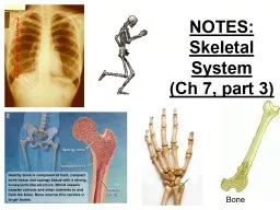
NOTES Skeletal System Ch 7 part 3 BONE FUNCTION Support and Protection bones shape and form body structures bones support and protect softer underlying tissues BONE FUNCTION ID: 772012

Share:

Link:

Embed:

Download Presentation from below link

Download Presentation The PPT/PDF document "NOTES: Skeletal System" is the property of its rightful owner. Permission is granted to download and print the materials on this web site for personal, non-commercial use only, and to display it on your personal computer provided you do not modify the materials and that you retain all copyright notices contained in the materials. By downloading content from our website, you accept the terms of this agreement.


Presentation Transcript

NOTES: Skeletal System(Ch 7, part 3)

BONE FUNCTION:  Support and Protection • bones shape and form body structures • bones support and protect softer, underlying tissues

BONE FUNCTION:  Body Movement • bones and muscles function together as LEVERS • a lever consists of: a rod, a pivot (fulcrum), a resistance, and a force that supplies the energy

BONE FUNCTION:  Blood Cell Formation (Hematopoeisis) • depending on stage of life, blood cell formation occurs in the : -yolk sac (embryo) -liver and spleen -bone marrow

• Marrow can be: -RED : makes RBCs, WBCs, and platelets -YELLOW: stores fatRatio changes over lifetime Child: all red marrowIncrease in yellow over lifetime

BONE FUNCTION:  Storage of Inorganic Salts • matrix of bone tissue contains large quantities of calcium phosphate ( makes bones hard) • bone also stores small amounts of magnesium, sodium, potassium, and carbonate

Individual bones are the organs of the skeletal system. A bone contains very active tissues.

Some of the cells that make up these tissues: Osteoprogenitor cells: unspecialized; can undergo mitosis & develop into osteoblastsOsteoblasts: cells that form bone matrix (no longer dividing); secrete collagen

OSTEOPROGENITOR  OSTEOBLAST 

Osteocytes: mature bone cells; exchange nutrients/waste with blood Osteoclasts: break down bone matrix OSTEOPROGENITOR  OSTEOBLAST  OSTEOCYTE

Throughout life, osteoclasts continually break down bone matrix and osteo blasts replace it; these opposing processes of resorption and deposition of calcium help to maintain calcium levels in the body.

Blood calcium levels vary; when blood calcium is: LOW: parathyroid hormone is released which causes osteoclasts to break down bone, releasing calcium salts HIGH : calcitonin is released which causes osteoblasts to form bone tissue and store calcium salts

CALCITONIN PARATHYROID HORMONE

Bones are classified by SHAPE: Long bonesFlat Irregular (ex: zygomatic, hyoid)Short (ex: carpals/tarsals)

BONE STRUCTURE:*Bone structure reflects its function.

Parts of a long bone: • EPIPHYSES: -enlarged portions at ends of a long bone;-covered with articular cartilage; -articulate (form JOINTS) with other bones.

Parts of a long bone: • DIAPHYSIS: -shaft of bone (located between the epiphyses)• PERIOSTEUM: -tough, vascular covering that encloses the entire bone except where the articular cartilage is Articular cartilage

**the center of the diaphysis is a hollow chamber ( MEDULLARY CAVITY), lined with a thin layer of cells ENDOSTEUM) and filled with soft connective tissue (YELLOW MARROW)

Types of Bone Tissue:  COMPACT BONE: has a continuous matrix with no gaps; found in the wall of the diaphysis

Types of Bone Tissue:  SPONGY BONE (a.k.a. cancellous bone): has irregular interconnecting spaces between bony plates that reduce the weight of bone.

**both compact and spongy bone are strong and resist bending

NOTES- Skeletal System Part 4: Microscopic Structure of Bone, Bone Development & Growth, & Bone Remodeling

Microscopic Structure of Bone: • Intercellular material = mostly COLLAGEN (gives bone its strength and elasticity) and inorganic salts (make bone hard and resistant to crushing). • The structural unit of compact bone is called the Osteon or Haversian System• Compact bone contains OSTEONS running in cylindrical rings cemented together which give long bones strength Transverse cut

In an osteon you would find…Inorganic matrix deposited in concentric layers called LAMELLAE Lamellae form concentric rings around a central or Haversian CanalContain nerves and blood vessels that supply osteocytesVolkmann’s Canals run at a right angle tocentral canals and connect the vascular and nerve supplies to the periosteum

In each lamella there are cavities called Lacunae which hold Osteocytes (mature bone cells)Lacunae also have tiny canals radiating out from them called CANALICULINutrients diffuse through these to reach osteocyte

AMGEN BONE VIDEO- CLICK!

Bone Development and Growth: Process by which bone forms is called ossificationBone starts out as hyaline cartilage in the right shape & then becomes calcified & gains a blood supply2 types: 1) Intramembranous Ossification(will become flat bones) 2) Endochondral Ossification

 Intramembranous Ossification • osteoblasts completely surrounded by matrix are mature OSTEOCYTES EXAMPLE: the broad, flat bones of the skull form in this way and fuse together at sutures.

Intramembranous: “within” “membrane”

Bone Development and Growth Endochondral Ossification: (“chondro” = cartilage) • Most bones in the body develop in this way • Bones develop in early embryos as hyaline cartilage “models”; later replaced by bone tissue 

endochondral: “within” “cartilage”

Endochondral Ossification • In the middle of the diaphysis, OSTEOCLASTS break down bone tissue and the resulting space becomes the medullary cavity, which later fills with marrow • The bone in the central regions of the epiphyses and diaphysis remain spongy bone • The hyaline cartilage that remains on the ends of the epiphyses persists throughout life as articular cartilage

Endochondral Bones • An epiphyseal plate remains between the primary and secondary ossification centers • Long bones continue to lengthen until the epiphyseal plate are ossified (hardened) in adolescence • A developing long bone thickens as compact bone is deposited beneath the periosteum ( appositional growth )

Growth occurs at the epiphyseal plate

Factors Necessary for Bone Growth: Growth hormoneGH: stimulate cartilage cells at epiphyseal plateThyroid hormoneTH: replace cartilage at plate; needed for GH Sex hormonesestrogens & androgens promote bone formationVitamins A & CVit. A: osteoclast/osteoblast activityVit. C: collagen synthesis **Genetics & nutrition play a role

Bone Remodeling: Bone continually renews itself Takes place at different rates in various body regionsDistal portion of femur replaced every 4 monthsOsteoclasts destroy matrix (resorption)Osteoblasts deposit new bone Approximately 20% of bone tissue is replaced annually by this process on a cyclical basis throughout the skeleton. The entire remodeling process occurs over approximately 4 to 8 months, with a range of 3 months to 2 years.

Osteoclasts are transported to the bone. The osteoclast cells release acids and enzymes that resorb the existing bone structure. After the osteoclasts resorb the bone, osteoblasts appear and coat the resorbed area with adhesive substances. Moreover, osteoblast cells produce bone proteins, such as collagen, to help calcium attach to the bone proteins. Only by attaching calcium to bone proteins, the new-bone can be formed.