/
Orbital Orbital

Orbital - PowerPoint Presentation

jane-oiler
jane-oiler . @jane-oiler
Follow
428 views
Uploaded On 2016-10-07

Orbital - PPT Presentation

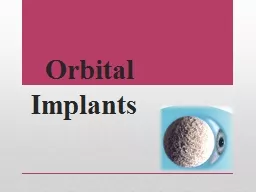
Implants Prior to 1885 orbital implants were not used The eye was removed by enucleation or evisceration and the socket was left to heal in its own The result was an unsightly sunken depression of the eyelids into the eye socket The use of an orbital implant was a major breakthrough in ID: 472577

eye implant orbital peg implant eye peg orbital porous socket hydroxyapatite tissue artificial implants infection sleeve fat sunken lid

Share:

Link:

Embed:

Download Presentation from below link

Download Presentation The PPT/PDF document "Orbital" is the property of its rightful owner. Permission is granted to download and print the materials on this web site for personal, non-commercial use only, and to display it on your personal computer provided you do not modify the materials and that you retain all copyright notices contained in the materials. By downloading content from our website, you accept the terms of this agreement.


Presentation Transcript

Slide1

Orbital ImplantsSlide2

Prior to 1885 orbital implants were not used. The eye was removed by

enucleation or evisceration and the socket was left to heal in its own. The result was an unsightly sunken depression of the eyelids into the eye socket. The use of an orbital implant was a major breakthrough in

anophthalmic

socket surgery. The implant improved postoperative cosmetic by filling orbital volume and also reducing the chance of socket contractions due to scar tissue formation

.

Over the last 100 years a variety of materials have been used for the orbital implant including: gold, silver, cartilage, bone, fat, cork, sponge, rubber, paraffin, wool, asbestos, as well as a variety of others in an attempt to find the most biocompatible

implant . Slide3

A variety of shapes and sizes have also been tried in an attempt to promote some motility to the socket. In 1985 a new concept in eye socket implants began to evolve when a researcher (Dr. Arthur Perry, San Diego, CA) began to study sea coral as an ocular implant. Through a patented hydrothermal process the calcium carbonate component of sea coral was turned into calcium phosphate and a substance known as hydroxyapatite was made. Hydroxyapatite has the same chemical structure and porous structure as human body tissue

.

Hydroxyapatite

orbital

implant

→Slide4

The implant material (

corralline hydroxyapatite) is biocompatible, non-toxic and non-allergenic. The body’s tissue recognizes the material as similar and because of the porous nature, tissue will grow into it. The implant becomes more fixed and therefore resists migration. The implant allows attachment of the

extraocular

muscles which in turn leads to improved orbital implant motility. The orbital implant can also be directly attached to the prosthesis through a peg, protruding from the implant (Figure 4b) allowing a wide range of prosthetic movement as well as the darting eye movements commonly seen when people are engaged in conversation. The increased range and fine darting movements allow a more life-like quality to the prosthetic eye.

The hydroxyapatite implant, also known as the Bio-Eye™ (integrated Orbital implants, San Diego) is called a “porous implant.” Since its introduction, a number of other “porous implants” have been introduced.Slide5

Another synthetic porous implant that has become increasingly popular is a type of porous plastic known as Porous Polyethylene (

Medpor

™ -

Porex

Surgical

Inc

, Cooledge Park, GA). This material is a synthetic man-made material that previously has been used in a wide range of cranio-facial reconstructive procedures and facial fractures, with few problems. Its properties demonstrate high tensile strength, malleability, biocompatibility and

fibrovascular

ingrowth. The porous polyethylene orbital implants are also cheaper than the original Bio-Eye™, and are available in spherical, egg, conical or mounded shapes

-Aluminum oxide (Al2O3) another man-made biomaterial that has been in use for more than 30 years as an implant in orthopedics and dentistry has also been studied extensively (Dr. D.R. Jordan - University of Ottawa Eye Institute, Ottawa, Ontario, Canada) and is emerging as an orbital implant in Canada, Europe and several other parts of the world. Slide6

The aluminum oxide implant, also known as the

Bioceramic Implant (FCI, Issy-Les-Moulineaux

,

Cedex

, France) looks identical to the Bio-Eye™ hydroxyapatite with multiple interconnected pores. Like the synthetic HA and porous polyethylene, it is less expensive than the Bio-Eye™. There is evidence that human osteoblasts and fibroblasts appear to grow better on aluminum oxide than hydroxyapatite, suggesting it may be more biocompatible than hydroxyapatite and better tolerated in the eye socket.Slide7

 Drawing of hydroxyapatite implant with muscles attached and a peg in

position .

 Synthetic hydroxyapatite implant.

Spherical

porous polyethylene.Slide8

Orbital Implant ComplicationsSlide9

Complications associated with hydroxyapatite that also apply to other porous orbital implants have gradually come to light since the introduction and widespread use of hydroxyapatite in the early 1990’s. Reported complications include: implant exposure,

conjunctival thinning, discharge, pyogenic granuloma formation (excess healing tissue formation), and rarely persistent pain or discomfort. The complication discussed most often is implant exposure with exposure rates ranging from 0 to 22% Slide10

Predisposing factors to exposure include: wound closure under tension, inadequate or poor wound closure technique, infection, mechanical or inflammatory irritation from the speculated surface of the HA implant and delayed ingrowth of

fibrovascular

tissue with subsequent tissue breakdown.

The most feared complication of porous orbital implants is infection within the implant. Porous orbital implants have multiple interconnected pores that fill with

fibrovascular

tissue over the first 6-12 months which theoretically should help resist infection. Prior to this time implant exposure can predispose the implant to entry of bacterial contamination and implant infection. Once an implant infection does occur, it may not be easy to recognize or treat. Hallmarks of implant infection are, recurrent discharge resistant to multiple drops, implant discomfort (to touch), and recurrent pyogenic granuloma (excess healing tissue) on the surface of the implant

.Slide11

Orbital

implant infection - the eye socket is very inflamed, there is lots of discharge and a recurrent pyogenic granuloma (black arrows).

 Exposure of a hydroxyapatite implant.Slide12

Implant infection does not respond to oral, intravenous and/or topical antibiotics and generally the implant has to be removed. Removal of an infected implant requires general

anaesthesia

and is traumatic to the eye socket as the implant has been partially integrated with the socket tissue. Following removal of the porous orbital implant it is not advisable to have another porous implant until a sufficient length of time (at least 6 months) has past to ensure tissue healing. A plastic nonporous sphere can be put in to maintain volume while waiting out the 6-month period and contemplating another porous implant.Slide13

Artificial Eye Motility : Implant Pegging ProceduresSlide14

a) 

To peg or not to peg? One of the many advantages of porous implants (hydroxyapatite, porous polyethylene, aluminum oxide) is the ability to integrate them with the overlying artificial eye through a peg system. By coupling the orbital implant to the artificial eye a wide range of prosthetic eye movements as well as darting eye movements commonly seen in conversational speech can occur. These movements impart a more life-like quality to the prosthetic eye. To peg or not to peg, is up to the surgeon and patient. They are certainly not for everyone and with all due respect, not all eye socket surgeons are equally skilled at putting them in. Before considering a peg the implant has to be fully vascularized (minimum of 6 months, in some this may take a year or more) and, the socket has to be a healthy one. Patients with diabetes, previous radiation, systemic disease such as Systemic Lupus

Erythematosis

(SLE) or, individuals on medications such as steroids, are not good candidates for pegging, because their socket tissue simply does not have good blood flow.Slide15

Pegs are not fool proof and do have their own inherent set of problems above and beyond those of the implant. A meticulous peg placement technique is required to obtain excellent results; a fact not appreciated by many. The pegs must be central and straight. They must also be flush with the implant with no exposed portion to ensure a good result. Once pegged, the individuals do require regular follow up initially to be sure the peg is sitting well. With time the follow-ups can decrease (yearly) if all is well. The most worrisome problem associated with pegging is introduction of infection to the implant, requiring implant removal.Slide16

Other potential peg problems include discharge, pyogenic granuloma (excess healing tissue) around the peg, peg falling out, poor transfer of movement, clicking,

conjunctival overgrowth, poor fitting or loose sleeve, part of sleeve shaft visible, peg drilled on an angle, peg drilled of center, HA visible around peg hole, and excess movement of peg

.

Thus, although pegs allow a more lifelike quality to the artificial eye, they are not for everyone.Slide17

b) 

Peg Variations the original peg was made of plastic (polycarbonate). A hole was drilled into the implant and a standard peg was put in place. To obtain a more secure fit between the orbital implant and peg, a peg and sleeve system was designed. Following drilling of a hole into the implant, a sleeve is screwed into the implant until it is tight and flush with the implant surface. A peg is then placed into the center of the sleeve. In more recent years titanium has replaced polycarbonate as a peg and sleeve material since it is better tolerated in the socket tissue `(more biocompatible).

Some companies produce pure titanium peg systems

whereas

others produce hydroxyapatite-coated

titanium

The hydroxyapatite coating results in significantly greater interface strength than uncoated titanium.Slide18

Figure

1:

 Polycarbonate peg system - original standard peg is to the right (black arrow); the peg and sleeve system to the left with screwdriver for sleeve below.

Figure

2:

 Pure titanium peg and sleeve system.

Figure

3:

 Hydroxyapatite-coated titanium sleeve with several titanium pegs.Slide19

Socket Reconstructive ProceduresSlide20

a) 

Volume Augmentation - some degree of sunkenness

is common in artificial eye patients. The appropriate selection of an adequate implant size at the time of

enucleation

or evisceration is the first step in decreasing the sunken appearance of the artificial eye patient. However, if there remains some

sunkenness

, techniques are available to decrease it. One option is to undergo a second surgical procedure to implant a second implant (sled or floor implant) into the eye socket, underneath and behind the first. A general anaesthetic

is required to put these volume augmentation implants into position. They are designed to slip in underneath and posterior to the first implant. They can be secured in position by glue, wire or a mini plate system used in facial reconstruction. The surgery is short (45 minutes) and patients are discharged with a patch in place on the day of surgery or the following morning. The artificial eye remains in position but may require an adjustment in the first few weeks. Pain is not a big factor as there is very little disruption to the socket

tissuesSlide21

Another technique to decrease a sunken appearance involves the use of “dermal fat” grafts. A graft of fat just beneath the skin (“dermal fat”) can be harvested from the hip of the patient. This fat is then implanted into the sunken appearing upper lid (sulcus). The hip incision lies beneath the underwear or bathing suit area. The fat is trimmed and implanted into the sunken appearing upper lid (sulcus) making a 1-inch long eyelid skin crease incision. A pocket is made for the fat graft, which is then implanted followed by skin closure. This procedure is routinely done under local or local stand-by

anaesthesia

(twilight

anaesthesia

) as a day patient. It is not painful and has good to excellent results. Over correction is required as some fat atrophy does occur in the first 3 months.

Lastly, to balance the sunken appearance of the artificial eye, removal of a small amount of skin and fat from the upper lid of the opposite seeing eye can be performed. This is a very simple and quick procedure that is routinely done as an outpatient. Sometimes it can be performed with nice results even prior to consideration of the “floor implants” or “fat graft technique” described.Slide22

Figure

1:

 Sunken socket appearance.

Figure

2:

 3 months following an orbital floor implant - the sunken appearance is gone.Slide23

b) 

Fornix Reconstruction - After many years of wearing an artificial eye, recurring socket infection and/or scarring secondary to trauma, the pocket behind the lower lid where the artificial eye sits (“inferior fornix”) can become shallow. This may be associated with a retracted appearing lower lid (a lower lid that looks too low) as well as artificial eye fitting problems with the artificial eye recurrently falling outSlide24

To correct this, an initial assessment by the

ocularist can be performed to determine if a modified custom made prosthesis might be of some benefit. If not, a fornix deepening surgical procedure is required. One such surgery involves borrowing some of the lining from the inside of the mouth (mucous membrane) and using it to create a deeper lower lid pocket. Hard palate mucosa (from the palate of the mouth) can also be used as well as ear cartilage or donor sclera. These surgeries are done under local stand-by

anaesthesia

(twilight

anaesthesia

) or general

anaesthesia. They generally take 1-1½ hours and are very well tolerated. If hard palate mucosa is used, hot foods and liquids may be difficult to tolerate for a few weeks.Slide25

 With a shallow inferior fornix the lower lid appears pushed down and the lashes are rolled upward toward the prosthesis.Slide26

Concluding Remarks About Artificial EyesSlide27

the artificial eye patient to maintain a natural, normal-appearing prosthetic eye. In recent years major developments have taken place in reconstruction of an eye socket following

enucleation

/evisceration or secondary implant surgery. The ideal orbital implant has been sought for more than a century. Porous materials (hydroxyapatite, porous polyethylene and aluminum oxide) are currently the preferred orbital implants primarily because of the vascularization and tissue integration that can occur. These implants are less likely to migrate than previously used plastic implant and are associated with a higher degree of motility especially when coupled to the overlying artificial eye through a peg system. Which implant is best is currently a matter of debate. The ideal porous implant is one that is biocompatible,

bioinert

, non-toxic, non-allergenic, inexpensive and stable over time.

 Slide28
Slide29
Slide30
Slide31

By :

Aisha

Lubbad

.

Montaha Ali .

Maryam

Alsarraj

.

Doaa

Tayeb

.

For :

Dr . Khaled Awad

. 2013 – 2014