/
 Rheumatology in Primary Care  Rheumatology in Primary Care

Rheumatology in Primary Care - PowerPoint Presentation

jane-oiler
jane-oiler . @jane-oiler
Follow
344 views
Uploaded On 2020-04-04

Rheumatology in Primary Care - PPT Presentation

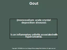
100319 Brian V Joachims MD Identify characteristics consistent with a diagnosis of Lupus Interpret ANA results Identify characteristics of Rheumatoid Arthritis Describe Spondyloarthropathy ID: 775440

lupus arthritis rheumatoid ana lupus arthritis rheumatoid ana anti methotrexate disease cutaneous patients erythematosus criteria ifa antibodies titer joints

Share:

Link:

Embed:

Download Presentation from below link

Download Presentation The PPT/PDF document " Rheumatology in Primary Care" is the property of its rightful owner. Permission is granted to download and print the materials on this web site for personal, non-commercial use only, and to display it on your personal computer provided you do not modify the materials and that you retain all copyright notices contained in the materials. By downloading content from our website, you accept the terms of this agreement.


Presentation Transcript

Slide1

Rheumatology in Primary Care

10/03/19

Brian V Joachims, M.D.

Slide2

Identify characteristics consistent with a diagnosis of LupusInterpret ANA resultsIdentify characteristics of Rheumatoid ArthritisDescribe Spondyloarthropathy

Objectives

Slide3

29 year-old Caucasian female presents with complaint of fatigueLab results:ANA IFA 1:80, speckled patternHgb 11.9TSH 1.6UA WNLHCG neg

Case 1

Slide4

Does our patient have lupus?A) yes, because she has a positive ANAB ) no, because the ANA isn’t high enoughC ) don’t know yet, need more informationD ) what’s lupus?

Lupus (?)

Slide5

Systemic Lupus Erythematosus (SLE)Chronic autoimmune disease of unknown cause that can affect virtually any organ of the bodyClinical manifestations:Constitutional symptomsCutaneous Arthritis / arthralgias / myalgiasRenalGIPulmonaryCardiacCNS (Neurologic / Psychiatric)

Lupus (!)

Slide6

FatigueMost common complaint (up to 100% at some point during the course of disease)FeverDistinguish from other causes (infection, drug reaction, malignancy)MyalgiasSevere muscle weakness or myositis uncommonWeight changeMay be related to disease or treatments

Lupus (Constitutional Symptoms)

Slide7

Acute cutaneous lupus erythematosus (ACLE)Localized (malar / butterfly rash)GeneralizedSubacute cutaneous lupus erythematosus (SCLE)AnnularPapulosquamousDrug-inducedChronic cutaneous lupus erythematosus (CCLE)Discoid lupusLupus panniculitis

Lupus (Skin)

Slide8

Acute Cutaneous Lupus Erythematosus

Slide9

ANA (Anti-Nuclear Antibody)The detection of antinuclear antibodies (ANA) in serum facilitates the diagnosis of patients with systemic lupus erythematosus (SLE) and related autoimmune diseasesThe absence of ANA in the serum of a patient with suspected SLE also provides important information in that it makes the diagnosis much less likely

Lupus (?)

Slide10

IFA – indirect immunofluorescence testHomogeneousSpeckledCentromereNucleolarSolid phase assays (ELISA) – ANA, directPanel of antigens

Antinuclear Antibody

Slide11

Titer matters1:40Approx 30% of normal controls97% of patients with SLE1:160Approx 5% of normal controls95% of patients with SLEPrevalence of ANA-associated diseases in the general population ~1%

ANA (IFA)

Slide12

ANA IFA

Slide13

ANA IFA

Slide14

Panel of antigens:Anti-dsDNA AbRNP AbSmith AbRheumatoid Arthritis factorSjogren’s Anti-SS-A (Ro Ab)Sjogren’s Anti-SS-B (La Ab)Antichromatin Ab

Lupus Panel (

Reichlin

)

Slide15

CriteriaClassification (not diagnostic)ACR – American College of RheumatologySLICC – Systemic Lupus International Collaborating Clinics

Diagnosing Lupus

Slide16

Clinical criteria (“in the absence of other causes…”)Acute cutaneous lupusChronic cutaneous lupusOral or nasal ulcersNon-scarring alopecia(not):Male patternHypothyroidismIron deficiencyArthritisSwelling or effusion – OR –Tenderness in 2 or more joints and at least 30 minutes of morning stiffness

SLICC Criteria

Slide17

Clinical criteria (cont.)SerositisPleurisy, pleural effusions or pleural rubRenalUrine protein-to-creatinine ratio representing 500 mg pro/24 hours- OR – RBC castsNeurologic

SLICC Criteria

Slide18

Hemolytic anemiaDAT / direct CoombsLeukopenia / LymphopeniaWBC <4000/mm3Lymph # <1000/mm3ThrombocytopeniaPlatelets <100,000/mm3

SLICC Criteria

Slide19

Immunologic criteriaANAAnti-dsDNAAnti-SmAntiphospholipid antibodyLupus anticoagulantAnticardiolipin antibodyAnti-2-glycoproteinFalse positive RPRLow complement (C3, C4 or CH50)Direct Coombs’ testIn the absence of hemolytic anemia

SLICC Criteria

Slide20

RheumaHelper

Slide21

Goals of therapy:ensure long-term survivalachieve the lowest possible disease activityprevent organ damageminimize drug toxicityimprove quality of lifeeducate patients about their role in disease management

Treatment

Slide22

SteroidsIMOral (prednisone)Disease-Modifying Anti-Rheumatic Drugs (DMARD’s) – “steroid-sparing” agentsHydroxychloroquineMethotrexateSulfasalazineAzathioprineLeflunomide

Medications

Slide23

FDA-approved:MalariaChemoprophylaxis, acute attackOff-label:LupusRheumatoid arthritisSjogren’s syndromePorphyriaQ fever

Hydroxychloroquine

Slide24

Marketed as Plaquenil in the U.S.$11.88 (per pill)Generic $1.83 - $4.36GoodRx: $38.67 for 60 tablets200-400 mg daily (200 mg tablets)“Typical” dose – 400 mg daily 5 mg/Kg (80 Kg person – 400 mg)Mechanism of action:Antimalarial, blocks toll-like receptorsImpairs complement-dependent antigen-antibody reactions

Hydroxychloroquine

Slide25

Adverse reactions:Retinal toxicity / maculopathyPotentially irreversible retinopathyAssociated with daily doses >5 mg/KgDuration of >5 yearsOphthalmologic exam – baseline / within the first year, followed by annual screening beginning after 5 years of use

Hydroxychloroquine

Slide26

“Bull’s-Eye” Maculopathy

Slide27

Adverse reactions (cont.):Hematologic effectsAnemiaLeukopeniaThrombocytopeniaNo prescribed surveillance lab (“CBC at baseline and ‘periodically’”)Skin discolorationDyschromia (skin and mucosal; black-blue color)

Hydroxychloroquine

Slide28

HCQ Dyschromia

Slide29

HCQ Dyschromia

Slide30

Slide31

Any-year-old any-ethnicity male/female presents with complaints of increasing joint pain, particularly in the hands, shoulders and ankles. The patient’s hands are typically swollen in the mornings and he/she has stiffness in most joints for about an hour after waking.

Case 2

Slide32

Lab results:Rheumatoid factor 65.4 (0.0-13.9)Anti-CCP 124 (0-19)ESR 44CRP 8.9 (0.0-3.2)

Case 2

Slide33

Does our patient have rheumatoid arthritis?A) yes

Rheumatoid Arthritis (?)

Slide34

Rheumatoid arthritis (RA) is a chronic, systemic, inflammatory disorder of unknown etiology that primarily involves synovial joints

Rheumatoid Arthritis

Slide35

Synovial Joint

Slide36

Synovitis

Slide37

Synovitis (clinically):Soft tissue swellingWarmth over a jointJoint effusionStiffness – “slowness or difficulty moving the joints when getting out of bed or after staying in one position too long, which involves both sides of the body and gets better with movement”Morning – at least 30 minutesGelling – stiffness with / after rest during the day

Rheumatoid Arthritis

Slide38

Morning stiffnessBetter with activitySwellingWarmthTendernessRednessSteroids helpful?

Rheumatoid Arthritis

Slide39

Rheumatoid Arthritis – Joint Distribution

Slide40

Rheumatoid factors are antibodies directed against the Fc portion of immunoglobulin G (IgG)Found in 75 to 80 percent of RA patients at some time during the course of their diseaseTiter matters:RF titer of 1:40 or greater was 28 percent sensitive and 87 percent specific for RAA titer of 1:640 or greater increased the specificity to 99 percent

Rheumatoid Factor (RF)

Slide41

Anti-citrullinated peptide antibodies (ACPA, which include anticyclic citrullinated peptides)More specific for rheumatoid arthritis than RFPearl: if low-titer RF (< 3 times upper limit of normal) and negative CCP, check for Hep C

CCP Antibodies

Slide42

Rheumatoid Arthritis

Slide43

Swan Neck Deformity

Slide44

Swan Neck

Slide45

Boutonniere Deformity

Slide46

Boutonniere

Slide47

Non-articular manifestationsOsteopeniaMyositisVasculitisSkin diseaseEye involvementLung diseaseCardiac diseaseKidney disease

Not Just the Joints…

Slide48

MethotrexateFDA-approved:ALLBreast cancerNon-Hodgkin’s lymphomaPsoriasisRheumatoid arthritisOff-label:Crohn’s diseaseDermatomyositis / PolymyositisSLE

Treatment

Slide49

Inhibits dihydrofolate reductase (DHFR)Can tightly bind to DHFR and inhibit DNA synthesis and cell proliferation7.5-15 mg weekly to beginLiterature indicates titrating to 20-30 mg weeklyAdjust gradually to optimum responseToxicity increased at doses >20 mg per weekConcomitant folic acid1-5 mg dailyAt least 5 mg per week

Methotrexate

Slide50

Adverse effects:AlopeciaStomatitisGI upsetIncreasing CrLiver enzyme abnormalitiesThrombocytopeniaAnemiaLeukopeniaPulmonary diseaseTeratogenic

Methotrexate

Slide51

• Pregnancy: [US Boxed Warning]: Methotrexate has been reported to cause fetal death and/or congenital abnormalities. Methotrexate is not recommended for women of childbearing potential unless there is clear medical evidence that the benefits can be expected to outweigh the considered risks. Pregnant women with psoriasis or rheumatoid arthritis should not receive methotrexate. Some products are contraindicated in pregnant women.

Methotrexate

Slide52

Methotrexate treatment should be discontinued for at least three months before attempting to become pregnant www.ACRPatientInfo.org © 2018 American College of Rheumatology

Methotrexate

Slide53

Slide54

39-year-old male with psoriasis presents with a 6-month history of progressive low back pain and stiffness which is most aggravating at rest. He indicates this is OK as long as he keeps moving.

Case 3

Slide55

In patients with chronic back pain >3 months:4 out of 5:Onset at age <40 (or 45) yearsInsidious onsetImprovement with exerciseNo improvement with restPain at night (with improvement upon getting up)

Inflammatory Back Pain

Slide56

Ankylosing spondylitis (AS)Nonradiographic axial SpA (nr-axSpA)Peripheral SpASpA associated with psoriasis or psoriatic arthritisSpA associated with Crohn disease and ulcerative colitisReactive arthritis (formerly called Reiter's syndrome)Juvenile-onset SpA

Spondyloarthritis

Slide57

Peripheral arthritisEnthesitis - inflammation around the enthesis, which is the site of insertion of ligaments, tendons, joint capsule, or fascia to bone, and is relatively specific to SpADactylitis (sausage digits)

Spondyloarthritis

Slide58

Enthesitis

Slide59

Enthesitis

Slide60

Enthesitis

Slide61

Dactylitis

(“Sausage Digit”)

Slide62

SacroiliitisHLA B27UveitisPsoriasisCrohn’s colitis (IBD)Good response to NSAID’sFam Hx of SpAElevated CRPPreceding infection (reactive arthritis)

Other Features

Slide63

TNF-inhibitorsHumira (Adalimumab)Human monoclonal antibodyEnbrel (Etanercept)Soluble receptor fusion proteinRemicade (Infliximab)Chimeric (mouse / human) antibody

Biologic DMARD’s

Slide64

Anti-IL-6Actemra (tocilizumab)Anti-IL-1Kineret (anakinra)JAK inhibitorsXeljanz (tofacitinib)

Biologic DMARD’s

Slide65

Anti-IL-17ACosentyx (secukinumab)Taltz (ixekizumab)Selective T-Cell Costimulation BlockerOrencia (abatacept)Phosphodiesterase-4 Enzyme InhibitorOtezla (apremilast)

Biologic DMARD’s

Slide66