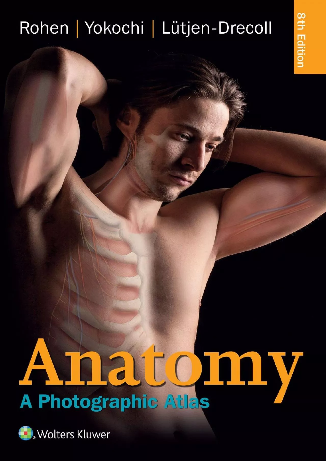
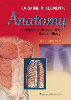
PDF-(BOOS)-Anatomy: A Photographic Atlas (Color Atlas of Anatomy a Photographic Study of the
Author : janisjacobo | Published Date : 2022-06-23
Prepare for the dissection lab and operating room with br Color Atlas of Anatomybr br 8ebr Featuring outstanding fullcolor photographs of actual cadaver dissections
Presentation Embed Code
Download Presentation
Download Presentation The PPT/PDF document "(BOOS)-Anatomy: A Photographic Atlas (Co..." is the property of its rightful owner. Permission is granted to download and print the materials on this website for personal, non-commercial use only, and to display it on your personal computer provided you do not modify the materials and that you retain all copyright notices contained in the materials. By downloading content from our website, you accept the terms of this agreement.
(BOOS)-Anatomy: A Photographic Atlas (Color Atlas of Anatomy a Photographic Study of the: Transcript
Prepare for the dissection lab and operating room with br Color Atlas of Anatomybr br 8ebr Featuring outstanding fullcolor photographs of actual cadaver dissections with accompanying schematic drawings and diagnostic images this proven text depicts anatomic structures more realistically than illustrations in traditional atlases Chapters are organized by region in the order of a typical dissection with each chapter presenting topographical anatomical structures in a systemic manner Authentic photographic reproduction of colors structures and spatial dimensions as seen in the dissection lab and on the operating table help you develop an understanding of the anatomy of the human bodyFunctional connections between single organs the surrounding tissue and organ systems are clarified to prepare you for the dissection lab and practical examsClinical cases and over 1200 images enhance your understandingDissections illustrate the topographical anatomy in layers from the outside in to better prepare you for the lab and operating roomTables of the nervous and muscular systems summarize key informationA userfriendly comprehensive subject index helps you find information and images of interest. High. . Dynamic . Range Software for Optimal Image Results. A Case Study using the. Hugh Morton Photograph Collection. A Photographic Journey brought to you by the Digital Production Center. Terminology. Dr. Tony Serino. “The nature of the body is the beginning of medical science.” . . Hippocrates ~350 . B.C.E. Intro: Topic Objectives. Be able to identify basic body plan that underlies all anatomy. Engine. . Hands. -on Session. Mobile Computing - CNT 5517-5564. Dr. . Sumi. . Helal. Professor . Computer & Information Science & Engineering Department. University of Florida, Gainesville, FL 32611. Lesson 1.1: The Language of Anatomy and Physiology. Lesson 1.2: Basic Physiological Processes. Lesson 1.3: How Forces Affect the Body. Lesson 1.4: Understanding Science. Lesson 1.1. The Language . of . Figure 1.1. Figure 1.2. Figure 1.3. MSI: Lab 6, Part C. Superficial Fibular N.. Lateral Cutaneous branch. of Superficial Fibular N.. Medial Cutaneous branch. of Superficial Fibular N.. Photo taken from: Color Atlas of Anatomy: A Photographic Atlas of the Human Body (2006). p. 493. Right leg and dorsum of foot, cutaneous nerves and veins (lateral aspect).. The Fifth Edition of Clemente\'s Anatomy features over 1,000 bright, realistically detailed full-color illustrations, plus a wealth of accompanying diagnostic images and numerous muscle tables. This classically organized regional atlas is based on the strikingly colorful yet realistic illustrations of the world-renowned Sobotta Atlas of Human Anatomy.This edition includes 68 new plates and 45 new clinical images, including cross-sectional CT scans. Of special note are 12 new cranial nerve plates and new diagrammatic drawings that help students learn the anatomy of the cranial nerves. This edition\'s numbering system has been improved to correlate figures more clearly with plates. The Fifth Edition of Clemente\'s Anatomy features over 1,000 bright, realistically detailed full-color illustrations, plus a wealth of accompanying diagnostic images and numerous muscle tables. This classically organized regional atlas is based on the strikingly colorful yet realistic illustrations of the world-renowned Sobotta Atlas of Human Anatomy.This edition includes 68 new plates and 45 new clinical images, including cross-sectional CT scans. Of special note are 12 new cranial nerve plates and new diagrammatic drawings that help students learn the anatomy of the cranial nerves. This edition\'s numbering system has been improved to correlate figures more clearly with plates. •Gross anatomy is the study of structures that can be seen with the naked eye.. •Microscopic anatomy is the study of structures that require a microscope to be seen. . Physiology is related to the functions of the body and all its parts, including cells, tissues and organs.. BY:. DR. Stella Selena. M.B.B.S., DGO.. Academy Director. Mother Teresa Institute Of. Pharmaceutical Education and Research. Two Branches Of Science. Anatomy. : Is the science of the body structures and their relationships among them. [DOWNLOAD] Anatomy: A Photographic Atlas
http://skymetrix.xyz/?book=1451193181 The topics covered by this resource include:. Informed consent. Ethical resource management. Further reading:. Cornwall J, Callahan D, Wee R. . Ethical issues surrounding the use of images from donated cadavers in the anatomical sciences. Charity Hilton MS. 1. , Saul Crumpton MS. 1. , Jon Duke MD, MS. 1,2. 1. Georgia Tech Research Institute, . 2. Georgia Institute of Technology. Background. OHDSI Atlas has long been an effective tool for developing rule-based cohort definitions in observational data. In the public version of Atlas, thousands of cohort definitions have been created. While patient record verification is a common method of cohort definition validation, it is not without difficulties, including but not limited to the need for clinical experts to access data, a tool to review all in-cohort patients, a method to gather review data, and a system of tabulation to determine in-cohort (case/no-case) participation or not. Figure 1.1. MSI: Lab 6, Part B. Figure 1.2. Figure 1.3. MSI: Lab 6, Part B. Medial View. Figure 1.4. Figure 1.5. Pes . Ansirinus. Semitendinosus m.. Sartorius m.. Gracilis. Tendon. Medial view. Photo taken from: Color Atlas of Anatomy: A Photographic Atlas of the Human Body (2006). p. 455. Flexors of the right thigh, superficial layer (dorsal aspect).. Anatomy & Physiology. Anatomy. (from the Greek . anatomia. ). : separate, apart from, and . temnein. , to cut up, cut open.) is a branch of biology and medicine that is the consideration of the structure of living things. .
Download Document
Here is the link to download the presentation.
"(BOOS)-Anatomy: A Photographic Atlas (Color Atlas of Anatomy a Photographic Study of the"The content belongs to its owner. You may download and print it for personal use, without modification, and keep all copyright notices. By downloading, you agree to these terms.
Related Documents