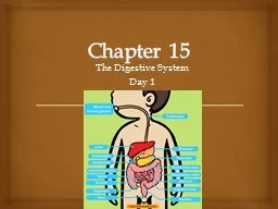
Day 1 Digestive System Made up of the alimentary canal which extends from the mouth to anus Includes Mouth pharynx esophagus stomach small intestine large intestine recturm and anus ID: 928119
Download Presentation The PPT/PDF document "Chapter 15 The Digestive System" is the property of its rightful owner. Permission is granted to download and print the materials on this web site for personal, non-commercial use only, and to display it on your personal computer provided you do not modify the materials and that you retain all copyright notices contained in the materials. By downloading content from our website, you accept the terms of this agreement.
Slide1
Chapter 15
The Digestive SystemDay 1
Slide2Digestive System:
Made up of the alimentary canal which extends from the mouth to anus
Includes:
Mouth, pharynx, esophagus, stomach, small intestine, large intestine,
recturm, and anusAccessory Organs:Salivary glands, liver, gallbladder and pancreasCovers 186 square meters
Introduction
Slide3Digestion:The chemical and mechanical breakdown of foods and the absorption of the resulting nutrients by cells.
Mechanical DigestionBreaks large pieces into smaller ones without altering the chemical compositionChemical Digestion:Breaks food into simpler chemicals
Intro Cont.
Slide4Alimentary CanalMuscular tube, roughly 8 meters in length
Structure of the Wall4 layersMucosa MembraneSubmucosa
Muscular Layer
Serosa
General Characteristics
Slide5Made up of epithelium, connective tissue and small amounts of smooth muscle
Contains folds or tiny projections that extend into the passageway called LUMENIncrease absorptive surface areSecrete mucus and digestive enzymesProtects tissues beneath it and
carries on secretion and absorption
Mucosa Membrane
Slide6Made up of loose connective tissue, glands, blood vessels, lymphatic vessels, and nerves
Nourishes surrounding tissues and carries away absorbed materialsSubmucosa
Layer
Slide7Produces movements of the tube
Contains two types of smooth muscleCircular FibersContraction causes decrease in tube’s diameterLongitudinal FibersContraction leads to shortening of tube
Muscular Layer
Slide8Outer coveringAlso called visceral peritoneum
Protect underlying tissues and secrete serous fluid which moistens and lubricates tube’s outer surfaceAllows other organs in abdominal cavity to slide freely against one another
Serosa Layer
Slide9Two types of motor movementsMixing Movements
Caused by smooth muscle contracts rhythmicallySegmentation: alternately contracting and relaxing the smooth muscle in nonadjacent segmentsOccurs in stomachPropelling movements
Peristalsis: wavelike motion that pushes contents of tube ahead
Occurs in esophagus and intestines
Movements of the Tube
Slide10Receives food and begins digestion by mechanically breaking up solid particles and mixing them with saliva
MasticationMouth
Slide11CheeksForm lateral walls of mouth
LipsContain sensory receptors that judge temp. and texture of food
Cheeks
and Lips
Slide12Fills
oral cavity
Responsible for mixing food with saliva and moving food towards pharynx during swallowing
Papillae
Rough projections on tongue that provide friction to help handle foodLingual FrenulumConnects tongue to floor of mouthLingual Tonsils: Posterior portionLymphatic tissue
Tongue
Slide13Forms roof of oral cavity
Anterior portion-hard palateMuscular posterior portion-soft palateUvulaCone-shaped projection in back of mouthPalatine Tonsils and Pharyngeal Tonsils-adenoids
Masses of lymphatic tissues that protect the body against infection
Commonly removed
Palate
Slide14Two Sets
Primary Teeth-deciduous20Erupt though gums in regular intervals from 6 months-2-4 years
of age
Usually shed in the same
order they erupted Secondary TeethPush primary teeth out of sockets32Erupt 6 years-17-25 years with wisdom teethTeeth
Slide15Role in digestion
Begin mechanical digestion by breaking
food into smaller pieces
Increases surface area of food to allow
for digestive enzymes to work more effectivelyParts to a toothCrown-projects beyond gum
Root-
below gums
Neck
-portion where crown and root meet
Enamel
-glossy white, covers crown, consists of calcium salts, hardest substance in body, never replaced when damaged
Dentin
-bulk of tooth beneath enamel
Pulp
-contains blood vessels, nerves, and connective tissue
Root Canals
-area where blood vessels and nerves reach pulp
Cementum
-bonelike material that helps attach tooth to jaw
Periodontal Ligament
-contains blood vessels and helps attached tooth to jaw
Teeth Cont.
Slide16Secrete saliva that helps moisten and bind food particlesBegins chemical digestion by enzyme amylase
Helps cleanse the mouth and teethSalivary Glands
Slide17Each gland has two types of secretory cells
SerousProduce a watery fluid that includes digestive enzyme salivary amylaseEnzyme splits carbohydratesMucous
Secretes thick liquid called mucus
Binds food particles and lubricates food during swallowing
Saliva is produced just by thinking of good foodsSaliva stops production in the presence of unpleasant smells or tastesSalivary Secretions
Slide18Three Pairs
Parotid GlandslargestLies anterior and somewhat inferior to earsFound between skin, cheeks, and masseter muscleSecretes clear, water fluid that is rich in amylase
Major Salivary Glands
Slide19Submandibular Gland
Located on floor of mouthSecretes both mucous and serous fluid
Major Salivary Glands
Slide20Sublingual glands
SmallestOn the floor of mouth inferior to the tongueSecretes primarily mucous type making their secretions thick and stringy
Major Salivary Glands
Slide21Cavity posterior to mouth which leads to esophagus
Do not digest food but functions in swallowingConnects nasal and oral cavities with the larynx and esophagusPharynx-The Crossroads
Slide22Three parts:Nasopharynx
-communicates with nasal cavity and provides a passageway for air during breathing, auditory tube connects to nasopharynxOropharynx-posterior to soft palate and inferior to nasopharynx
, allows food and air to pass through
Laryngopharynx- inferior to oropharynx, passage way to esophagus
Pharynx Cont.
Slide233 Stages
1. Food is mixed with saliva, tongue rolls mixture into a mass called bolus, forces bolus into oropharynx2. Once food reaches oropharynx, swallowing reflex kicks in (See steps on Page 409)3. Peristalsis transports food into esophagus and on to the stomach
Swallowing Mechanism
Slide24Straight, collapsible tube about 25 cm long that allows food to pass from mouth to stomach
Mucous glands are scattered throughout Lower Esophageal Sphincter/Cardiac SphincterCloses the entrance to stomach preventing contents from regurgitating into esophagus.Peristaltic waves allow food to enter stomach.
Esophagus
Slide25Which organs constitute the digestive system?
Describe the wall of the alimentary canal.Name the two basic types of movements in the alimentary canal.How does the tongue function as part of the digestive system?Where are the tonsils located?
How do primary teeth differ from secondary teeth?
Describe the structure of a tooth.
Explain how a tooth is attached to the bone of the jaw.What is the function of saliva?What stimulates salivary glands to secrete saliva?Where are the major salivary glands?Describe the regions of the pharynx.List the major events of swallowing.What is the function of the esophagus?
Review Questions