/
Tumour  of the  stomach Gastric carcinoma Tumour  of the  stomach Gastric carcinoma

Tumour of the stomach Gastric carcinoma - PowerPoint Presentation

jasmine
jasmine . @jasmine
Follow
27 views
Uploaded On 2024-02-02

Tumour of the stomach Gastric carcinoma - PPT Presentation

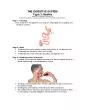
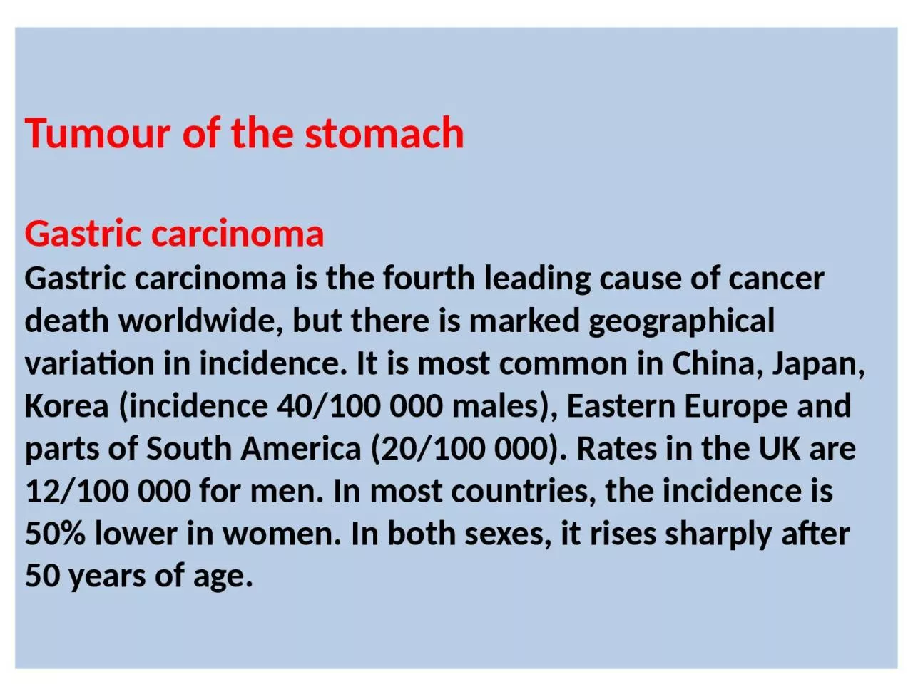
Gastric carcinoma is the fourth leading cause of cancer death worldwide but there is marked geographical variation in incidence It is most common in China Japan Korea incidence 40100 000 males Eastern Europe ID: 1043472

acute pancreatitis patients chronic pancreatitis acute chronic patients severe gastric pancreatic cases common disease pain diagnosis cancer serum pancreas

Share:

Link:

Embed:

Download Presentation from below link

Download Presentation The PPT/PDF document "Tumour of the stomach Gastric carcinom..." is the property of its rightful owner. Permission is granted to download and print the materials on this web site for personal, non-commercial use only, and to display it on your personal computer provided you do not modify the materials and that you retain all copyright notices contained in the materials. By downloading content from our website, you accept the terms of this agreement.


Presentation Transcript

1. Tumour of the stomachGastric carcinomaGastric carcinoma is the fourth leading cause of cancerdeath worldwide, but there is marked geographical variation in incidence. It is most common in China, Japan, Korea (incidence 40/100 000 males), Eastern Europe and parts of South America (20/100 000). Rates in the UK are 12/100 000 for men. In most countries, the incidence is 50% lower in women. In both sexes, it rises sharply after 50 years of age.

2. PathophysiologyInfection with H. pylori plays a key pathogenic role. It isassociated with chronic atrophic gastritis, gastric mucosal atrophy and with gastric cancer . It has been estimated that H. pylori infection may contribute to the occurrence of gastric cancer in 60–70% of cases and that acquisition of infection at an early age may be important.There is strong evidence that H. pylori eradication, especially if achieved before irreversible pre-neoplastic changes have developed, reduces the risk of cancer development in high-risk populations.Diets rich in salted, smoked or pickled foods and the consumption of nitrites , Diets lacking fresh fruit and vegetables, as well as vitamins C and A, may also contribute.

3.

4. Risk factors for gastric cancer• H. pylori• Smoking• Alcohol• Dietary associations (see text)• Autoimmune gastritis (pernicious anaemia)• Adenomatous gastric polyps• Previous partial gastrectomy (> 20 yrs)• Hereditary diffuse gastric cancer families (HDC-1 mutations).• Familial adenomatous polyposis

5. Clinical featuresEarly gastric cancer is usually asymptomatic but may bediscovered during endoscopy for investigation of dyspepsia.Two-thirds of patients with advanced cancers have weight loss and 50% have ulcer-like pain. Anorexia and nausea occur in one-third, while early satiety, haematemesis,melaena and dyspepsia alone are less common. Dysphagia occurs in tumours of the gastric cardia which obstruct the gastro-oesophageal junction. Anaemia from occult bleeding is also common.

6. InvestigationsUpper gastrointestinal endoscopy is the investigationof choice and should be performed promptly in dyspeptic patient with ‘alarm features’. Multiple biopsies from the edge and base of a gastric ulcer are required.TreatmentSurgeryResection offers the only hope of cure, and this can beachieved in about 90% of patients with early gastric cancer. For the majority of patients with locally advanced disease, total gastrectomy with lymphadenectomy is the operation of choice. Proximal tumours involving the oesophago-gastric junction also require a distal oesophagectomy.

7. Palliative treatmentIn patients with inoperable tumours, survival can beimproved and palliation of symptoms achieved withchemotherapy using 5-fluorouracil and cisplatin.

8. Acute pancreatitis

9. The exocrine pancreas is necessary for the digestion of fat, protein and carbohydrate. Inactive proenzymes are secreted from acinar cells in response to circulating gastrointestinal hormones .Bicarbonate-rich fluid is secreted from ductular cells to produce an optimum alkaline pH for enzyme activity.

10.

11.

12. Acute pancreatitis accounts for 3% of all cases of abdominal pain admitted to hospital. Despite recent advances in management, mortality has remained unchanged at 10%. About 80% of all cases are mild with a mortality of less than 5%; 98% of deaths occur in the 20% of severe cases and about one-third of these occur within the first week.

13. PathophysiologyAcute pancreatitis occurs as a consequence of premature intracellular trypsinogen activation, releasing proteases which digest the pancreas and surrounding tissue. The normal pancreas has only a poorly developed capsule, and adjacent structures, including the common bile duct, duodenum, splenic vein and transverse colon, are commonly involved in the inflammatory process.Acute pancreatitis is often self-limiting, but insome patients with severe disease, local complications,such as necrosis, pseudocyst or abscess, occur, as well assystemic complications that lead to multi-organ failure.

14.

15.

16. Clinical features:Severe, constant upper abdominal pain which radiates to the back in 65% of cases builds up over 15-60 minutes. Nausea and vomiting are common. There is marked epigastric tenderness, but in the early stages (and in contrast to a perforated peptic ulcer) guarding and rebound tenderness are absent because the inflammation is principally retroperitoneal. Bowel sounds become quiet or absent as paralytic ileus develops. In severe cases the patient becomes hypoxic and develops hypovolaemic shock with oliguria. Discoloration of the flanks (Grey Turner's sign) or the periumbilical region (Cullen's sign) is a feature of severe pancreatitis with haemorrhage

17. Cullen’s signGrey turner’s sign

18. Adverse prognostic factors in acute pancreatitis (Glasgow criteria)• Age > 55 yrs• PO2 < 8 kPa (60 mmHg)• White blood cell count (WBC) > 15 × 10^9/L• Albumin < 32 g/L (3.2 g/dL)• Serum calcium < 2 mmol/L (8 mg/dL) • Glucose > 10 mmol/L (180 mg/dL)• Urea > 16 mmol/L (45 mg/dL) (after rehydration)• Alanine aminotransferase (ALT) > 200 U/L• Lactate dehydrogenase (LDH) > 600 U/L

19. Differential diagnosis of acute pancreatitisMild attack*Biliary colic or acute cholecystitis*Complicated peptic ulcer disease*Acute liver conditions*Incomplete bowel obstruction*Renal disease*Lung disease (for example, pneumonia or pleurisy)Severe attack*Perforated or ischaemic bowel*Ruptured aortic aneurysm*Myocardial infarction

20. InvestigationsThe diagnosis of acute pancreatitis is usually established by the detection of an increased level of serum amylase Values 3 fold or more above normal virtually clinch the diagnosis However, there appears to be no definite correlation between the severity of pancreatitis and the degree of serum amylase elevation. After 48-72 h, even with continuing evidence of pancreatitis, total serum amylase values tend to return to normal because it is efficiently excreted by the kidneys . However, pancreatic isoamylase and lipase levels may remain elevated for 7 to 14 days. A persistently elevated serum amylase concentration suggests pseudocyst formation.

21. Serum lipase activity increases in parallel with amylase activity, and measurement of both enzymes increases the diagnostic yield. Ultrasound scanning confirms the diagnosis, although in the earlier stages the gland may not be grossly swollen. The ultrasound scan is also useful because it may show gallstones, biliary obstruction or pseudocyst formation. Contrast-enhanced pancreatic CT is very useful in diagnosis and follow up

22. Pancreatic necrosis. Lack of vascular enhancement of the pancreas during contrast-enhanced CT indicates necrosis (arrow). The presence of gas suggests that infection has occurred.

23.

24.

25. ManagementManagement comprises several related steps:• establishing the diagnosis and disease severity• early resuscitation, according to whether the diseaseis mild or severe• detection and treatment of complications• treating the underlying cause.The initial management is based upon analgesia using Opiate analgesics( e.g.pethidine) and correction of hypovolaemia using normal saline and/or colloids. All severe cases should be managed in a high-dependency or intensive care unit.Hyperglycaemia is corrected using insulin, but it is not necessary to correct hypocalcaemia by intravenous calcium injection unless tetany occurs.

26. Nasogastric aspiration is only required if paralyticileus is present. Enteral feeding, if tolerated, should bestarted at an early stage in patients with severe pancreatitis (because they are in a severely catabolic state and need nutritional support).Prophylaxis of thromboembolism with subcutaneous low-molecular-weight heparin is also advisable.The use of prophylactic, broad-spectrum intravenous antibiotics to prevent infection of pancreatic necrosis is ofunproven benefit but they are often given empirically.

27. ERCP in acute pancreatitis: In patients with actual or predicted severe pancreatitis of suspected biliary origin, emergency ERCP with biliary sphincterotomy improves outcome and is best undertaken within 72 hours of onset. Patients who present with cholangitis or jaundice in association with severe acute pancreatitis should undergo urgent ERCP to diagnose and treat choledocholithiasis, but In less severe cases of gallstone pancreatitis, biliary imaging (using MRCP or EUS) can be carried out after the acute phase has resolved.

28. Cholecystectomy should be undertaken within 2 weeks following resolution of pancreatitis-and preferably during the same admission-to prevent further potentially fatal attacks of pancreatitis.

29. Chronic pancreatitis

30. Chronic pancreatitis is a chronic inflammatory disease characterized by fibrosis and destruction of exocrine pancreatic tissue. Diabetes mellitus occurs in advanced cases because the islets of Langerhans are involved.

31. Causes of chronic pancreatitis:Toxic–metabolic• Alcohol • Tobacco • Hypercalcaemia• Chronic renal failureAutoimmune• Isolated or as part of multi-organ problem (chronic pancreatitis associated with : Sjögren’s syndrome, IBD , Primary biliary cirrhosis).Recurrent and severe acute pancreatitis• Recurrent acute pancreatitis • Post-necrotic.Obstructive• Ductal adenocarcinoma • Intraductal papillary mucinous neoplasia• Pancreas divisum • Sphincter of Oddi stenosis.

32. Clinical features Chronic pancreatitis predominantly affects middle-aged alcoholic men. Almost all present with abdominal pain. Pain may be relieved by leaning forwards or by drinking alcohol. Approximately one-fifth of patients chronically consume opiate analgesics. Weight loss is common.Steatorrhoea occurs when more than 90% of the exocrine tissue has been destroyed.Overall, 30% of patients are diabetic, but this figure rises to 70% in those with chronic calcific pancreatitis.Physical examination reveals a thin, malnourished patient with epigastric tenderness. Skin pigmentation over the abdomen and back is common and results from chronic use of a hot water bottle (............................................).

33. Erythema ab igne

34. Erythema ab igne

35. Investigations in chronic pancreatitisTests to establish the diagnosis*Ultrasound *CT (may show atrophy, calcification or ductal dilatation) *Abdominal X-ray (may show calcification) *MRCP *Endoscopic ultrasound

36. Imaging in chronic pancreatitis. (A) CT scan showing a grossly dilated and irregular duct with a calcified stone (arrow A). Note the calcification in the head of the gland (arrow B). (B) MRCP of the same patient showing marked ductal dilatation with abnormal dilated sidebranches (arrows A). A small cyst is also present (arrow B).A

37. Complications of chronic pancreatitis*Pseudocysts and pancreatic ascites, which occur in both acute and chronic pancreatitis *Extrahepatic obstructive jaundice due to a benign stricture of the common bile duct as it passes through the diseased pancreas *Duodenal stenosis *Portal or splenic vein thrombosis leading to segmental portal hypertension and gastric varices *Peptic ulcer

38. TreatmentA range of analgesic drugs, particularly NSAIDs, are valuable, but the severe and unremitting nature of the pain often leads to opiate use with the risk of addiction.Oral pancreatic enzyme supplements suppress pancreatic secretion and their regular use reduces analgesic consumption in some patients.Coeliac plexus neurolysis or minimally invasive thoracoscopic splanchnicectomy sometimes produces long-lasting pain relief.

39. Malabsorption:This is treated by dietary fat restriction (with supplementary medium-chain triglyceride therapy in malnourished patients) and oral pancreatic enzyme supplements.

40. Autoimmune pancreatitis (AIP) This is a form of chronic pancreatitis that can mimic cancer but which responds to corticosteroids. AIP is characterised by abdominal pain, weight loss or obstructive jaundice, without acute attacks of pancreatitis.

41. Blood tests reveal increased serum immunoglobulin G (IgG) or IgG4, and the presence of other autoantibodies. Imaging shows a diffusely enlarged pancreas, narrowing of the pancreatic duct and stricturing of the lower bile duct on ERCP. AIP may occur alone or in association with other autoimmune disorders such as Sjögren's syndrome, primary sclerosing cholangitis (PSC) or inflammatory bowel disease.

42. Treatment:The response to steroids is usually excellent but some patients require azathioprine.

43.