/
115J Obstet Gynecol India Vol 56 No 2  MarchApril 2006      Pg 11 115J Obstet Gynecol India Vol 56 No 2  MarchApril 2006      Pg 11

115J Obstet Gynecol India Vol 56 No 2 MarchApril 2006 Pg 11 - PDF document

jordyn
jordyn . @jordyn
Follow
342 views
Uploaded On 2022-08-20

115J Obstet Gynecol India Vol 56 No 2 MarchApril 2006 Pg 11 - PPT Presentation

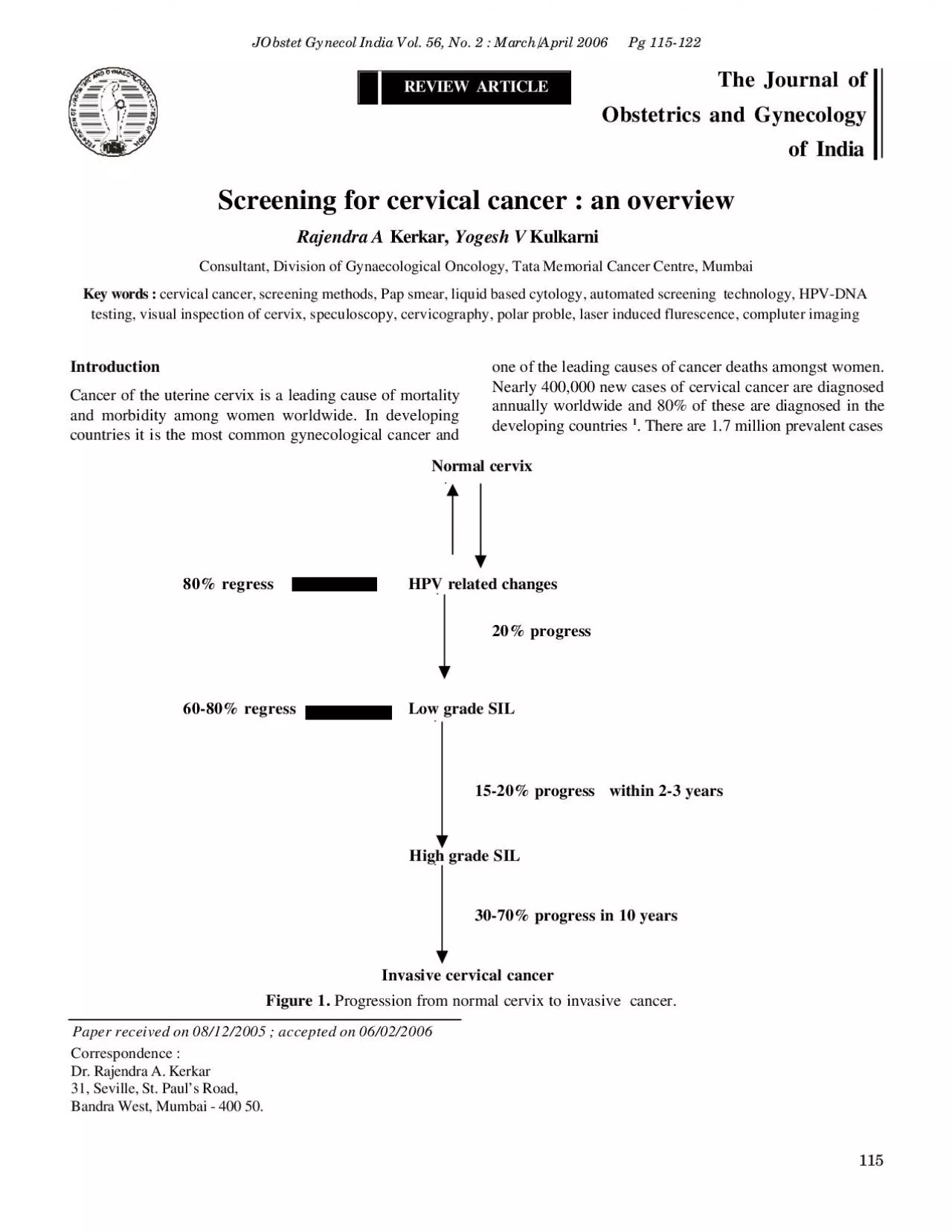
REVIEW ARTICLEThe Journal ofObstetrics and Gynecologyof India Screening for cervical cancer an overviewRajendra A Kerkar Yogesh V KulkarniConsultant Division of Gynaecological Oncology Tata Memor ID: 938861

screening cervical hpv cancer cervical screening cancer hpv pap smear cytology testing cervix women test dna technics program colposcopy

Share:

Link:

Embed:

Download Presentation from below link

Download Pdf The PPT/PDF document "115J Obstet Gynecol India Vol 56 No 2 M..." is the property of its rightful owner. Permission is granted to download and print the materials on this web site for personal, non-commercial use only, and to display it on your personal computer provided you do not modify the materials and that you retain all copyright notices contained in the materials. By downloading content from our website, you accept the terms of this agreement.


Presentation Transcript

115J Obstet Gynecol India Vol. 56, No. 2 : March/April 2006 Pg 115-122 REVIEW ARTICLEThe Journal ofObstetrics and Gynecologyof India Screening for cervical cancer : an overviewRajendra A Kerkar, Yogesh V KulkarniConsultant, Division of Gynaecological Oncology, Tata Memorial Cancer Centre, MumbaiKey words : cervical cancer, screening methods, Pap smear, liquid based cytology, automated screening technology, HPV-DNA one of the leading causes of cancer deaths amongst women.Nearly 400,000 new cases of cervical cancer are diagnosedannually worldwide and 80% of these are diagnosed in thedeveloping countries 1. There are 1.7 million prevalent cases Normal cervix 80% regressHPV related changes 20% progress 60-80% regressLow grade SIL15-20% progress within 2-3 years High grade SIL30-70% progress in 10 years Invasive cervical cancer Paper received on 08/12/2005 ; accepted on 06/02/2006Correspondence :Dr. Rajendra A. Kerkar31, Seville, St. Paul’s Road,Bandra West, Mumbai - 400 50.Figure 1. Progression from normal cervix to invasive cancer. 116in the developing world and as many as 5-13 million womenhave precancerous lesions 2. As the female population in thedeveloping countries continues to rise with an increase in lifeexpectancy, the proportion of older women will alsonecessarily rise. It is expected that the number of cases ofcancer cervix will rise further in the years to come.Natural history of cervical carcinomaThe natural history of cervical cancer represents a stepwiseprogression from a histologically normal cervix to frankinvasive cancer 3 (Figure 1).Why is screening for cervical cancer effective?An ideal screening test is one that is minimally invasive,easy to perform, acceptable to the subject, cost-effectiveand efficacious in diagnosing the disease process in itspreinvasive or early invasive state when the diseaseprocess is more easily treatable and curable.In all probability cervical cancer is the only gynecologicalcancer that satisfies the well recognized WHO criteria forimplementation of a screening program –Existence of well defined premalignant lesions?Long latent period in which premalignant change oroccult cancers can be detected and effectively treatedthereby altering the natural history of the disease?A clearly defined viral etiology which could beincorporated as a marker in mass screening program?Easy and direct access of the uterine cervix forexamination and sampling?Effective treatments available for the premalignantchanges.creening for cervical carcinomaScreening programs for cervical cancer have been institutedin developed countries for decades and over a period of timehave been shown to be effective in reducing the overallmortality from this disease. Such programs however canonly be made to work provided the necessary infrastructureand funds are available 4Following its introduction by Papanicolau in 1927 5 exfoliativecervicovaginal cytology has been extensively investigated andused as a screening test for cervical cancer. Over the yearsit has been found that this test has well recognisedlimitations. A better understanding of the natural history ofcervical cancer as also increasing evidence for the putativerole of the human papilloma virus (HPV) in its causation hasnow prompted investigators to find viable alternatives toconventional exfoliative cytology.A variety of screening tests have therefore been developedin an attempt to overcome the innate limitations ofconventional cytology. These are currently under evaluationand it is hoped that they may improve upon the accuracy ofconventional screening cytology.Screening tests for cervical carcinomaScreening technics for cervical cancer include 6 –Conventional exfoli

ative cervicovaginal cytology i.e. thecervical (Pap) smear?Fluid sampling technics with automated thin layerpreparation (liquid based cytology)?Automated cervical screening technics?Neuromedical systems?HPV testing?Polar probe?Laser induced fluorescence?Visual inspection of cervix after applying Lugol’s iodine(VILI) or acetic acid (VIA).?Exfoliative cytology (conventional Pap smear)Exfoliative cervicovaginal cytology has been regarded as thegold standard for cervical cancer screening programs. Despitethe apparent success of the Pap smear in detecting preinvasivecancer, the expected beneficial impact vis-à-vis mortalityreduction has not been significant 7. The standard technicfor Pap smear collection is to sample the portio vaginalis ofthe cervix and the endocervical canal using a cervical spatulaand endocervical brush. The collected sample is smeared ona slide and then fixed immediately with cytology fixative.Most clinicians are concerned with reducing sampling errorsby focusing on the technic of smear acquisition and eliminatingdrying artifacts through rapid fixation.There are various problems associated with conventionalcytology –Incorrect and inadequate sampling in 5-10% of cases 8Only upto 20% of harvested cells are transferred on theslide leading to a reduction in the sensitivity of the test 8Mean sensitivity of only 55-60% 9Reported false negative rates varying from 25 to 50% 10Reported false positive rates varying from 15 to 20 % 11Interobserver variation in the interpretation of cytologicalabnormality making reporting subjective and poorlyreproducible 12Equivocal smears and mildly irregular Pap results have aRajendra A Kerkar 117low yield of underlying high grade pathology and representa significant cost in terms of specialist referral and followup 7Epidemiological data suggest that the current method ofPap smear testing is unlikely to prevent more than 60%of the cases of cervical cancer 13Owing to these problems, several technics have been recentlydeveloped in an attempt to automate the various steps of Papsmear preparation and processing in order to try and improvethe sensitivity and specificity of conventional cervicalcytology.Fluid sampling technics with automated thin layerpreparationRecently liquid based cytological technologies have beendeveloped and have gained popularity because in preliminarystudies the use of such technics was associated with areduction in the incidence of inadequate cervical smears 14,15Two such technics that have been extensively tested areThinPrep (Cytyc Corp, Booxborough, MA) and Autocyte(TriPath Imaging, Burlington, NC). These fluid samplingdevices have been approved by the USFDA.A special sampling device is used for sampling the cervix inthe usual manner as in the traditional Pap smear. The samplingdevice is then directly placed in a vial containing a specialpreservative with additional hemolytic and mucolytic agents.The general idea is to provide a well preserved sample that isautomatically transferred to a slide as a coin sized thin layer.In the laboratory, the cells are collected either by extractionacross a special filter (ThinPrep) or by layering onto a densityreagent.Comparisons of the conventional Pap smear with thin layerfluid preparations have shown a marked improvement in theadequacy of the specimen as evidenced by a more evendistribution of cells, and reduction in cellular debris and RBCs.This in turn leads to a decrease in the incidence of falsepositive diagnosis of cytological atypia and an excellentcorrelation with the detection of squamous abnormalities.The ThinPrep test was approved for marketing in the USAbased upon studies showing an increase in the detection ofLSIL or worse, from a rate of 8% by the conventiona

l Papsmear to 9.4% with the ThinPrep 16Bernstein et al 17 performed a metaanalysis of 25 prospectivestudies of the ThinPrep method with a total of 5,33,039patients. They found that the overall sample adequacyimproved with the ThinPrep test (Odds ratio2.11; 95% CI2.07-2.15), but the incidence of diagnosis of ASCUS wasnot reduced (Odds ratio1.05; 95% CI 95-1.16). The overallsensitivity of the screening test was also found to be increasedwith ThinPrep.It is now generally accepted that the improvement in thedetection rates with these tests is more marked in centerswith a low risk population as compared to those catering tohigh risk populations (65% improvement as against 6%).Hartmann et al 18 performed an extensive review of samplingtechnics and found that most studies of the thin layer technicsdid not have a proper control group thus hindering the abilityto assess the true sensitivity, specificity and predictive valueof the technics. They concluded that the current evidence isnot adequate enough to recommend that the ThinPrep test issuperior to conventional Pap smear testing. They alsosuggested that the cost-effectiveness of these tests needs tobe assessed very carefully as these tests appear to have alower specificity than conventional Pap smear testing, thusleading to more specialist referrals.Automated screening technologyThe effectiveness of any cervical cancer screeningprogram that relies on cervical cytology is the qualitycontrol of the cytological review of Pap smears. This isessential for reducing the false positives and falsenegatives that invariably result from inter- and intra-observer variation.Automated screening technics have recently beendeveloped that can not only perform this quality controlrescreening but also can be used for primary screeningof cervical smears.The following automated screening technics that rely largelyon neural network technology and are based on thecomputerized imaging and identification of abnormal cervicalcells are available –Autopap300 (TriPath Imaging, Burlington NC)?PAPNET (Neuromedical systems).Of these, only the Autopap300 is approved by the USFDAfor primary and secondary cervical screening while thePAPNET is only approved for secondary screening.The Autopap300 system utilizes a specialized high speed videomicroscope, image interpretation software, and speciallydesigned field of view computers to image, analyze andclassify abnormal cervical cells. The screened slides are givena score and adequacy statement. Cases scoring a total of 30or more are then rescreened by a cytotechnologist for furtherevaluation.The PAPNET 19 is a semi-automated system, which consistsReview Article 118of two phases, a scanning phase and a review phase for cervicalsmears. After identification of 128 cells with the highest networkscore, the cytologists are required to only review those cells.Wilbur et al 20,21 found that considering a 10% review threshold,Autopap300 successfully selected 77% of HSIL slides, whichwas substantially more than the approximately 10% expectedon the basis of a random review. A large multicentric trial hasalso shown the superiority of the Autopap300 system for theidentification of cervical cellular abnormalities at the level ofASCUS or higher.HPV-DNA TestingThe etiopathological role of HPV in the development of cervicalcancer has been proved beyond doubt. HPV 16, 18, 31, 33, 35,39 45, 51, 52, 56, 59 and 68 are known to be frequentlyassociated with HSIL and invasive cancers of the cervix. Testingfor the presence of HPV-DNA in the cervical cells is thus apotentially useful screening method, which could be incorporatedin cervical cancer screening programs. There are various technicsavailable for HPV-DNA testing of which Southern Blothybridiz

ation is regarded as a laboratory gold standard. This ishowever unsuitable for clinical use as it is laborious, tediousand requires fresh tissue. Currently the Hybrid capture II assay(Digene, Silver Spring, MD) is the most useful technic for HPV-DNA testing. This utilizes nonradioactive RNA probes in amodified ELISA procedure to report the presence or absenceof 13 strains of high risk HPV-DNA 11,22. The specimen forHPV-DNA testing can be obtained in two ways, either by usinga cell suspension from liquid based cytology or by using theendocervical cytobrush.The Bethesda system and current status of HPV-DNA testingThe Bethesda system for the classification and reporting ofabnormal cervical cytology was initially developed in 1988.This was recently revised in 2001 during the Annual Meetingof the American Society for Colposcopy and CervicalPathology (ASCCP). New guidelines were also developedfor the evaluation and management of women with abnormalcervical cytology 12Although the Bethesda system was originally designed to identifyall precancerous lesions of the cervix, the focus has now shiftedtowards facilitating the detection and treatment of high gradecervical intraepithelial lesions (HSIL). This is based on theunderstanding that most of the low grade lesions (LSIL) especiallyin young women are associated with self-limiting HPV infections.Following the development of HPV-DNA testing, thisapproach has been extensively evaluated for its putative rolein the triage and treatment of women with abnormal cervicalcytology. This test is currently being used as an adjunct tothe conventional Pap smear in the follow-up of patients withASCUS abnormalities.Since the majority of such patients are asymptomatic and mostof these lowgrade lesions (LSIL) either regress spontaneouslyor do not progress, the clinical meaning and implication of thistest is not fully understood. It has been suggested that in womenwith ASCUS abnormalities, this test will help to separate thosewith a true infection needing colposcopy, from those withreactive changes. The test also helps to identify those patientswho require aggressive follow-up. Two studies have been criticalin identifying and demonstrating role of HPV-DNA testing insuch patients.Manos et al 23 compared the follow-up with HPV-DNA testingwith a routine follow-up with a repeat Pap smear with liquidbased cytology. The HPV-DNA test had greater sensitivity forthe detection of HSIL and invasive cancer (89%) as comparedto a repeat follow-up Pap smear (76%). The number of specialistreferals for colposcopy (40%) were approximately the same inboth the groups.The ASCUS/LSIL Triage Study - The ALTS trial 26 - followedup 3488 women and reported that HPV-DNA testingdemonstrated a sensitivity of over 96% for severe CIN lesions,referring 54% women for colposcopy.In both the studies HPV-DNA testing for women with ASCUSabnormalities was more sensitive and resulted in significantlyfewer colposcopy referrals. HPV-DNA testing thus appears tobe most useful in determining the appropriate triage of womenwith ASCUS abnormalities.It is now accepted that 31-60% of all women with ASCUS willtest positive for high risk HPV-DNA 25. According to the newerguidelines these women must be referred for an immediatecolposcopy. With a negative predictive value of 98.5% or more,a negative test result proves to be more definitive in reassuringboth the patient and the doctor.The current Bethesda System that is recommended as also thealgorithm for the triage and management of women with ASCUSabnormalities is given in Table1 and Figure 2.Table 1. The Bethesda System.Epithelial cell abnormalityASC-USAtypical Squamous Cells of UndeterminedSignificanceAtypical Squamous Cells - cannot exclude HSIL.LSILLow Gra

de Squamous Intraepithelial Lesion (LSIL)?HPV related changes?Mild dysplasia? CIN IHSILHigh Grade Squamous Intraepithelial Lesion (HSIL)?Moderate and severe dysplasia?CIN II/III Rajendra A Kerkar 119Refer for HPV testingNegative for HPV Positive for HPVRoutine screening Refer for colposcopy +ve for CIN -ve for CINFollow guidelines for Repeat cytology Repeat HPVabnormal histologyafter 6-12 months andfollow-up accordinglyFigure 2. Triage of women with ASCUS abnormality. Low cost screening strategies for cervical cancerThe screening strategies mentioned above though applicableto the developed world may not be cost effective enough forwidespread application in the third world countries. Currently,cervical cytology is widely regarded as the gold standard forcervical cancer screening in all developed countries. It ishowever not feasible to implement a systematic cytology basedscreening programme in a country like India. This is mainlydue to severe restrictions on the availability of infrastructure,resources, and funding.There is therefore a need to develop low cost screeningstrategies for cervical cancer. This will necessarily involvethe use of a very simple technics that can be easily taught toand practiced by paramedical personnel in the rural areas.Such technics will need to be cost effective while retainingadequate sensitivity and specificity to perform as practicalscreening technics.Visual inspection of the cervix with acetic acid (VIA)Visual inspection of the cervix both unmagnified and magnifiedhas been shown to be effective in reducing the morbidityassociated with cervical cancer.The technic is very simple and consists of an examinationof the cervix after acetic acid application. After obtainingthe clinical history and performing a general examination,the cervix is exposed using a bivalve speculum. A 4%dilute solution of acetic acid is then applied to the cervixand any excess liquid is aspirated from the posterior vaginalfornix.The cervix is inspected after two minutes. Lesions whichstain acetowhite are regarded as positive for VIA. Thosewith dull white plaques and faint borders are consideredlow grade VIA while those with sharp borders areconsidered high grade VIA. The test is regarded as beingnegative if no acetowhite lesions are detected. Studieshave shown that VIA is a reliable, sensitive and costeffective alternative to conventional Pap smear testing,particularly in low resource settings (Table 2).Review Article 120Table 2. Cervical screening using VIANumberpredictivevalueShankarnarayan et al 26300092%17%97%Zimbabwe /JHPIEGO 27Phase I8731 NANA73.3%Phase II220364.1%18.6%96.3%44.3%90.6%33.3%93.9% Speculoscopy involves inspection of the cervix followingthe application of 5% acetic acid with chemiluminiscentlight and a low power magnification (4x – 6x).Published data on speculoscopy appear to suggest thatthe results with this test are not convincing 28,29. Wertekeet al 28 examined the impact of speculoscopy in 5692women in the primary health care setting and found thatthe addition of speculoscopy to negative Pap smearresulted in the detection of 11 HSIL, 154 LSIL, 123reparative changes and 35 normal cervical biopsies.However this does not address the basic issue as towhether the routine addition of speculoscopy to a Papsmear in all cases will improve the outcome by reducingthe mortality from cervical cancer. It is however clearthat speculoscopy results in a significant increase in thenumber of women requiring a referral for colposcopy andcervical biopsy, who may well not benefit from thisprocedure. This implies an increase of 30 colposcopiesand cervical biopsies per case diagnos

ed as HSIL.CervicographyCervicography involves taking photographs of the cervixusing a special camera following the application of 5%acetic acid during a routine pelvic examination and Papsmear collection. The photographs are then developed andthe slide is projected on a 2x2 meter screen and read byan expert in colposcopy.The reported sensitivity of cervicography ranges from 44 to95% 30 and specificity ranges from 58 to 99%. Similar to allnewer technologies it is not clear whether the addition ofcervicography improves the outcome desired by a screeningprogram for cervical cancer over Pap smear alone. In areasof the world where screening programs are not in place, thistechnic could possibly have an impact 30Investigational strategies for cervical cancer screeningPolar probeThis technology is based upon the fact that the tissueimpedance to electrical stimulation differs between normaland abnormal tissues. Investigators have tried to utilizespectral and electrical stimulation of the cervical tissuesas an adjunct to conventional Pap smear testing.The concordance between the findings of a Polar probeassessment and colposcopy/histology ranges from 85%for LGSIL to 90% for HGSIL and to 99% for invasivecancer 31. To date, there is a paucity of data to supportand recommend this technic outside of a research protocol.Laser induced fluorescenceVarious investigators have shown that low powered laserillumination can induce endogenous tissue fluorescence.This depends upon the chemical and morphologicalcomposition of individual tissues. The spectroscopicdifference if detectable can be used to differentiate normaland diseased tissues 32,33. This technology is not availablefor widespread use but may have a role to play in future.Computer imagingThe diagnosis of precancerous changes is primarily a taskof visual discrimination and sorting of graphicalinformation. Recently there has been a lot of focus on theuse of computers to assist this process. This is very similarto cervicographic technics except that a computer replacesthe colposcopy expert. A prototype of this technic hasbeen described by Craine and Craine 34 with furtherdevelopment by Crisp et al 35. However a lot of researchneeds to be done to critically evaluate this technologybefore it can be incorporated into a screening program.Rajendra A Kerkar 121Towards developing a national screening program forcervical cancerThe easy accessibility of the cervix for clinical examinationand the existence of clearly defined, easily detectable andeasily treatable precursor stages would suggest that cervicalcancer can be effectively prevented even in developingcountries. What has been lacking in low resource settingshas been a realistic match between the resources that areavailable, cost effective and efficacious methods of detection,and universal and uniform availability of facilities for treatment.While contemplating the implementation of a screeningprogram that is easily accessible to a large at risk population,it is important to try and identify those factors that mightconstitute major obstacles to the effective implementation ofsuch a screening program. Having done that, the next stepwould be the development of a program that is customizedto specifically address those issues.The following are some of the issues that will need to beconsidered –Limited public awareness?Limited availability of screening services?Inadequate service provider training?Inadequate cytological services?Inadequate infrastructure, funding, and resources?Difficulty in patient follow-up?Inadequate follow-up services?Inadequate treatment centers?High cost.Any practical screening program must incorporate publichealth awareness to address what is truly a public healthproblem. L

ocal policy makers, clinicians, cytopathologists,women’s groups and health administrators must join handsto develop a program that suits available health resourcesand medical infrastructure.The following key components will need to be consideredwhile developing a national screening program –Age at primary screening?Screening frequency?Selection of an appropriate screening test?Approach to the management of an abnormal screeningresult.It may be worthwhile to keep the following recommendationsof the United States Preventive Services Task Force(USPSTF) in mind while considering the above mentionedissues –All women who are sexually active should be offeredscreeningScreening should begin at 21 years or 3 years withinstarting sexual activity, whichever is earlier?Screening is recommended every 3 years?Screening is not recommended in women more than 65years, provided they have been regularly screened beforeand are not at a high risk for cervical cancer?Current evidence is not conclusive to recommend for oragainst the use of newer technologies?Current evidence is insufficient to recommend for oragainst the use of routine HPV testing for screening forcervical cancer.ConclusionsCytology based screening for cervical cancer is undoubtedlyone of the major success stories in the history of medicineand since its inception it has emerged as the gold standardfor cervical cancer screening in the developed countries.In recent years several newer technologies have beendeveloped to try and overcome the acknowledged limitationsof conventional Pap smear testing and to improve itssensitivity, specificity and predictive values. It is howeververy clear that we cannot adopt these technologies as a routineuntil we provide robust evidence in favor of these technicsby conducting large multiinstitutional studies.A major challenge for the countries of the third world is toformulate a screening program that is based upon availableresources and which is easily available to a large section ofsociety, particularly the rural populations. It is also importantto set clear and realistic long term goals. With the activeparticipation of medical personnel, paramedical workers andthe local population, a cost effective screening program forcervical cancer needs to be formulated and implemented.ReferencesParikh DM, Pisani P, Ferlay J et al. Estimates of the world wideincidence of cervical cancer. Int J Cancer 1993; 54: 594-606.2.Bishop A, Sherris TJ. Cervical dysplasia treatment: key issues fordeveloping countries. Bull Pan Am Health Organ 1996;30:378-86.3.Maria ES, Lynne G, Paul DB. Cervical cancer screening in developingcountries. Prim Care Update Ob/Gyn 2000;7:118-23.4.Solomon D, Davey D, Kurman R et al. The 2001 Bethesda system:Review Article 122Terminology for reporting of cervical cytology. JAMA 2002;287:2114-9.Papanicolaou GN. New cancer diagnosis. Proceedings of the ThirdRace Betterment Conf. 1928:528.6.Cheryl LR, Clair WM, Kevin R et al. Prevention of cervical cancer.Critical reviews in Oncology/Haematology 2000;33:169-857.Cuzick J, Sasieni P, Davies P et al. A systematic review of the role ofHPV testing within a cervical cancer screening programme: summaryand conclusions. Br J Cancer 2000;83:561-5.8.Janicek MF, Averette HE. Cervical cancer: Prevention, diagnosis andtherapeutics. CA Cancer J Clin 2001;51:92-114.9.Apgar BS. New tests for cervical cancer screening. Am Fam Physician2001;64:729-31.Mandelblatt JS, Lawrence WF, Womack SM et al. Benefits andcosts of using HPV testing to screen for cervical cancer. JAMABovicelli A, Bristow RF, Montz FJ. HPV testing: Where are we?Anticancer Res 2000;20:4673-80.12.Wright TC, Cox JT, Massad LS et al. The 2001 consensusguidelines for the management of women with cerv

icalcytological abnormality. JAMA 2002;287:2120-9.13.Sasieni PD. HPV screening and cervical cancer prevention. J AmMed Wom Assoc 2000;44:538-49.14.Howell LD, Bartley RH, Rosenthal DS et al. The autocytepreparation system for gynaecological cytology. Acta Cytol 1998;42:171-7Bishop JW, Bisner SH, Colgan TJ et al. Multicentre maskedevaluation autocyte and thinprep thin layer with matchedconventional Pap, including biopsy results. Acta Cytol 1998;42:189-97.Lee KR, Ashfaq R, Birasong GG A comparison of conventionalPap smear and fluid based technique for cervical cancer screening.Obstet Gynecol 1997;90:278-84.17.Bernstein SJ, Sachez–Ramos L, Naubisi B. Liquid - basedcytological smear study and conventional Pap smears: Ametaanalysis of prospective studies. Am J Obstet Gynecol2001;185:308-17.Hartmann EK, Nanda K, Hall S et al. Technological advances forcervical cancerscreening –Is newer better? Obstet Gynecol Surv2001;56:765-74.19. Manso LJ. Neuromedical systems. Acta Cytol 1998;81:322-4.20. Wilbur DC, Bonfisilo TA, Rutkwoski MA et al. Sensitivity of theAutopap300 system for cervical cytologicalabnormality andbiopsy data confirmation. Acta Cytol 1996; 40:127-32.21. Wilbur DC, Prey MU, Miller WM et al. The autopap system forprimary screening in cervical cytology: comparing the results ofa prospective intended-use study with routine manual practice.Acta Cytol 1998;42:214-20.22.Ouzick J, Sasieni P, Davies P et al. A systematic review of HPVtesting within a cervical cancer screening program. BrJ Cancer2000;13:561-5.Manos MM, Kinney WK, Hurley LB et al. Identifying womenwith cervical neoplasia using HPV-DNA testing for equivocal Papsmear results. JAMA 1999;281:1605-10.24.Zuna RE, Wang SS, Rosenthal DL et al. ALTS GroupDeterminants of human papilloma virus-negative, low gradesquamous intraepithelial lesions in the atypical squamous cells ofundetermined significance /low grade squamous intraepitheliallesions. Triage study (ALTS). Cancer 2005;10:253-62.25.Sherman ME, Schiffman MJ, Cox JT. ASCUS Lesion Triage Study- Effect of age and HPV viral load on colposcopy triage. J NatlCancer Inst 2002;94:102-07.26.Sankaranarayanan R, Wesley R, Thara S et al. Visual inspectionof cervix after application of acetic acid for the detection ofcervical carcinoma and its precursors. Cancer 1998;83:2150-6.27.Visual inspection with acetic acid for cervical cancer screening :test qualities in a primary care setting. University of Zimbabwe/JHPIEGO Project. Lancet 1999;353:869-73.28.Wertlake P, Francus K, Newkirk G et al. Effectiveness of Papsmear and speculoscopy as compared with the Pap smear alone.A community based clinical trial. Obstet Gynecol 1997;90:421-27.29.Massad L, Lonky N, Mutch D et al. Use of speculoscopy in theevaluation of women with atypical Pap smears. Improved costeffectiveness by selective speculoscopy. J Reprod Med 1993;38:163-9.Schneider A, Zahm D. New adjunctive methods for cervicalcancer screening. Obstet Gynecol Clin North Am1996;23:657-73.31.Coppelson M, Skalndev V, Darylmple J. An electronic approachto the detection of pre-cancer and cancer of the uterine cervix. Apreliminary evaluation of the Polar probe. Int J GynecolCancer1994;4:79-83.Schomaker K, Frisoli J, Compton C et al. Ultraviolet laserinduced fluorescence of colonic tissue. Basic biology and diagnosticpotential. Lasers Surg Med 1992;12:63-78.33.Lam S, Hung J, Kennedy S et al. Detection of dysplasia andcarcinoma in situ by radio flurometry. Am Rev Resp Dis 1992;146:1458-61.Craine BI, Craine ER. Digital imaging colposcopy. Basic techniqueand its application. Obstet Gynecol 1993;82:869-73.35.Crisp WE, Craine BL, Craine EA. A computerized digital imagingcolposcope - future directions. Am J Obstet Gynecol1990;162:1364-5.Rajendra A