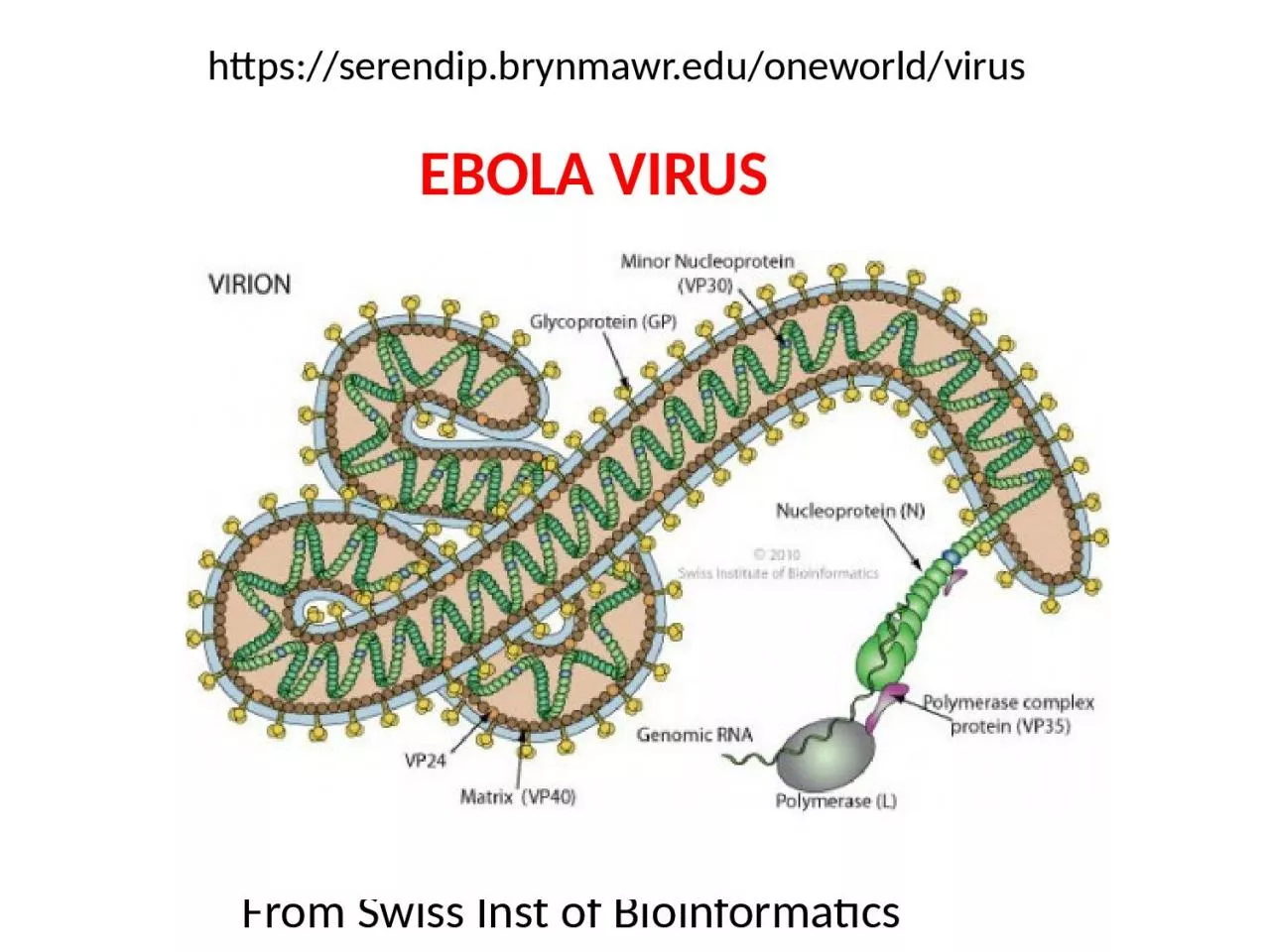
/
From Swiss  Inst  of Bioinformatics From Swiss  Inst  of Bioinformatics

From Swiss Inst of Bioinformatics - PowerPoint Presentation

jordyn
jordyn . @jordyn
Follow
0 views
Uploaded On 2024-03-13

From Swiss Inst of Bioinformatics - PPT Presentation

https serendipbrynmawredu oneworld virus EBOLA VIRUS Ebola Virus Discovery 1976 Inject in mice Electron microscopy Transmission of EBOV and search for reservoir species Microbes and Infection 2005 ID: 1047283

binding ebola amp virus ebola binding virus amp 2014 cell drugs vp35 ebov viral anti stat1 immune binds aug

Share:

Link:

Embed:

Download Presentation from below link

Download Presentation The PPT/PDF document "From Swiss Inst of Bioinformatics" is the property of its rightful owner. Permission is granted to download and print the materials on this web site for personal, non-commercial use only, and to display it on your personal computer provided you do not modify the materials and that you retain all copyright notices contained in the materials. By downloading content from our website, you accept the terms of this agreement.


Presentation Transcript

1. From Swiss Inst of Bioinformaticshttps://serendip.brynmawr.edu/oneworld/virusEBOLA VIRUS

2. Ebola Virus Discovery1976Inject in miceElectron microscopy

3.

4. Transmission of EBOV (and search for reservoir species)Microbes and Infection, 2005,The Natural History of Ebola Virus in AfricaSome infections occurFollowing bat sightings.Related viruses have bat reservoirsExperiments have failed toreproducibly infect a wide varietyof putative reservoir species with EBOV Susceptible to EBOVGorillaChimpanzeeDuiker(laboratory mice)

5.

6. Ghana 2008-2011 SurveyMethodology—surveyed fever patients Central/North Ghana—18 hospitals/285 patientsPCR to amplify EBOV L geneELISA (hepatitis) SequencingNo EbolaMajor illness Hepatitis B & C (adults), A (kids)

7. NE J. Med.4/27/14TEMContact tracing

8. Diagnosis –PCRRT-PCR to find high viral loadsGenome sequencingViral Growth cell culture/FL-anti Ebola AbEMphylogeny

9.

10. Eurosurveillance, Volume 19, Issue 36, 11 September 2014 Rapid communicationsEarly transmission dynamics of Ebola virus disease (EVD), West Africa, March to August 2014 H Nishiura , G ChowellMath modellingTransmission RatesRt need to be<1 to control epidemicRt= number of peopleInfected by each patient

11. Science Express, 28 Aug. 2014

12.

13. Pathogenesis of Ebola-Hartman et al, CDCFilovirusesHemorraghic fever/vascular dyfunctionEbola (Zaire & Sudan)MarburgReston & Ivory Coast (animals)4-10 day incubation period; 50-90% fatalNon-specific initial symptoms (fever, pains, nausea)Virus and antibodies detectable with symptoms

14. Ebola pathogenesisCoagulation connection—TF (tissue factor) levels increased Ebola macrophages engulfed in fibrin but Protein C level decreased (decreases coagulation)Animal fatality reduced by anti-coagulation factor Endothelial leakageshock, hemorrhage Glycoprotein GP1, GP2 connection not clear

15. Ebola pathogeneisFatal patients—bleeding, coagulation defects, high viral levels many organs, low antibody levelsTransmission body fluids, not aerosolEntry—mucous tissue, cuts, common receptor, replicate many cell typesImmunosuppression—enters dendritic (immature immune) cellslymph system. Dendritic cells coordinate innate/adaptive immune response—cytokine signalling (VP35), T-cells, interferonLymphocyte apoptosis

16. Ebola GoalsHistory and GeographyPathogenesisBiochemistry & Molecular Biology (and limitations & unknowns)Nucleic Acids7 ProteinsImplications for understanding biologyTherapeutic implications

17. Annual Rev Genetics, 1998, Conzelman

18. Replication/Transcription

19.

20. “universal” procedure for RT-PCRExtract RNA from 140 uL serum 20 min reverse transcriptase 50°C95°C 5 min10 “precycles” 95°C 5 s, 60-55°C 5s, 72°C 25 sec40 cycles 95°C (denature), 56°C 10 s (anneal), 72°C 10 s (elongate)$$$ equipment/hoursUnder development—10 min test for “field”

21. Advertising e-mail arrived Aug. 8, 2014

22. Comprehensive Functional Analysis of N-Linked Glycans on Ebola Virus GP1—Lenneman et al MBIO Aug. 1, 2014Endosome proteolytic processing

23. Why study GP1/GP2?Viral entry—RBD=receptor binding siteVaccine developmentUnderstand role of conserved glycosylation—Mutate conserved N, Y so GP1 will be expressed –sugarPseudovirus in tissue cultureResultsSugar removal allows protein production, increases viral entry (including macrophages), cell binding, protease susceptibility, cathespin B independence, but decreases binding NPC1 receptor Ca 2+ binds receptor lectins that bind glycans has some reduced binding when glycosylation is removedNear complete sugar removal allows greater anti-sera recognition for GP1 core onlyAuthors suggest GLYCAN SHIELD more important than entry reduction as reason evolutionary conservation.

24. Ebola Vaccine DevelopmentWant anti-GP antibodies (acute infection)Want longer term protection (CD8/T cell/cytokine)Adenovirus can deliver GP, but many are immune to human vectorsChimp adenovirusUse non-replicating form of virusBooster with MVA protects 100% Macaques

25. Nature Medicine, 7 Sept. 2014

26. 10 month challenge with lethal Ebola Dose

27. Ebola Polymerase L associates withTopoisomerase I

28. Topoisomerase IKnown to be important in viral replication/transcriptionStrand breaking is importantInhibition of Top I results in less Ebola replicationEbola changes localization of TopITherapeutic implications??J. Virology Aug. 2013, Takahashi et al

29. L gene—RdRpRNA dependent RNA polymerase2000 amino acidsTwo domainsPolymeraseTranscription factorTemplate is RNPHumans don’t have this enzymeGood therapeutic targetLike HIV RT???Why so little research?

30. Co-factor

31. Potential PPIs for VP35(based on VP35 biochemistry/structure)

32. NMR—solution experimentVerifies that drugs bind.Perturbs chemical shift.Binding pocket mutants haveno chemical shift change

33. Do drugs disrupt NP/VP35-IID interaction? Stop polymerase?Pull down assay withamylose beads.If VP35/IID binds NP,an NP-His band is visible.2 drugs inhibit bindingEbola polymerase complexIncludesEBOV L, VP30, VP35, NPSeveral drugs show doseDependent inhibition

34. Do drugs stop virus?Some drugs reduce viralinfectivityandviral release

35. Ebola Virus Modulates Transforming Growth Factor Signaling andCellular Markers of Mesenchyme-Like Transition in HepatocytesProteomics/Kinomics ApproachHow does EBOV affect global signalling?How are phosphorylation patterns changed?Therapeutic targets?Kindrachuk et al J. Virology September, 2014

36. Kinome Array300 peptide targets in arrayexpose to active kinases in lysateP1P300PEPTIDE TARGET + cognate kinase PEPTIDE TARGET—PPattern of target phosphorylation kinase IDWhat pathways are activated 1 hr, 6 hr, 24 hr?

37. ResultsTGF-b pathway up-regulated (secretion TGF-b and VEGF confirmed by ELISA)Use inhibitors of pathway TGF-b, P13K/AKT, MAPK/EFK, raf, JNK, PKCEarly use of some inhibitorsReduced mouse fatalities

38. EMT (epithelial to mesenchyme transition)TGF-b normal roles—wound healing, cell growth/differentiation, migration, immune responseWhat is happening to EBOV-infected cells?Epithelial tissues first infected—adherens/tight junctions disassembled. Gene expression pattern changes to reduce epithelial and increase mesenchyme cytoskeletal expression

39. Western blots/phosphorylationEMT protein TGF-b pathway P antibodiesArticle error—figure legend switched 8&9

40. EMT—local plus systemic effects

41.

42. From Medical Microbiology 4th ededited by Baron (1996)

43. Interferon protects uninfected cellsby changing gene expression

44. Ebola Virus VP24 Targets a Unique NLS Binding Siteon Karyopherin Alpha 5 to Selectively Competewith Nuclear Import of Phosphorylated STAT1 Xu et al Cell Host & Microbe, 2014

45. Proposed ModelSTAT1 must enter nucleus for interferon responseSTAT1 binds to KPNA for nuclear transporteVP24 binds strongly to KPNAeVP24 competes with STAT1 for same KPNA binding siteeVP24 and STAT1 binding sites overlap and are distinct from binding sites for normal transported moleculeseVP24 prevents normal interferon response by blocking STAT1 transport to nucleus, but does allow entry other molecules

46. Evidence for modelCrystallography structure—shows hydrophobic and H-bondingat interface very strong bindingeVP24/KPNAKPNA can be severely truncatedand still bind eVP24

47. More evidence for modelImmunoprecipitation using anti-FLAG antibodiesTo pull down eVP24 plus “normal” cargo or STAT1WT eVP24 prevents STAT1 but not DMC1 from binding

48. Last evidence for modelAssay tests whether interferoncan induce promoterWT eVP24 reducesExpression by >90%Marburg Virus mVP24has different sequencein key positions anddoes not block STAT1nuclear entry or interferonresponse.