/
Gynaecological  Examination of vagina Gynaecological  Examination of vagina

Gynaecological Examination of vagina - PowerPoint Presentation

kimberly
kimberly . @kimberly
Follow
0 views
Uploaded On 2024-03-15

Gynaecological Examination of vagina - PPT Presentation

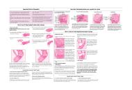
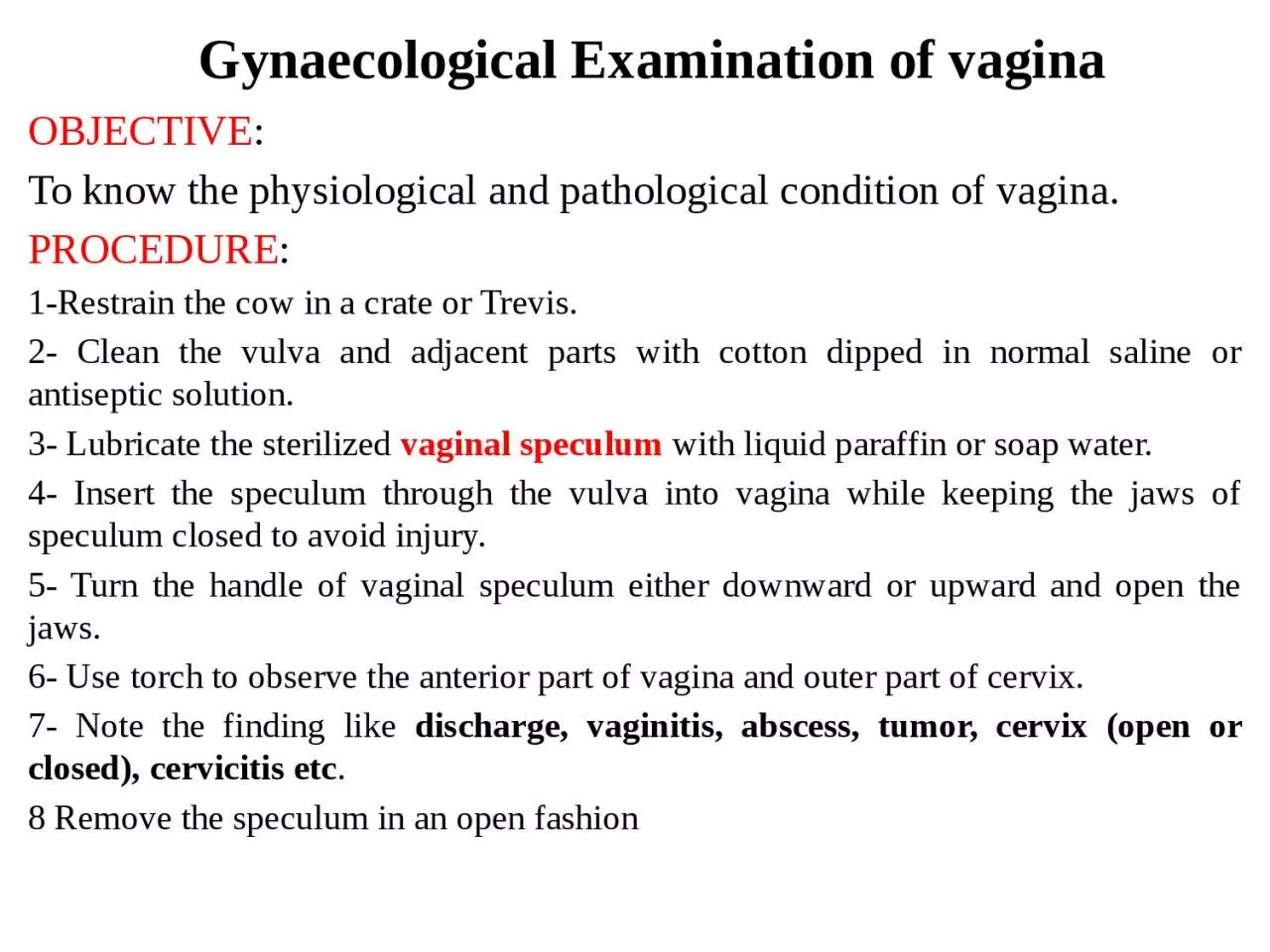
OBJECTIVE To know the physiological and pathological condition of vagina PROCEDURE 1Restrain the cow in a crate or Trevis 2 Clean the vulva and adjacent parts with cotton dipped in normal ID: 1048498

gun cervix vagina vaginal cervix gun vaginal vagina semen uterine speculum external catheter hand insert rectal open examination vulva

Share:

Link:

Embed:

Download Presentation from below link

Download Presentation The PPT/PDF document "Gynaecological Examination of vagina" is the property of its rightful owner. Permission is granted to download and print the materials on this web site for personal, non-commercial use only, and to display it on your personal computer provided you do not modify the materials and that you retain all copyright notices contained in the materials. By downloading content from our website, you accept the terms of this agreement.


Presentation Transcript

1. Gynaecological Examination of vagina OBJECTIVE: To know the physiological and pathological condition of vagina.PROCEDURE:1-Restrain the cow in a crate or Trevis.2- Clean the vulva and adjacent parts with cotton dipped in normal saline or antiseptic solution.3- Lubricate the sterilized vaginal speculum with liquid paraffin or soap water.4- Insert the speculum through the vulva into vagina while keeping the jaws of speculum closed to avoid injury.5- Turn the handle of vaginal speculum either downward or upward and open the jaws.6- Use torch to observe the anterior part of vagina and outer part of cervix.7- Note the finding like discharge, vaginitis, abscess, tumor, cervix (open or closed), cervicitis etc.8 Remove the speculum in an open fashion

2. Technique of Intra-uterine therapyOBJECTIVE: To introduce the drugs into the uterus to overcome the infection in various disease conditions.PROCEDURE:1- Clean vulva and perineal region with dry cotton.2- Insert the left hand in the rectum and remove the fecal material by back racking.4- Spread vulva apart and insert the instrument (catheter or gun apparatus) up to fornix.5- Hold the cervix between two fingers through rectal wall and keep thumb on the external os.6- The catheter is initially inserted pointing upwards at an angle of about 300 to avoid entering into the external urethral opening and is then moved horizontally until it is engaged in the external os of the cervix.

3. Vaginal speculum to ewe vaginal examination Vaginal scope to mare vaginal examination

4. Technique of Intra-uterine therapy7- Entry into the external os is accompanied by a characteristic 'gritty‘ sensation.8 There after, introduce the catheter through convoluted cervical canal by manipulation of the cervix through rectal wall.9- Place one finger over the internal os of the cervix, so that the tip of the catheter can be palpated when it passes the cervical canal10- As soon as, the catheter is passed, the drug should be pushed through syringe into the body of uterus not in uterine horn.11- In this way, drug is equally distributed between the two uterine Horns.

5. Because the rumen displaces the reproductive tract to the right, it is much easier to locate and manipulate the tract with your left hand.The opening into the cervix protrudes back into the vagina

6. The cervix is located on the floor of the pelvic cavity near the anterior end of the pelvic bone.As you insert the breeding gun into the vagina, keep your gloved hand even with the gun tip.

7. Keep your open hand flat against the floor of the rectum,allowing manure to pass over the top of your hand and armTo relax rectal constriction rings, insert two fingers through the center of the ring and massage back and forth

8. Grasp the cervix and push it forward to straighten vaginal folds.Grasp the external opening to the cervix with the thumb on top and the forefingers underneath to close the fornix and guide the gun tip into the cervix

9. Using the flexibility of your wrist, twist and bend the cervix until you feel the second ring slide over the gun tipUse your index finger to check gun placement (1/4 inch past the end of the cervix) before depositing semen.

10. Push the plunger slowly so that drops of semen fall directly into the uterine body.With proper A.I. technique and gun placement, semen will be deposited in the uterine body and contractions will transport spermatozoa forward to the horns and oviducts

11. If the gun is more than 1 inch through the cervix, all the semen will be deposited into only one horn.If you encounter cervical mucous which feels thick and sticky on the gun in a cow that has been previously inseminated, she may be pregnant. In this case, deposit the semen halfway through the cervix

12. Make sure you push in with the plunger and do not pull back on the gun. Pulling back may result in much of the semen dose being deposited in the cervix and vaginaAfter 21 days rectal examination this cow after not return to estrous to confirmation pregnancy