/
Florid cemento osseous  dysplasia a case report Florid cemento osseous  dysplasia a case report

Florid cemento osseous dysplasia a case report - PDF document

kittie-lecroy
kittie-lecroy . @kittie-lecroy
Follow
365 views
Uploaded On 2017-09-04

Florid cemento osseous dysplasia a case report - PPT Presentation

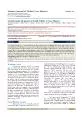
Med Oral Patol Oral Cir Bucal 2007 12E34850 Florid cementoosseous dysplasias Med Oral Patol Oral Cir Bucal 2007 12E34850 Florid cemento ID: 90079

Med Oral Patol Oral Cir

Share:

Link:

Embed:

Download Presentation from below link

Download Pdf The PPT/PDF document "Florid cemento osseous dysplasia a case..." is the property of its rightful owner. Permission is granted to download and print the materials on this web site for personal, non-commercial use only, and to display it on your personal computer provided you do not modify the materials and that you retain all copyright notices contained in the materials. By downloading content from our website, you accept the terms of this agreement.


Presentation Transcript

E348 Med Oral Patol Oral Cir Bucal 2007; 12:E348-50. Florid cemento-osseous dysplasias Med Oral Patol Oral Cir Bucal 2007; 12:E348-50. Florid cemento-osseous dysplasia Florid cemento-osseous dysplasia: A case report Saadettin Da i stan 1 , Ümmühan Tozo lu 2 , Mustafa Göregen essentially, the same pathological process. They are usually classified, depending on their extent and radiographic ap - pearances, into three main groups: periapical (surrounds the periapical region of teeth and are bilateral), florid (sclerotic symmetrical masses) and focal (single lesion) cemental dysplasias. Florid cemento-osseous dysplasia clearly appears to be a form of bone and cemental dysplasia that is limited to jaws. Patients do not have laboratory or radiologic evidence of bone disease in other parts of the skeleton. For the asymptomatic patient, the best management consists of regular recall examinations with prophylaxis and RO Florid cemento-osseous dysplasia is more commonly seen in middle-aged black women (in some series, more than 90 per - cent of patients), although it also may occur in Caucasians and Asians (1). The reason for this racial and gender pre - dilection is unknown (2). In some cases, a familial tendency has been reported, but most examples appear to represent isolated instances (3). The lesions show a marked tendency for bilateral and often quite symmetric involvement, and - lly in the tooth-bearing regions. They are often confined within the alveolar bone (5). Histologically, these lesions are composed of anastomosing bone trabeculae and layers of cementum-like calcifications embedded in a fibroblas - tic background. Management of florid cemento-osseous dysplasia may be difficult and not very satisfactory. The disease may persist for indefinite periods of time without causing any symptoms. For the asymptomatic patient, the best management consists of regular recall examinations with prophylaxis and reinforcement of good home hygiene E349 Med Oral Patol Oral Cir Bucal 2007; 12:E348-50. Florid cemento-osseous dysplasias Med Oral Patol Oral Cir Bucal 2007; 12:E348-50. Florid cemento-osseous dysplasia A 47-year-old Caucasian female presented for dental care. She was systemically healthy and extra-oral examination was within normal limits. Intraoral examination revealed a partially edentulous area and the overlying gingiva and mucosa were normal without any clinical signs of inflam - mation. Panoramic and periapical radiographs were obtai - ned. A biopsy is not required to confirm the diagnosis as this is usually established radiographically. Indeed biopsy increases the risk of infection or fracture of the jaw and it is not normally justified to surgically remove these lesions, as this often requires extensive surgery. In the panoramic radiograph, multiple sclerotic masses with radiolucent bor - ders were found in mandibula (Fig1). Periapical radiographs, periapical regions of lower right first molar and premolar and lower left first molar and premolar teeth, confined within the alveolus at a level corresponding to the roots of the teeth, above the inferior alveolar canal (Fig 2). D Florid cemento-osseous dysplasia was first described by Melrose et al. in 1976 (1).This condition has been interpreted as a dysplastic lesion or developmental anomaly arising in tooth-bearing areas. These lesions exhibit a sclerotic ap - pearance similar to that of other lesions on conventional radiographs. Histopathologically, it is a benign fibro-osseous lesion that must be differentiated from other benign fibro- osseous lesions such as fibrous dysplasia, ossifying fibroma, Paget’s disease, and sclerosing osteomyelitison on the basis of combined clinical, radiographic, and histological features. However, this lesion do not appear to be developmental in nature such as fibrous dysplasia (7), nor do it show the characteristics of neoplasia such as ossifying fibroma (8,9). Paget’s disease of the bone may have a cotton-wool appea - rance. However, this condition affects the bone of the entire mandible and shows loss of lamina dura, whereas florid cemento-osseous dysplasia is centered above the inferior Fig. 1 . In the panoramic radiograph, multiple sclerotic masses with radiolucent borders were found in mandibula. Fig. 2 . Periapical radiographs, periapical regions of lower right first molar and premolar and lower left first molar and premolar teeth, confined within the alveolus at a level corresponding to the roots of the teeth, above the inferior alveolar canal.