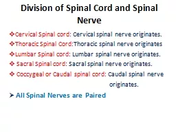
Atlantoaxial system anatomy and pathology Articulation of C1 atlas with C2 axis is complex comprising several joints Median atlantoaxial joint Two lateral atlantoaxial joints These joints are held in place and supported by several ligaments ID: 214258
Download Presentation The PPT/PDF document "Spinal involvement in Morquio A" is the property of its rightful owner. Permission is granted to download and print the materials on this web site for personal, non-commercial use only, and to display it on your personal computer provided you do not modify the materials and that you retain all copyright notices contained in the materials. By downloading content from our website, you accept the terms of this agreement.
Slide1
Spinal involvement in Morquio A Slide2
Atlantoaxial system: anatomy and pathology
Articulation of C1 (atlas) with C2 (axis) is complex, comprising several joints
Median atlantoaxial jointTwo lateral atlantoaxial joints These joints are held in place and supported by several ligamentsMajor stabilizing ligaments are the transverse and alar ligamentsIncompetent ligaments and/or dens hypoplasia may cause excessive independent movement between the C1 anterior arch and the dens to result in atlantoaxial subluxation and instability During flexion, spinal cord compression at the C1-C2 level results from indentation by the C1 posterior arch and posterior tilting of the dens Upward translation of the dens may also result from transverse ligament failureVertical subluxation can lead to compression of the medulla, paralysis and death
Solanki et al, J Inherit Metab Dis, 2013
Competent transverse and alar ligaments maintain the integrity of the C1-C2 articulation by limiting posterior translation of the dens (odontoid process) Slide3
Top image courtesy of Michael Beck, MD, and Christina Lampe, MD
Bottom image courtesy of Christina Lampe, MD
Spinal involvement is a major cause of morbidity and mortality in Morquio A SyndromeSpectrum of spinal involvement:Bony anomaliesCervical spine subluxation and instabilitySpinal canal stenosisSpinal cord compression Spinal problems predispose patients to myelopathy, paralysis, and premature deathSolanki et al, J Inherit Metab Dis, 2013; Montano et al, J Inherit Metab Dis, 2007; Tomatsu et al, Curr Pharm Biotechnol, 2011Slide4
Harmatz et al
, Mol Genet Metab
, 2013MorCAP baseline dataSpinal involvement is common in Morquio An = 325 Morquio A subjects (mean age = 14.5 years)Data based on medical history reviewsSlide5
Dens hypoplasia
PlatyspondylyAnterior beakingPosterior scalloping Thoracolumbar kyphosisSolanki et al, J Inherit Metab Dis, 2013Bony anomalies: Dysostosis multiplexSlide6
Etiology:
dens hypoplasia
ligamentous laxityAtlantoaxial (C1-C2) subluxation:ADI > 5 mm or PADI < 14 mmInstability is present when ADI difference between flexion/extension views > 2 mmSolanki et al, J Inherit Metab Dis, 2013Cervical spine subluxation and instability Risk of cord compression and neurological compromise especially in presence of cervical spinal canal stenosisSlide7
Etiology
Diffuse
stenosis:Generalized thickening of the posterior longitudinal ligament and the ligamentum flavum due to GAG accumulation Most likely to result in compression at C4-C7 and T10-L1Focal stenosis:CCJ: thickening of the membrana tectoria and apical and occipito-atlantal ligamentsC1-C2: thickening of the peri-odontoid tissue and transverse atlantoaxial ligament + C1 posterior archC3-C7: bulging discsThoracolumbar and upper thoracic spine: kyphosisSolanki et al, J Inherit Metab Dis, 2013Spinal canal stenosisSlide8
Etiology:
Thickened ligaments
Cervical instabilityCartilaginous and ligamentous hypertrophy at the C1-C2 jointSpinal canal stenosisDisc protrusionKyphosisSpinal canal stenosis or a combination of stenosis and instability may be predictive of spinal cord compression Spinal stenosis with concomitant loss of CSF flow on MRI signifies spinal cord compressionUntreated cord compression can lead to cord damage and myelopathySolanki et al, J Inherit Metab Dis, 2013Spinal cord compression Slide9
Image courtesy of Kenneth Martin, MD
Diagnostic and monitoring tools:
Neurological examinationImagingRadiographyComputed tomography (CT)Magnetic resonance imaging (MRI)Other diagnostic examinationsFunctional testing (e.g. 6 minute walk test)Sleep studiesUrodynamics Solanki et al, J Inherit Metab Dis, 2013Early recognition and diagnosis of spinal problems can minimize morbidity and mortality Slide10
Neurological examination can identify patients
at early stages of spinal cord compression
Presenting symptoms include loss of endurance, diminished walking distance, gait instability, leg weakness, paresthesia (legs and/or arms)Hyperreflexia, raised muscle tone, pyramidal tract signs (ankle clonus, Babinski sign) and proprioceptive deficits may be observed upon examination Limitations: Morquio A patients may be difficult to assess neurologically due to lower limb joint involvementneurological signs and symptoms may underestimate the severity of spinal cord compression seen on MRIdetermination of the responsible level is challenging in patients with multi-segmental myelopathySolanki et al, J Inherit Metab Dis, 2013Slide11
Images courtesy of Kenneth Martin, MD
Goals of imaging:
Detect treatable spinal cord compressionStratify risk to spinal cord prior to permanent loss of functionAssist in surgical planningAssess efficacy of surgical and medical treatmentSystematic and careful imaging involves:Plain radiography, including instability imagingMRI of the spinal cordCT may be requiredClinical and neurological findings should be correlated with imaging studiesSolanki et al, J Inherit Metab Dis, 2013Imaging is critical for risk assessment and diagnosis of spinal cord compressionSlide12
Strengths
Limitations
Assess bone malformationAssess spinal canal stenosisAssess malalignment
Flexion-extension instability Rapid
Inexpensive
Poor soft tissue discrimination
Limited by overlapping structures
Ionizing radiation
Limited to ossified structures
Radiography
Solanki et al,
J Inherit Metab Dis
, 2013Slide13
Strengths
Limitations
Rapid (may obviate need for anesthesia) Multiplanar imaging of bony structuresAlternative method for assessing flexion-extension instability in difficult cases (recommend low radiation dose protocol) Can assess some soft tissue components of canal
stenosis and cord compression with appropriate filtering
Preoperative planning
Suboptimal for visualizing soft tissues and the spinal cord
Ionizing radiation
More expensive and less accessible than plain film radiography
CT
Solanki
et al,
J Inherit
Metab
Dis
, 2013Slide14
Strengths
Limitations
Multiplanar imaging Ideal for soft tissue imaging Preferred method for assessing spinal cord compression and myelomalacia Flexion-extension imaging directly visualizes spinal cord
Demonstrate venous collaterals
Non-ionizing radiation
Long imaging times
May require anesthesia
Metal and motion artifacts
Limited access
Expensive
MRI
Solanki
et al,
J Inherit
Metab
Dis
, 2013Slide15
Myelomalacia
is diagnosed by an increase in T2 signal coupled with volume loss in regions of cord compressionMRI sequences:T1T2CisternographyCSF FlowDiffusionSpectroscopyMR venographySolanki et al, J Inherit Metab Dis, 2013MRI is the single most useful tool for assessing spinal cord compressionSlide16
Natural history of cord compression
Solanki
et al, Mol Genet Metab, 2012 - Threshold for critical cord compression - Slide17
Assessment
At diagnosis
FrequencyNeurological exam
Yes
6 months
Plain radiography cervical spine (AP, lateral neutral and flexion-extension)
Yes
2-3 years
Plain radiography spine (AP, lateral
thoracolumbar
)
Yes
2-3 years if evidence of kyphosis
or scoliosis
MRI neutral position, whole spine
Yes
1 year
Flexion-extension of cervical spine by MRI
Yes
1-3 years
CT neutral region of interest
Preoperative planning
Solanki et al,
J Inherit
Metab
Dis
, 2013
Regular assessments are recommended
for improved patient outcomesSlide18
White
, Curr Orthop Prac
, 2012Ain et al, Spine, 2006Indications include:Neurological deficits + instabilityCord compression with signal change on MRICervical spine:Posterior fusion for C1-C2 subluxation and instability, often with posterior occipito-cervical fixationIf subluxation is irreducible and cord compression is present, decompression + fusion is indicatedProphylatic fusion recommended by someThoracolumbar kyphosis:Decompression, segmental instrumentation and fusionAnterior discectomy and fusion strongly recommended to augment posterior fusion in cases of rigid kyphosisSolanki et al, J Inherit Metab Dis, 2013; White, Curr
Orthop Prac, 2012; Ain et al, Spine (Phila PA 1976)
, 2006; Ransford et al, J Bone Joint Surg Br, 1996; Lipson, J Bone Joint Surg Am, 1977
Surgical interventionsSlide19
Morquio patient 26 years post-surgery:
complete resolution of
quadriparesis achieved and neurological function maintained 26 years after C1-C2 decompression and stabilization White, J Bone Joint Surg Am, 2009Short-term post-operative outcomes generally goodPossible post-surgical complications:Late instability below fusion site may necessitate multiple fusions Halo pin tract infection → Long-term monitoring is importantLong-term outcomes beyond 5 years are less known – few studiesSolanki et al, J Inherit Metab Dis, 2013; White, J Bone Joint Surg Am, 2009; Ain et al, Spine (Phila PA 1976), 2006; Dalvie et al, J Pediatr Orthop B, 2001; Holte et al, Neuro-Orthopedics,1994; Houten et al, Pediatr Neurosurg, 2011; Lipson, J Bone Joint Surg Am, 1977; Ransford et al,
J Bone Joint Surg Br, 1996; Stevens et al, J Bone Joint Surg Br 1991; Svensson and Aaro, Act Orthop Scand,
1988. Surgical outcomesSlide20
Morquio
A patients are at high risk of anesthesia-related morbidity and mortality due to:
Cervical instability and myelopathyCompromised respiratory functionUpper and lower airway obstructionRestrictive lung diseaseCardiac abnormalitiesAny elective surgery requires:Thorough pre-operative ENT, pulmonary and cardiac evaluationsPre-operative radiological evaluation of the cervical spine Skilled personnel in airway managementSpectrum of airway management equipment Morquio A patients should be managed by experienced anesthesiologists at centers familiar with MPS disordersTheroux et al, Paediatr Anaesth, 2012; Solanki et al, J Inherit Metab Dis, 2013; Walker et al, J Inherit Metab Dis
, 2013; McLaughlin et al, BMC Anesthesiol, 2010; Morgan et al,
Paediatr Anaesth, 2002; Shinhar et al, Arch Otolaryngol Head Neck Surg, 2004;
Belani
et al,
J
Ped
Surg
, 1993; Walker et al,
Anaesthesia
, 1994
Airway and anesthetic management of Morquio A patients presenting for surgery is challenging